DEVELOPMENT AND DISEASE
RESEARCH ARTICLE 2849
Development 136, 2849-2860 (2009) doi:10.1242/dev.035246
Wash functions downstream of Rho and links linear and branched actin nucleation factors Raymond Liu, Maria Teresa Abreu-Blanco, Kevin C. Barry, Elena V. Linardopoulou, Gregory E. Osborn and Susan M. Parkhurst* Wiskott-Aldrich Syndrome (WAS) family proteins are Arp2/3 activators that mediate the branched-actin network formation required for cytoskeletal remodeling, intracellular transport and cell locomotion. Wasp and Scar/WAVE, the two founding members of the family, are regulated by the GTPases Cdc42 and Rac, respectively. By contrast, linear actin nucleators, such as Spire and formins, are regulated by the GTPase Rho. We recently identified a third WAS family member, called Wash, with Arp2/3-mediated actin nucleation activity. We show that Drosophila Wash interacts genetically with Arp2/3, and also functions downstream of Rho1 with Spire and the formin Cappuccino to control actin and microtubule dynamics during Drosophila oogenesis. Wash bundles and crosslinks F-actin and microtubules, is regulated by Rho1, Spire and Arp2/3, and is essential for actin cytoskeleton organization in the egg chamber. Our results establish Wash and Rho as regulators of both linear- and branched-actin networks, and suggest an Arp2/3-mediated mechanism for how cells might coordinately regulate these structures. KEY WORDS: Arp2/3, Rho1 GTPase, Spire, Wash, Wiskott Aldrich Syndrome, Actin nucleation, Drosophila
Division of Basic Sciences, Fred Hutchinson Cancer Research Center, 1100 Fairview Avenue North, Seattle, WA 98109, USA. *Author for correspondence (e-mail:
[email protected]) Accepted June 5 2009
activators, the Wiskott-Aldrich Syndrome (WAS) protein family, has been shown to function downstream of Rho GTPases to mediate the branched-actin network formation required for cytoskeletal remodeling, intracellular transport and cell locomotion (Ben-Yaacov et al., 2001; Campellone et al., 2008; Linardopoulou et al., 2007; Stradal et al., 2004; Takenawa and Suetsugu, 2007; Zallen et al., 2002). WASP and SCAR/WAVE, the two founding subclasses of the family, are activated by the GTPases Cdc42 and Rac, respectively (Stradal et al., 2004; Takenawa and Suetsugu, 2007). Two new WAS subclasses, WASH and WHAMM, have recently been reported (Campellone et al., 2008; Linardopoulou et al., 2007) and have been shown to exhibit Arp2/3-mediated branched nucleation activity. Which GTPases might regulate them, however, is not known. Here, we report that Drosophila Wash functions downstream of Rho1 and interacts with Spire and Capu to regulate actin and microtubule organization during Drosophila oogenesis. We show that Wash nucleates actin in an Arp2/3-dependent manner, and exhibits F-actin and microtubule bundling and crosslinking activity that is regulated by a pathway involving Rho1, Spire and Arp2/3. We find that Wash genetically interacts with Rho1, Capu, Spire and Arp2/3, and is essential for actin cytoskeleton organization during oogenesis. Our results establish Wash and Rho as regulators of both linear- and branched-actin networks, and suggest an Arp2/3-mediated mechanism of cytoskeletal control through which cells might coordinately regulate linear and branched architectures. MATERIALS AND METHODS Fly strains and genetics
Flies were cultured and crossed on yeast-cornmeal-molasses-malt and maintained at 25°C. Alleles used in this study were: wash⌬185, Rho11B, capuEY12344, spirex18, spirRP, wimp and Sop2Q25st. All of these alleles have been previously described (Hudson and Cooley, 2002; Linardopoulou et al., 2007; Magie and Parkhurst, 2005; Parkhurst and Ish-Horowicz, 1991; Rosales-Nieves et al., 2006), except for spirex18, which is described in Fig. S2A in the supplementary material. Fly lines for our RNAi studies were obtained from the VDRC Stock Center (Wash RNAi stocks 24642 and 39769) and were driven with a maternal-Gal4 line (7063; Bloomington Stock Center).
DEVELOPMENT
INTRODUCTION The actin cytoskeleton consists of linear and branched filament networks required for processes ranging from cell division to migration (Chhabra and Higgs, 2007; Faix and Grosse, 2006; Goley and Welch, 2006). How these two networks function and are coordinated is of major interest, as their misregulation results in infertility, immunodeficiency, and tumor metastasis in humans (Bione et al., 1998; Burns et al., 2004; Yamaguchi and Condeelis, 2007). Linear actin filament networks, required for cytokinesis and filopodia formation, are regulated by nucleators and bundling proteins, which enhance filament formation rates and control filament organization, respectively (Chhabra and Higgs, 2007; Faix and Grosse, 2006; Goode and Eck, 2007; Wallar and Alberts, 2003). Examples include Spire and the formin Cappuccino (Capu), which exhibit both nucleation and bundling activities and are essential for oocyte development during Drosophila oogenesis (Chhabra and Higgs, 2007; Kerkhoff, 2006; Manseau and Schupbach, 1989; Quinlan et al., 2005; Quinlan et al., 2007; Rosales-Nieves et al., 2006; Theurkauf, 1994; Wang and Riechmann, 2008). Both Spire and Capu are regulated by the GTPase Rho1 of the Rho family of small GTPases, which is upstream of other linear nucleators, such as Diaphanous, and is considered a key regulator of linear filament formation (Goode and Eck, 2007; Wallar and Alberts, 2003). Branched or dendritic actin filament networks, which are required for phagocytosis and lamellipodia formation, are primarily regulated by the Arp2/3 complex and by nucleationpromoting factors that associate with Arp2/3 and actin monomers to nucleate daughter filaments off of existing mother filaments (Goley and Welch, 2006; Takenawa and Suetsugu, 2007). Like Spire and Capu, Arp2/3 is essential for Drosophila oogenesis, specifically for maintaining proper nurse cell cyto-architecture and function (Hudson and Cooley, 2002). One family of Arp2/3
Development 136 (16)
Microscopy and image analysis
RESULTS Wash activates Arp2/3 to nucleate branched actin, and bundles F-actin and microtubules The mammalian homologs of Wash (WASH), Wasp, Scar/WAVE, and the most recently identified WHAMM subclasses bind Arp2/3 and actin monomers through their C-terminal VCA domains to nucleate actin filaments (Campellone et al., 2008; Linardopoulou et al., 2007; Stradal et al., 2004; Takenawa and Suetsugu, 2007). Using pyrene-actin polymerization assays, we show here that the Drosophila Wash VCA activates Arp2/3 to nucleate actin in vitro, with similar activity to Drosophila Wasp and Scar VCA at equimolar concentrations (Fig. 1A,D). Mammalian homologs of several WAS family proteins have been shown to be regulated by auto-inhibition (Faix and Grosse, 2006; Stradal et al., 2004; Takenawa and Suetsugu, 2007). This does not appear to be the case in flies, as bacterially expressed and purified full-length Drosophila Wash, Wasp and Scar stimulated actin nucleation, requiring molar concentrations of less than threefold that of their VCA fragments to achieve similar levels of activity (Fig. 1B,D). Interestingly, this difference between mammalian and fly actin nucleators has also been observed with the Drosophila formin protein Capu, as it lacks the auto-inhibitory regulation found in its mammalian counterparts (Rosales-Nieves et al., 2006). Drosophila Wash, Wasp and Scar are thus either constitutively active, or inhibited in trans, as has been suggested for Scar and Capu (Quinlan et al., 2007). Wash stimulation of actin nucleation was both concentration and Arp2/3 dependent, and was not affected by the addition of Rho1GTP or Rho1GDP (Fig. 1C; data not shown). Whamy (CG12946; the closest Drosophila homolog of both WHAMM and JMY) VCA did not exhibit actin nucleation activity, which is consistent with the Drosophila homolog lacking the conserved tryptophan residue that is required for mammalian WHAMM nucleation (Campellone et al., 2008) (data not shown). We previously demonstrated that the linear-actin filament nucleators Capu and Spire, in addition to catalyzing actin polymerization, have the ability to directly bundle and crosslink Factin and microtubules (MTs) (Rosales-Nieves et al., 2006). We were interested in determining whether these last-mentioned biochemical properties apply to Wash, and performed in vitro Factin and stabilized MT crosslinking/bundling assays with bacterially purified full-length Wash alongside Wasp, Scar and Whamy for comparison. In the absence of additional proteins, Factin and taxol-stabilized microtubules were not bundled together, but appeared as relatively uniform haystacks of individual short filaments across the field of view (Fig. 1E-E⬙). The addition of Wash bundled these filaments together to form intertwined aggregate strands of F-actin and MTs, with the concomitant loss of the diffuse haystack appearance (Fig. 1F,G). Wash also crosslinks both F-actin and MTs, as shown by the overlap of F-actin and MT bundles in reactions containing both filaments (Fig. 1H-H⬙). In contrast to Wash, we found that Wasp bundles only MTs (Fig. 1I-I⬙), whereas Scar bundles neither (Fig. 1J-J⬙). Whamy bundled MTs and, to a lesser extent, F-actin (Fig. 1K-K⬙), consistent with a recent study showing that mammalian WHAMM binds MTs and G-actin (Campellone et al., 2008). We examined the F-actin and MT bundles mediated by Wash by electron microscopy (Fig. 1L-T). In the absence of Wash, individual F-actin and microtubule filaments were scattered in a random pattern (Fig. 1L-R). The addition of Wash induced these filaments to align parallel to each other and form bundles (Fig. 1M-Q). Cross-linking of these filaments by Wash was also observed (Fig. 1S,T). To quantify the extent of bundling in these assays, we performed low-speed co-sedimentation of the proteins
Immunofluorescence, confocal microscopy, and live imaging of egg chambers were performed as previously described (Rosales-Nieves et al., 2006). Movies 1-5 in the supplementary material are provided at 200⫻ real time; Movie 6 in the supplementary material is provided at 45⫻ real time. Swirling speeds were analyzed with ImageJ by individually tracking the distance traveled per unit time of 10 yolk granules in an active swirling region of mutant oocytes, or in a random region of a wild-type oocyte. Average speeds were calculated for at least three oocytes, and oocytes from capuEY12344 and spirex18/spirRP females were used for comparison. For electron microscopy, 10 μl of an F-actin/microtubule crosslinking reaction was pipetted onto a glow-discharged 200 mesh formvar/carbon-coated grid, washed with 0.1 M cacodylate buffer and water, stained with 1% uranyl acetate, and desiccated overnight. Images were acquired with a JEOL 1230 transmission electron microscope (TEM). Plasmids and constructs
Constructs used in this study have been previously described (Linardopoulou et al., 2007; Rosales-Nieves et al., 2006). Additional constructs used were: Wash FL (full-length Wash, amino acids 1-500), Wash A (amino acids 1-124), Wash B (amino acids 125-212), Wash C (amino acids 213-322), Wash D (amino acids 323-352), Wash E (amino acids 353-500), Wash VCA (amino acids 363-500), Wasp (full-length Wasp, amino acids 1527), Wasp VCA (amino acids 401-527), Scar (full-length Scar, amino acids 1-613), Scar VCA (amino acids 496-613), Whamy VCA (amino acids 419515) and Whamy FL (full-length Whamy, amino acids 1-515). Protein expression and biochemistry
Protein expression, bacterial lysate preparation, protein purification, GST pulldowns, and immunoprecipitations were performed as previously described (Linardopoulou et al., 2007; Rosales-Nieves et al., 2006). For expression and purification of full-length Wash, Wasp, Scar and Whamy, constructs were cloned into a modified ‘double-tag’ pGEX (GE, Piscataway, NJ, USA) vector containing a GST protein epitope tag at the 5⬘ end and a His6 protein epitope tag at the 3⬘ end of the protein of interest within internal PreScission protease (PP) cleavage sites (GST>PP>multiple cloning site>PP>His6). For His-tag and or double-tag purification, supernatant from lysates prepared as previously described (Linardopoulou et al., 2007; Rosales-Nieves et al., 2006) was coupled to Fastflow nickel-sepharose 6 (GE) for 3 hours at 4°C, washed with T100G5 buffer [50 mM Tris (pH 7.6), 100 mM NaCl, 5 mM DTT, 5% glycerol] with 50 mM imidazole, and eluted with T100G5 buffer containing 400 mM to 1M imidazole. For double-tag purification, eluates were then diluted with T100G5 buffer and coupled to glutathione-sepharose 4B (GE) for 3 hours at 4°C, washed with T100 buffer [50 mM Tris (pH 7.6), 100 mM NaCl, 1 mM DTT), treated with PreScission protease (GE) according to manufacturer’s instructions, dialyzed into either buffer A [2 mM Tris (pH 8), 0.1 mM CaCl2, 1 mM DTT, 5% glycerol] or storage buffer [50 mM Tris (pH 7.6], 50 mM NaCl, 1 mM DTT, 5% glycerol], concentrated as necessary using Centricon centrifugal filter devices (Millipore, Billerica, MA, USA), flash frozen and stored at –80°C. Antibody production and characterization
BALB/c BYJ Rb(8.12) 5BNR/J mice (Jackson Laboratories) were immunized with GST-Wash. The P3H3 monoclonal line was generated in the Fred Hutchinson Cancer Research Center (FHCRC) Hybridoma Production Facility as described (Magie and Parkhurst, 2005). Western blotting was used to test antibody specificity against in vitro translated Wash, Wasp and Scar, and for the detection of endogenous protein in whole-cell, nuclear and ovary extracts (see Fig. S3 in the supplementary material). Wild-type Drosophila whole-cell (from 0- to 2-hour embryos) and nuclear (from 0- to 12-hour embryos) extracts were gifts from Toshi Tsukiyama (FHCRC). Actin and microtubule assays
Pyrene actin-polymerization and F-actin/MT-crosslinking assays were performed as previously described (Rosales-Nieves et al., 2006), using a SpectraMax M5 Fluorescence Spectrophotometer (Molecular Devices, Sunnyvale, CA, USA). Actin, MTs and Arp2/3 were obtained from Cytoskeleton (Denver, CO, USA).
DEVELOPMENT
2850 RESEARCH ARTICLE
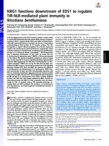
Fig. 1. See next page for legend.
RESEARCH ARTICLE 2851
DEVELOPMENT
Rho1, Spire and Arp2/3 regulate Wash function
Fig. 1. Wash activates Arp2/3 to nucleate actin, and bundles Factin and microtubules. (A-C) Pyrene-actin polymerization assays. Wash, Wasp and Scar VCA fragments (A) and full-length (FL) proteins (B) nucleate actin in the presence of Arp2/3. Wash nucleation activity is concentration dependent and requires Arp2/3 (C). All polymerization assays were performed a minimum of three times. (D) Coomassiestained protein gels of full-length and VCA fragments of Wasp family proteins used in this study. (E-K⬙) Stabilized F-actin (1 μM; top) and MTs (1 μM; middle) were incubated with WAS family proteins and observed by confocal microscopy: (E) no protein added; (F) Wash with actin only; (G) Wash with MTs only; (H-H⬙) Wash; (I-I⬙) Wasp; (J-J⬙) Scar; (K-K⬙) Whamy. (L-T) Electron micrographs of negatively stained samples from the indicated F-actin and MT bundling/crosslinking assays above. Two examples are shown for each assay with Wash (M-T). (U,V) Quantification of F-actin and MT bundling/crosslinking efficiency of indicated reactions by low-speed pelleting assays. The crosslinking/bundling reactions described above were centrifuged to pellet actin and microtubule bundles. Supernatants (S) and pellets (P) were separated and analyzed by Coomassie staining. Percentages of protein, actin and microtubules in each fraction are given. All assays were performed a minimum of three times. Final protein concentrations: Wash, 300 nM; Wasp, 300 nM; Scar, 300 nM; Whamy, 300 nM. Scale bars: 10 μm for E-K⬙; 500 nm for L-T.
following bundling assays (Fig. 1U,V). Unbundled actin and MT filaments did not pellet efficiently (1% and 12% in pellet fractions, respectively) and remained in the supernatant following centrifugation (Fig. 1U, actin+MTs lane). F-actin bundles and MT bundles pelleted efficiently (96-99% actin, 85-99% MT) in the presence of Wash, as did Wash itself when combined with F-actin (94%), MTs (90%), or both (95%; Fig. 1U, Wash lanes). To rule out the possibility that Wash pellets F-actin and MTs by forming aggregates that trap F-actin or MTs, we performed assays on Wash alone, and found that Wash did not pellet in the absence of F-actin or MTs. Similar quantifications were performed for Wasp, Scar and Whamy bundling/crosslinking (Fig. 1V). Members of both linearand branched-actin filament nucleator classes, therefore, can bundle F-actin and MTs. Wash is essential for oogenesis in Drosophila Taken together, our results suggest that Wash participates in both branched-filament (Arp2/3) and linear-filament (bundled F-actin) regulation. To examine the role of these two functions of Wash in vivo, we tested whether the removal of Wash disrupted Arp2/3 and actin/MT bundling-dependent processes during Drosophila oogenesis. It is not possible to completely eliminate maternal wash function, as germ lines homozygous for the washΔ185 mutant allele (Linardopoulou et al., 2007) cannot be generated because of cell inviability (data not shown). Therefore, in order to modulate the level of Wash activity, we used the wimp mutation, which reduces transcription at the wash locus, in trans to the loss-of-function wash allele, thereby mimicking a hypomorphic mutation (female genotype is washΔ185/+; wimp/+; designated reduced wash) (see Fig. S1A,B in the supplementary material) (Parkhurst and Ish-Horowicz, 1991). We found that reduced wash females are sterile, laying eggs with fused dorsal appendages (49%; Fig. 2B,C) or smaller eggs with no dorsal appendages (36%; n=1048; Fig. 2D). Interestingly, the egg phenotypes we observed are reminiscent of phenotypes associated with mutants lacking Spire, Capu or Arp2/3. Fused dorsal appendages are a manifestation of improper
Development 136 (16)
dorsoventral patterning, and are associated with spire and capu mutants, in which premature ooplasmic streaming in the oocyte (described below) results in the mislocalization of polarity markers (Manseau and Schupbach, 1989; Theurkauf, 1994). Small eggs are a phenotype previously associated with Arp2/3 mutants, in which defects in nurse cell cyto-architecture and ring canal formation result in incomplete cytoplasmic transfer from nurse cells to the oocyte (a process called nurse cell dumping), which leads to smaller eggs (Hudson and Cooley, 2002). Our results suggest that Wash plays a role in these two processes. Wash genetically interacts with Rho1 and the linear-actin nucleators Capu and Spire We first examined whether Wash regulates ooplasmic streaming. Up to stage 10 of oogenesis, the oocyte gradually increases in size as nurse cells transport their cytoplasmic content into the oocyte. During stage 10 of development, subcortical arrays of microtubules reorganize along the oocyte periphery, and motor proteins begin to drive the coordinated movement of cytoplasmic material to redistribute the contents, in a process termed ooplasmic streaming. Premature onset of swirling results in mislocalized polarity markers and patterning defects. We previously demonstrated a complex role for Rho1, Capu and Spire in the coordinated regulation of actin and microtubule cytoskeleton dynamics to control the timing of ooplasmic streaming, and proposed that the bundling and crosslinking properties of Capu and Spire are essential for the process (Rosales-Nieves et al., 2006) (see also Discussion and Fig. S2 in the supplementary material). Given that Wash also has F-actin and microtubule bundling activity, we examined stage 7 oocytes from ovaries of reduced wash flies and found that these oocytes exhibited premature ooplasmic streaming compared with wild type (Fig. 2E,F; see also Movies 1 and 2 in the supplementary material). Yolk granules in wild-type oocytes exhibited uncoordinated, saltatory movements (20.9±2 nm/second), whereas the classical swirling mutants capu and spire exhibited higher rates of directed granule movements (165.5±29 nm/second and 41.5±3.2 nm/second, respectively). Yolk granules in reduced wash oocytes exhibited speeds that were almost twice those observed in wild type (38.6±1.7 nm/second, P