International Journal of
Molecular Sciences Article
Valproate Attenuates Endoplasmic Reticulum Stress-Induced Apoptosis in SH-SY5Y Cells via the AKT/GSK3β Signaling Pathway Zhengmao Li 1,† , Fenzan Wu 2,† , Xie Zhang 3 , Yi Chai 4 , Daqing Chen 5 , Yuetao Yang 5 , Kebin Xu 1 , Jiayu Yin 1 , Rui Li 1 , Hongxue Shi 1 , Zhouguang Wang 1 , Xiaokun Li 1,6 , Jian Xiao 1, * and Hongyu Zhang 1, * 1
2 3 4 5 6
* †
Key Laboratory of Biotechnology and Pharmaceutical Engineering, School of Pharmaceutical Sciences, Wenzhou Medical University, Wenzhou 325035, China;
[email protected] (Z.L.);
[email protected] (K.X.);
[email protected] (J.Y.);
[email protected] (R.L.);
[email protected] (H.S.);
[email protected] (Z.W.);
[email protected] (X.L.) Science and Education division, Cixi People’s Hospital, Wenzhou Medical University, Ningbo 315300, China;
[email protected] Ningbo Medical Treatment Center, Li Huili Hospital, Ningbo 315000, China;
[email protected] Department of neurosurgery, The second Affiliated Hospital, Nanchang University, Nanchang 330006, China;
[email protected] Emergency Department, The Second Affiliated Hospital, Wenzhou Medical University, Wenzhou 325035, China;
[email protected] (D.C.);
[email protected] (Y.Y.) Institute of Life Sciences, Wenzhou University, Wenzhou 325035, China Correspondence:
[email protected] (J.X.);
[email protected] (H.Z.); Tel.: +86-577-8577-3087 (J.X. & H.Z.) These authors contributed equally to this work.
Academic Editor: Anthony Lemarié Received: 27 September 2016; Accepted: 27 January 2017; Published: 8 February 2017
Abstract: Endoplasmic reticulum (ER) stress-induced apoptosis plays an important role in a range of neurological disorders, such as neurodegenerative diseases, spinal cord injury, and diabetic neuropathy. Valproate (VPA), a typical antiepileptic drug, is commonly used in the treatment of bipolar disorder and epilepsy. Recently, VPA has been reported to exert neurotrophic effects and promote neurite outgrowth, but its molecular mechanism is still unclear. In the present study, we investigated whether VPA inhibited ER stress and promoted neuroprotection and neuronal restoration in SH-SY5Y cells and in primary rat cortical neurons, respectively, upon exposure to thapsigargin (TG). In SH-SY5Y cells, cell viability was detected by the 3-(4,5-dimethyl-2-thiazolyl)-2,5-diphenyl-2-H-tetrazolium bromide (MTT) assay, and the expression of ER stress-related apoptotic proteins such as glucose-regulated protein (GRP78), C/EBP homologous protein (CHOP), and cleaved caspase-12/-3 were analyzed with Western blot analyses and immunofluorescence assays. To explore the pathway involved in VPA-induced cell proliferation, we also examined p-AKT, GSK3β, p-JNK and MMP-9. Moreover, to detect the effect of VPA in primary cortical neurons, immunofluorescence staining of β-III tubulin and Anti-NeuN was analyzed in primary cultured neurons exposed to TG. Our results demonstrated that VPA administration improved cell viability in cells exposed to TG. In addition, VPA increased the levels of GRP78 and p-AKT and decreased the levels of ATF6, XBP-1, GSK3β, p-JNK and MMP-9. Furthermore, the levels of the ER stress-induced apoptosis response proteins CHOP, cleaved caspase-12 and cleaved caspase-3 were inhibited by VPA treatment. Meanwhile, VPA administration also increased the ratio of Bcl-2/Bax. Moreover, VPA can maintain neurite outgrowth of primary cortical neurons. Collectively, the neurotrophic effect of VPA is related to the inhibition of ER stress-induced apoptosis in SH-SY5Y cells and the maintenance of neuronal growth. Collectively, our results suggested a new approach for the therapeutic function of VPA in neurological disorders and neuroprotection.
Int. J. Mol. Sci. 2017, 18, 315; doi:10.3390/ijms18020315
www.mdpi.com/journal/ijms
Int. J. Mol. Sci. 2017, 18, 315
2 of 16
Keywords: ER stress; valproate; apoptosis; neurological disorders; neurite outgrowth
1. Introduction The endoplasmic reticulum (ER), an important subcellular organelle in eukaryotic cells, is the major site for protein folding, synthesis, trafficking and calcium storage, and it plays essential roles in multiple cellular processes that are required for cell survival and normal cellular functions [1]. Many external factors, such as oxidative stress, protein inclusion bodies, ischemia-reperfusion injury, spinal cord injury, disturbance of calcium homeostasis, and the inhibition of protein glycosylation, can disturb homeostatic ER function, leading to ER stress [2–4]. When the adaptive capacity of ER fails, the misfolded proteins accumulate in the ER lumen, and the unfolded protein response (UPR) is triggered [5]. ER stress triggers an evolutionarily conserved series of signal transduction events, such as enhancing the ability of proteins to fold properly, accelerating protein degradation, and increasing the probability of cell survival [6]. These signaling events aim to attenuate the accumulation of unfolded proteins in the ER. However, either under severe conditions or when ER stress is activated excessively, these events can induce cell death [7]. Several ER stress-associated transcription factors play important roles in regulating ER stress and any related apoptosis. Glucose-regulated protein 78 (GRP78), a member of the heat shock protein (HSP) family, is regarded as the marker of ER stress. It has been reported that GRP78 is released from IRE1 to support protein folding, and the expression GRP78 can be up-regulated by ER stress [8,9]. The activating transcription factors (ATF6) have been deemed to be a sensor of ER stress. When cleaved from the Golgi membrane, ATF6 enhances its localization to the nucleus, in which the transcription of UPR target genes were up-regulated, leading to apoptosis [10]. Activated IRE1 can promote the splicing of X-box-binding protein 1 (XBP-1) messenger RNA (mRNA), and mature XBP-1 promotes the transcription of UPR target genes such as C/EBP homologous protein (CHOP), leading to apoptosis [11]. UPR also results in intracellular calcium release, leading to cell death via caspase-12 and caspase-3 pathways [12]. Although ER stress has been demonstrated to play an important role in neuronal cell death, the correlative mechanism still requires further research. Matrix metalloproteinase-9 (MMP-9) is involved in the stability of the extracellular matrix (ECM). In the brain, MMP-9 is critical for neuronal network remodeling and the integrity of the blood-brain barrier. One study reported that up-regulation of MMP-9 is related to the downstream ERK/JNK pathway [13]. c-Jun N-terminal kinases (JNKs) are a family of protein kinases that play an important role in many neurological disorders. JNKs are involved in apoptosis, neurodegeneration, cell differentiation and proliferation [14]. Mounting evidence suggests that the JNK pathway plays a major role in apoptosis in many cell death paradigms. It was reported that JNK can phosphorylate and directly activate apoptosis-related proteins such as BIM (homologous to BAX) [15]. Huili Zhu et al. reported that liraglutide exerts neuroprotection against ischemia-induced apoptosis through the activation of mitogen-activated protein kinase (MAPK) pathways as well as inhibition of the phosphorylation of c-Jun-N-terminal kinase (JNK) and p38 [16]. Whether Valproate (VPA) can also regulate the expression of MMP-9 and JNK still needs further study. Valproate (VPA), a nonselective histone deacetylase inhibitor, is widely used for the treatment of seizures and bipolar mood disorder. Currently, it has been reported to exert a neuroprotective role in neurological disorders such as Amyotrophic lateral sclerosis (ALS), Parkinson’s disease (PD) and spinal cord injury (SCI) [17–19]. However, whether VPA can protect against apoptosis induced by ER stress (as well as the therapeutic mechanism involved) remains unclear. In addition, Zhang C et al. reported that VPA could be neuroprotective via activation of the extracellular signal-regulated kinase (ERK) and serine/threonine kinase (AKT) signaling pathways after traumatic brain injury (TBI) [20]. The phosphatidylinositol-3 kinase/protein kinase B (PI3K/AKT) signaling pathway is involved in the regulation of cell functions such as proliferation, differentiation, apoptosis, and glucose
Int. J. Mol. Sci. 2017, 18, 315
3 of 16
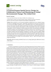
transport [21,22]. When AKT is phosphorylated, glycogen synthase kinase (GSK3β), which is a functional downstream target of AKT, is deactivated [23]. Whether VPA can regulate the AKT/GSK3β signaling pathway to exert a protection in SH-SY5Y cells is still unclear. In the present study, we show that thapsigargin (TG) can cause apoptosis in SH-SY5Y cells via activation of ER stress. Moreover, we identify that VPA can attenuate TG-induced apoptosis in SH-SY5Y cells via suppression of ER stress-related proteins such as CHOP and caspase-12 and -3, activation of the AKT/GSK3β signaling pathway, and inhibition of JNK phosphorylation and MMP-9 expression. In addition, we show that TG can lead to the outgrowth stagnation of primary cortical neurons. However, VPA can maintain the development of neurons. Our findings demonstrate that VPA protects neurons against TG-induced apoptosis via inhibition of ER stress in SH-SY5Y cells, and against TG-induced outgrowth stagnation of primary cortical neurons. Thus, we provide new evidence for VPA in the treatment of neurological disorders. 2. Results 2.1. VPA Reduces TG-Induced Apoptosis in the SH-SY5Y Human Neuroblastoma Cell Line Thapsigargin (TG) is a classic ER stress inducer that can initiate the stress condition in different cells [24]. When cells are exposed to TG, ER stress can be induced through inhibiting Ca2+-ATPase transporters at the ER membrane [25], which results in apoptosis when the degree of endoplasmic reticulum stress is overwhelming [26]. To determine whether VPA can inhibit cell apoptosis in SH-SY5Y cells treated with TG, the MTT assay was used as previously described [4]. After medium replacement, SH-SY5Y cells were treated with VPA 2 h prior to the administration of TG (Figure 1A). Compared with the control group, cell viability in the TG group was significantly decreased, while cell viability in the TG and VPA group was increased compared to the TG group, no change was seen in the VPA group compared to the control group (Figure 1B; p < 0.05). To investigate whether TG affects ER stress, we detected the expression of GRP78, the maker of ER stress. In the TG group, the expression of GRP78 was increased significantly. This result suggested that TG can efficiently cause ER stress. However, the expression of GRP78 was also increased in the TG and VPA group (Figure 2A,B; p < 0.05). Immunofluorescence microscopy was used to assess the expression of GRP78. Similar to the aforementioned results, the optical density of GRP78 was increased in the TG group, and the optical density of GRP78 in the TG and VPA group was also elevated (Figure 2C). These results suggest that VPA may maintain the degree of ER stress and enhance the ability of proteins to fold properly in the initial stage, thereby exerting a neuroprotection against cell death induced by TG in human SH-SY5Y cells. 2.2. VPA Attenuates TG-Induced ER Stress in SH-SY5Y Cells To evaluate the effect of VPA against ER stress induced by TG in SH-SY5Y cells, Western blot analysis was used to quantify the levels of the ER stress-associated proteins ATF6 and XBP-1. The levels of ATF6 and XBP-1 were significantly increased in the TG group compared to the control group. In the TG and VPA group, the levels of ATF6 (p < 0.01) and XBP-1 were reduced compared to the TG group (Figure 3A–D). Immunofluorescence microscopy was used to assess the level of XBP-1. Similar to the Western blot results, the optical density of XBP-1 was up-regulated in the TG group. However, compared with the TG group, the optical density of XBP-1 in the TG and VPA group was down-regulated (Figure 3E). These results demonstrated that VPA can efficiently reduce ER stress induced by TG in SH-SY5Y cells.
Int. J. Mol. Sci. 2017, 18, 315
4 of 16
Int. J. Mol. Sci. 2017, 18, 315
4 of 17
Int. J. Mol. Sci. 2017, 18, 315
4 of 17
1. Valproate (VPA) reduces thapsigargin (TG)-induced apoptosis in SH-SY5Y cells. (A) The FigureFigure 1. Valproate (VPA) reduces thapsigargin (TG)-induced apoptosis in SH-SY5Y cells. (A) The protocol cell viability assay performed in this(TG)-induced study. Cells were divided into fourcells. groups: the Figure 1.ofValproate (VPA) reduces thapsigargin apoptosis in SH-SY5Y (A) The protocol of cell viability assay performed in this study. Cells were divided into four groups: the control control TG group; TG and in VPA VPA group; (B) into MTTfour assay result the of protocolgroup; of cellthe viability assay the performed thisgroup; study. the Cells were divided groups: group; the TG group; the TG and VPA group; the VPA group; (B) MTT assay result of VPA-treated VPA-treated SH-SY5Y exposed to and TG. *VPA p < 0.05 versus group,(B) n =MTT 6; (C) Induction control group; the TGcells group; the TG group; the the VPATGgroup; assay result of of SH-SY5Y cells exposed toSH-SY5Y TG. * pcells < 0.05 versus the TG group, n = 6; (C) Induction of apoptosis in apoptosis in human was byversus flow cytometry after treatment TG, TG VPA-treated SH-SY5Y cells exposed to determined TG. * p < 0.05 the TG group, n = 6; (C) with Induction of humanand SH-SY5Y cells was determined by flow cytometry after treatment with TG, TG and VPA, and VPA, in and VPA; SH-SY5Y (D) The percentage of apoptoticbycells the treatment waswith calculated, apoptosis human cells was determined flowincytometry after groups treatment TG, TG VPA; (D) percentage of cells treatment was calculated, ** p < 0.01 versus ** pThe