DRAFT Applications of open source frameworks for advanced medical image processing Polina Mihova 1, Georgi Petrov 2, Filip Andonov 3 1
Health
and
social
work
Department,
New
Bulgarian
University
Bulgaria, Sofia 1618, Ovcha Kupel 2, Montevideo 21 Str., NBU ll.2, fl.2, office 210 2
Telecommunication
Department,
New
Bulgarian
University
Bulgaria, Sofia 1618, Ovcha Kupel 2, Montevideo 21 Str., NBU ll.2, fl.6, office 609 3
Informatic
Department,
New
Bulgarian
University
Bulgaria, Sofia 1618, Ovcha Kupel 2, Montevideo 21 Str., NBU ll.2, fl.6, office 610 1
[email protected] ; 2
[email protected] ;
[email protected] 3
Acknowledgments Curent work was implemented using MIT Pydicom pure python package for working with DICOM
files,
and
analysed
Medical
Image
Samples
were
downloaded
http://www.barre.nom.fr/. We express our appreciation to d-r Turchev for providing dental archival material.
from
Abstract This publication focuses on the opportunities for application of open software libraries for processing medical images stored in DICOM format, and in particular is presented solution for application of three-dimensional histograms (3D) of image sequences to create methods for rapid pretreatment comparison. The method, described here, uses an original algorithm for multidimensional non-parametric statistical analysis of semantically related images used in security systems and video processing. Authors focus on specific method features and the necessary changes in the algorithm, which allow to be used in the field of medical image processing. Keywords: 3D image histograms; DICOM; pydicom; medical image comparrison Introduction The widespread use of digital imaging systems in medicine created conditions for accumulation of huge amount of patient records and related information. Simultaneously, the development and introduction of Digital Imaging and Communications in Medicine (DICOM) [1] standard allow essential lowering the cost of medical images transfer, which facilitated and contributed to weakening and improving the quality of medical services. Substantial progress has been made in the development of algorithms for reconstruction, processing and analysis of medical images related to the discovery of diseases in their early stages. Despite that, it should be noted that most hospitals nevertheless growing information infrastructure archived these records in classic file servers, which hampers it`s future, when medical experts need to exchange good practices in the treatment and diagnosis of disease between hospitals and medical experts. Development of closed systems for processing and storage of such medical records is extremely expensive and proved ineffective due to the inability of doctors to use the latest methods and techniques of digital processing and computer-assisted analysis of medical images. That is why a number of leading universities in the world created frameworks were integrated solutions enable the realization of a systems approach for processing and analysis of medical images.
Short literature review For the realization of this example, we selected to use of three advanced open source software systems, enabling rapid application development, which are widespreaded among leading universities in the field of information and communication technologies.
Python Python is a modern general-purpose and high-level scripting programming language. It was founded on the philosophy of readability, elegance and minimalism of the code. The syntax of Python allows implementation of algorithms with less amount of written code than in C ++, for example. Python is fully object-oriented, imperative language with dynamic types and automatic memory management. It has a huge standard library. It is important to note that the reference implementation of Python - Cpython e free open source software. Some large organizations use Python: Google, Yahoo !, CERN, NASA, Industrial Light and Magic. There are numerous libraries, allowing the use of Python in scientific calculations NumPy, SciPy, Matplotlib. In many OS Python is a standard component and can be used from the command line. pydicom Pydicom is pure python library for reading DICOM files and their attributes. The library provides integration with various reproduction programs and packages as matplotlib, Python Imaging Library and Tkinter. Especially important parameter of the language is the shortest time for absorption by non-specialists. SimpleCV SimpleCV is open source framework code, designed to build applications for computer vision. It provides easy access to powerful libraries such as OpenCV. Purpose of the paper In connection with the need to develop systems for automated comparative analysis of different patient records, combined with the necessity to derive knowledge related to the treatment of patients and the exchange of good practices, here we offer implementation of an original model [2] for multidimensional probabilistic statistical processing of semantically
related sequences of images, developed for automated segmentation and motion systems analysis in digital video. The application of 2D histogram of adjacent pixels in each image is often used method for comparing and finding similarities between digital photos. The modification of the algorithm applies 3D histograms of adjacent pixels of the series semantic prototypical image sequences. As usual 3D medical images, deriving or obtained from MRI, CT, US, etc. can be viewed as sequences of semantically related video frames, a similar method can be used to detect similarities and fast pre-processing and comparison of biomedical images stored in DICOM file format. According to the specificity of medical file formats and large amounts of data associated with the requirement for greater dynamic of the signal, some major changes in the original processing algorithm will be discussed. We have provided initial experimental results and discussed some important areas of the method’s application. Material and methods In practice there is huge amount of analytical methods and automated segmentation of movement (methods for detection of changes), used in systems for massive video processing. One basic characteristic of the proposed model is that through the use of 2D and 3D histograms, it can be enclosed to block any kind of segmentation of digital data represented as 2D and 3D matrices. Non-parametric approach uses well known in practice criteria: Kolmogorov, Pearson and complex criterion, which are enough reliable to detect minor changes and differences between two randomly selected images for comparison. We believe that the method of 3D probabilistic statistical analysis and segmentation of movement can be effectively implemented and successfully used in systems for automated comparison and preliminary processing of large amounts of biomedical images, stored in DICOM file format. This process may precede from the application of methods for data mining and knowledge in various healthcare institutions, training and exchange of good practice between physicians and between engineers, associated with the development of biomedical information systems. Systems for massive video processing, installation and automatic segmentation of videos are optimized for fast processing of large information amounts, but they are mainly focused on the processing of video images with low brightness dynamics (8 bit RGB images mostly). Simultaneously, these systems suffer from some significant disadvantages, which are
generally the lack of precision and the presence of errors between the original image and already compressed footage, making them inapplicable directly to the needs of medical image processing. The base of the model, proposed here, is using 2D histograms related pairs of pixels in the images, which is well described in the cited below series of publications [3], [4], [5]. The basis of the method is related to the use of a variety of known metrics for comparing statistical data, obtained for an image to the same calculated statistics of another image (ie. 2D and 3D histograms). The application of non-parametric method, unlike other methods, such as fast Fourier and Wavelet transformation allows easy adaptability of the proposed algorithms without imposing specific restrictions on data that they can be applied on. Discussion The implementation of such a model in the digital processing, segmentation and analysis of medical images is meeting several problems that do not allow direct implementation. 1. Dynamics of medical images varies based on the system type through which are made
and the quality of incision series. That does not allow to apply processing standard full-coulor images and videos. Also, it doesn’t allow the direct use of algorithms to process standard digital photo and video images, as they bring a lot of excessive noise and error in the processed images. At the same time it should be maintained a high level of understanding of the effects of certain treatment methods, their areas of application and the results, obtained with them by medical personnel. Management system should resemble the activities that a person does for manual collation and analysis of images. 2. The medical images vary and with respect to the spatial resolution 256x256 or 512x512,
proposing even higher resolutions. Unlike the multimedia information, in medical imaging is important only the object of study, which requires to be made precise object segmentation separation only of the patient's body from the rest of the background image and removing artefacts, caused by concrete performance study (Fig. 1.) Many of the modern automated equipment provides a function to eliminate these artefacts, but this is not always possible with conventional X-ray machines.
Fig. 1 Artefacts of X-ray induced mechanical
parts fixing the position of the patient.
3.
Series
image
records,
used
in
3D
reconstruction, are not comparable between patients due to purely physiological differences between models of research, who have their own specificity and the results, obtained for one patient, although it will have statistical determinativeness, can not be directly compared with data for another. I.e., the presented above method of comparing directly series of multidimensional histograms for different patients, made with different devices, is directly applicable. The next challenge is to develop methods for results normalization, concerning the series of images to make them comparable with results from the same patient, made by another system. More serious is the problem, concerning the collation of data for many patients. 4.
Owing to most medical images (above 12-14 bit resolution) the output irregular
volume statistics will exceed several times the volume of the particular image, which creates problems with the processing speed – solution is described in [6] and additionally, several problems, related to the minimization of these data, where can be used multiple entropy segmentation before subsequent analysis are presented in citing [7]. Fig. 2 is a 3D histogram of the image of Fig. 1, where, in order to minimize the data, is made a gross normalization of luminance to 256, and the effective dynamics of the signal is above 2048 levels. Without minimizing data, the direct implementation of the method
would
generate
over
64GB
probabilistic statistical information for each image size and resolution 512x512 with 16 bits per pixel, which is untenable.
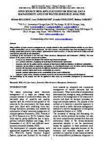
Fig. 2 3D histogram of Fig.1, can be seen in those areas regarding the outline of the object from the background, and image artifacts
Results and possible applications of the proposed model Firstly, the most appropriate application of the proposed multidimensional statistical probability model for processing and analysis of medical images is to trace disease dynamics, because the method allows observation of those areas of the image, that hasn’t yet clearly changed from recordings, made at various treatment stages. Possible problems here would occur by changing the geometry of the internal and external organs of the body after surgery, but they could be fixed by software methods for manual adjustment of images. Generally, the method would allow to track hardly noticeable changes in detail as in periodic monitoring of rise and dental implants or other implants, where it is important to follow the degree of scarring or the absorption by the body. The method also would find appropriate use in comparison to prior segmented 3D models of tissues, organs and systems, while can not be applied directly, since the current approach ignores some of the details. The method can very well be applied for comparative analysis of images, relating to the same patient in the treatment process, thereby reliably could be compared with the entries made by variety of apparatus. Often, in the process of treatment, patients visit different hospitals and even in the same hospital can be made 3D recordings with different techniques. This poses some problems, connected to the opportunities for objectively comparison of differences between these records over the time. By the proposed model using 3D histograms and Kolmogorov criteria, we reliably could define the dynamics of change between the individual pictures. The main objective of suggested methods is to support the work of the medical expert in searching and comparison of object content from multiple DICOM images. Modification techniques for multiple entropy segmentation Entropy is a well known measure, used in the analysis and comparing images. The application of multiple segmented entropy functions at the same image, finds realization and implementations, thus can be achieved sufficiently objective minimization of data without significant information loss. This model makes possible one medical image with very high dynamics to submit and process image with far less performance but better segmentation (Fig. 3.) Such techniques may include pure histogram groupings, because they are equally
applicable, both for the entire image, as well as locally to specific areas of it. It should be noted that the computer analysis could only be used as a supporting factor in the medical experts work and diagnosis.
Fig. 3 segmentation of
magnetic
resonance image of the head - 1, segmented image recognition probability distribution of pixels of different authorities - 2 3D segmentation by entropy mask, histogram of the image - 1 and the corresponding no matches in the probability distribution of the various bodies and pre- estimated average statistical deviation - A, B, C, D, E
Complex methodology for improved visualisation As another example of the application of open software products it could be pointed the realisation of a complex methodology for processing and coloration of black and white images. Under this approach, the perception of images with high dynamics is preserved, as they are pre-treated and coloured. The actual colouring can be done with so-called Correlation tables, where it is known what grey-scale possess a concrete object and all the
objects in the original image, which correspond most likely to it can be shown in certain colour from a pre-selected range or dynamically. The second image is processed by grouping in it’s 3D histogram, thus the homogeneous areas are better highlighted.
Fig. 4. Original coloured grayscale image and pre-processed image by grouping homogeneous zones with 3D histogram (the effect is similar to that obtained by filtration local mods, but it is globally applicable
Conclusions The presented experiments show that the application of the modified methods for 3D histogram processing of medical images can be used in the process of comparing medical records and following disease progress, and particularly where one of the objects – for example implant, should not tolerate changes in their physical parameters and size. The implementation of the method, however, encounter a number of problems, which are mainly related to the need for very serious computing system in terms of nominal volumes memory, which can equally well be used for analysis and comparison of medical images with different dynamics and spatial resolution. Despite these limitations, we offer a technique, known as entropy segmentation of multiple individual images, which aims to minimize the preliminary data set prior to the accumulation of statistical information, concerning specific study, comparison or experiment. At the same time, the different techniques for segmentation of medical images, widely used today, give radically different results and can be used equally well by untrained and non-medical staff. At present, the method application could be limited to the study and implementation of number of experimental and comparative analysis for plurality of images, relating to situations requiring tracking adaptation of the organism to the prostheses of variable geometry, whose basic form is 100% pre-known. The additional field research should be sought broad form of cooperation with leading medical organizations and clinics in the field of imaging, thus will increase the capacity of
specialists from both areas in the concrete implementation of open software tools for computer assisted processing and analysis of medical images. References [1]. RNJ Graham, RW Perriss, AF Scarsbrook, " DICOM Demystified: A Review of digital file
formats and Their Use in Radiological practice ", Clinical Radiology (2005) 60, 1133-1140 [2]. P. Iliev, P. Tzvetkov, G.Petrov, "Motion Detection Using 3D Image Histogram Sequence Analysis", IEEE International Workshop on Intelligent Da TA Acquisition and Advanced Computing Systems: Technology and Applications, 5-7 September 2005, Sofia, Bulgaria ISBN 0-7803-9446-1, Library of Congress 2005931175 [3]. L. Cinque, G. Ciocca b, S. Levialdi a, A. Pellicano a, R. Schettini, Color-based image retrie val using spatial-chromatic histograms, Image and Vision Computing 19 (2001) 979 ± 986, Elsevier [4]. G.Petrov, P. Iliev, P. Tzvetkov, Global Histogram Comparison of Methods for 2D and 3D Entropy Based Image Segmentation 9th WSEAS International Conference on Computing utionary Evol (EC '08), Sofia, Bulgaria, May 2-4, 2008 [5]. K. Koonsanit, C. Jaruskulchai, A. Eiumnoh, A Simple Anomaly Detection for Spectral Imagery Using Co-occurrence Statistics Techniques, International Journal of Computer and Electrical Engineering, V OL. 4, No. 4, August 2012 [6]. AD Bosakova-Ardenska, LD Bosakov, "Performace of mean filter on different NVIDIA platforms", Scientific works 2012 - food science, engeneering and technologies [7]. Collignon, Andr é, et al. "Automated Multi-modality image regis tration Based on information Theory. " Information Processing in Medical Imaging . Vol. 3. No. 6. 1995.