Asian Pacific Journal of Cancer Prevention, Vol 14, 2013 7433 ... Nasopharyngeal carcinoma (NPC) is a rare tumor in most countries. However, it is very ...
DOI:http://dx.doi.org/10.7314/APJCP.2013.14.12.7433 EA-D p45-IgG as a Potential Biomarker for Nasopharyngeal Carcinoma Diagnosis
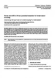
RESEARCH ARTICLE EA-D p45-IgG as a Potential Biomarker for Nasopharyngeal Carcinoma Diagnosis Hao Chen1&,Yao-Ling Luo1&, Lin Zhang1, Li-Zhen Tian2, Zhi-Ting Feng3, WanLi Liu1* Abstract Aim: To identify new biomarkers for NPC diagnosis with an anti-EBV Western blot test kit. Methods: Serum samples from 64 NPC patients and healthy subjects with four specific VCA-IgA/EA-IgA profiles were tested with an anti-EBV Western blot test kit from EUROIMMUN AG. Proteins were quantified with scores of intensity visually assigned to the protein bands. The markers which showed statistical differences between the NPC and non-NPC subjects were further evaluated in another 32 NPC patients and 32 controls in comparison with established biomarkers including VCA-IgA, EA-IgA, EBV-related protein IgG, and EBV DNA. Results: Among the markers screened, EA-D p45-IgG showed a statistically significant difference (p < 0.05) between NPC and non-NPC subjects with VCA-IgA positivy. In 32 VCA-IgA positive NPC patients and 32 control subjects, the diagnostic accuracy of EA-D p45-IgG was 78.1% with a positive predictive value of 77.8% and a negative predictive value of 78.6%. In the verification experiment, the specificity and sensitivity of EA-D p45-IgG were 75.0% and 90.6 %, respectively. Conclusions: EA-D p45-IgG might be a potential biomarker for NPC diagnosis, especially among VCA-IgA positive subjects. Keywords: EBV - NPC - Western blot - viral capsid antigen - early antigen - EBV nuclear antigen - diagnosis Asian Pac J Cancer Prev, 14 (12), 7433-7438
Introduction Nasopharyngeal carcinoma (NPC) is a rare tumor in most countries. However, it is very common in southern regions of China, especially in Guangdong Province, with the prevalence of 10 to 30 per 100 000 population (Su et al., 2002; Yau et al., 2006; Cao et al., 2011; Wei et al., 2011). Diagnosis in the early stage is not easy because of its anatomical location and mild early symptoms (Seong et al., 2000; Lin et al., 2003). Since treatments at late stages are often less effective (Yau et al., 2006) early diagnosis of NPC is critical for achieving good treatment results (Ji et al., 2011). One important feature of NPC is that it is associated with Epstein-Barr virus (EBV) infection. The majority of NPC patients have a variety of EBV antigens, and anti-EBV antibody serological testing has become an important tool for NPC diagnosis (Henle et al., 1970; Henle et al., 1973; Ho et al., 1976; Lynn et al., 1985; Brooks et al., 1992; Cochet et al., 1993; Ng et al., 2006; Ai et al., 2012). Traditional assays of anti-EBV antibodies have been very useful in clinical diagnosis of NPC, however, high false-positive rates may occur in endemic areas (Chang et al., 2008). VCA-IgA and EA-IgA antibody tests are most
commonly used in clinical diagnosis and screening of NPC (Henle et al., 1973; De et al., 1988; Zong et al., 1992). However, the lack of specificity of the VCA-IgA assay and sensitivity of the EA-IgA assay limits their effectiveness in NPC diagnosis. For example, while more than 90% of NPC patients are VCA-IgA positive (Cao et al., 2006) 90% of people who are VCA-IgA positive do not have NPC (Zhang et al., 2002). VCA-IgA positive patients often need further examinations such as the CT/MR review which is quite costly. Thus the suboptimal performance of the traditional biomarker assays affect the accuracy and cost of NPC diagnosis. EBV DNA is considered to be a state-of-the-art quantitative blood biomarker in current NPC research. However, it has been reported that EBV DNA load is independent of serological parameters and does not reflect the number of intact tumor cells (Sheen et al., 1998; Lin et al., 2001). In addition, EBV DNA load quantified is expensive. Therefore, the EBV DNA load as a primary screening assay for the diagnosis of NPC is limited (Stevens et al., 2005). In our study, we detected the EBV-related proteins with a western blot test kit, determined the scores of each test strip according to the color depth (Paramita et
Department of Clinical Laboratory, Sun Yat-sen University Cancer Center; State Key Laboratory of Oncology in South China; Collaborative Innovation Center for Cancer Medicine, 2Department of Clinical Laboratory, Guangzhou Drug Administration Hospital, 3First Affiliated Hospital of Guangdong College of Pharmacy, Guangzhou, Guangdong, China &Equal contributors *For correspondence: liuwl @sysucc.org.cn 1
Asian Pacific Journal of Cancer Prevention, Vol 14, 2013
7433
Hao Chen et al
Table 1. Scores of Some Representative Samples Sample DP-NPC 16 DP-NPC 15 DP-NPC 14 SP-NPC 16 SP-NPC 15 SP-NPC 14 SP-NON-NPC 16 SP-NON-NPC 15 SP-NON-NPC 14 DN-NOR 16 DN-NOR 15 DN-NOR 14
p22
p27
p33
p40,41,42
1 4 0 0 4 1 1 4 1 2 3 0 1 3 0 1 3 0 0 0 0 0 1 0 0 0 0 0 0 0 3 0 0 0 3 0
p43
p45
p65
p79
p93
p125
3 3 2 2 4 1 1 2 2 3 2 2 0 1 3 3 1 1 4 1 2 2 2 0 1 0 0 0 0 0 1 1 0 1 0 2 2 3 1 0 0 0 1 0 2 0 0 1 0 1 0 0 0 1 1 0 2 1 1 0 0 1 0 0 0 0 1 0 0 0 1 0 0 1 0 0 0 1 1 1 0 1 0 0
VCA-p22, VCA-p33, VCA-p65, VCA-p125, VCA-p42, VCA-p41, and VCA-p40 as VCA makers, EA-R p93, EA-D p45, and EA-D p43 as EA markers, and p79 as EBNA1 marker and p27 for further antigens
7434
Asian Pacific Journal of Cancer Prevention, Vol 14, 2013
Anti-EBV Western Blot (IgG) Test Assay EBV-IgG was measured using anti-EBV western blot (IgG) test kit from EUROIMMUN AG, Lübeck, Germany (lot numbers of s100520cv-27, s100520cv-28, and s100621cv-75). The EUROIMMUN anti-EBV western blot test strip is an EBV whole extract including VCA-p22, VCA-p33, VCA-p65, VCA-p125, VCA-p42, VCA-p41, and VCA-p40 as VCA makers, EA-R p93,
30.0
51.1 30.0
30.0
33.1
Chemotherapy
Remission
Persistence or recurrence
Newly diagnosed with treatment
0 Real-Time Quantitative PCR Real-time quantitative PCR of EBV DNA was carried out at SunYat-sen University Cancer Center (Lo et al., 1999). The real-time quantitative PCR system was developed for EBV DNA detection toward the BamHI-W region. The system consisted of the amplification primers W-44F (5’-AGT CTC TGC CTC CAG GCA-3’) and W-119R (5’-ACA GAG GGCCTGTCCACC G-3’) and the dual-labeled fluorescent probe W-67T (5’-[FAM] CACTGTCTGTAAAGTCCAGCCTCC [TAMRA]-3’). Sequence data for the EBV genome were obtained from the GenBank sequence data base. Newly diagnosed without treatment
Specimen Collection Sera samples were collected from 64 NPC patients and healthy subjects (HS) with four specific VCA-IgA /EAIgA profiles (16 NPC patients with VCA-IgA positive and EA-IgA positive, 16 NPC patients with VCA-IgA positive and EA-IgA negative, 16 HS with VCA-IgA positive and EA-IgA negative, and 16 HS with VCA-IgA negative and EA-IgA negative). In addition, sera and plasma samples were collected from 32 patients with newly diagnosed NPC, eight subjects with other tumors, and 24 healthy subjects to verify the screened markers. The ratio of males to females in each group was 3:1, with ages from 23 to 76, at an average of 38. Among the 32 newly diagnosed NPC patients, there were 26 cases of undifferentiated non-keratinizing carcinoma and six cases of differentiated non-keratinizing carcinoma. In accordance with the clinical nasopharyngeal ‘92 Fuzhou staging, the patients were classified as one case of stage I, two cases of stage II, 26 cases of stage III, and three cases of stage IV. NPC Patients were diagnosed at SunYat-sen University Cancer Center from October 2010 to October 2011.
12.8
None
The patient samples were taken after the diagnosis was confirmed but before treatment was received. All diagnoses of NPC had been confirmed by biopsy, and the patients had undergone routine check-ups including head and neck MRIs and chest x-rays. The healthy subjects had undergone routine health examination. Healthy subjects with VCA-IgA positive did not develop NPC during the 3 years after the VCA-IgA test. Venous blood was collected and the sera samples were separated by centrifugation. The samples were stored at Figure 1. The Results of Some Samples Detected by 100.0–80 °C for later testing. Anti-EBV Western Blot (IgG) Test Assay Evaluation 6.3 10.1 20.3 Protocol: A membrane strip of the same lot was incubated Immunoenzymatic Assays with a positive reference serum in every test kit. Each protein 25.0 antibodies band was visually assigned a score of 0, 1, 2, 3, or 4 according75.0 Serological tests for VCA-IgA and EA-IgA were taken with IEA (Yi et al., 1980) method supplied to their intensity 46.8of Biological Products. IEA were by Shanghai 56.3 Institute al., 2008), and compared scores of the VCA-IgG, EAprepared from B95 cell line 54.2 for VCA, and Raji cell line IgG, and EBNA1-IgG antibodies in four subject groups 50.0for EA. Plasma were screened at a dilution 31.3 of 1:10, and with specific VCA-IgA/EA-IgA profiles. The results followed by two-fold serial dilutions. The antibody titer demonstrate that EA-D p45-IgG is a potential biomarker was the reciprocal of the highest dilution clearly showing for NPC diagnosis. 25.0brown color within 15% of the cells. Levels of VCA IgA 38.0 and EA-IgA with the cut-off 31.3were determined by titration, 31.3 23.7 and 1:10 for EA-IgA. values setting at 1:40 for VCA-IgA Materials and Methods
DOI:http://dx.doi.org/10.7314/APJCP.2013.14.12.7433 EA-D p45-IgG as a Potential Biomarker for Nasopharyngeal Carcinoma Diagnosis
Table 2. Scores of the Six Markers (IgG) in the Four Study Groups
Index
DP-NPC (χ±SD)
Total 16.31±5.88 EA 6.44±2.83 EA+EBNA 7.75±3.44 VCA 7.88±2.25 p45 2.88±1.26 p43 2.81±1.33
SP-NPC SP-NON-NPC DN-NOR (χ±SD) (χ±SD) (χ±SD) 8.13±3.77 2.69±2.06 3.63±2.22 4.44±1.63 1.31±0.95 1.31±1.08
5.94±2.32 1.19±1.33 2.00±1.37 3.94±1.18 0.38+0.62 0.69±0.70
3.65±1.59 2.88±2.90 1.88±1.20 1.06±1.34 0.31±0.60 1.33±1.33
Data are presented as mean ± SD
Table 3. The Differential Diagnosis of EA-D p45IgG and EA-IgG + EBNA1-IgG in 32 NPC Patients (n=16) and Healthy Subjects (n=16) with VCA-IgA Single Positive
Index Accuracy(%) Positive predictive value(%)
Negative predictive value(%)
ED-D p45-IgG 78.1(25/32) 77.8(14/18) 78.6(11/14) EA-IgG+EBNA1-IgG 65.6 (21/32) 72.7(8/11) 61.9(13/21)
Figure 2. Scores of the Six Screened Markers in NPC Patients and Non-NPC Subjects by Anti-EBV Western Blot (IgG) Test Assays EA-D p45, and EA-D p43 as EA markers, p79 as EBNA1 marker, and p27 for further antigens. The intensity of the antigen bands correlates with the antibody titer. The results were presented as scores of 0, 1, 2, 3, and 4, respectively, according to the intensity of each band in comparison with the evaluation protocol (Figure 1 and Table 1). The study was carried out by three individuals. One person was responsible for collecting specimens and performing assays, and the other two people assigned the scores of each strip test under blind conditions. Calculation and Statistical Analysis All statistical analyses were performed using SPSS software version 19.0 (Chicago, IL, USA). Sensitivity = the positive cases of NPC group/ the total cases of NPC group × 100%.Specificity = the negative cases of healthy group/ the total cases of healthy group × 100%. Receiver operating characteristic (ROC) curves were plotted by sensitivity versus (1 - specificity). Comparisons between groups were made using one-way analysis of variance (ANOVA) with least significant difference t-test for post hoc analysis. All statistical tests were two-sided, and a p-value of 1, 81.3%, 68.8%; >2, 75.0%, 78.1%; >3, 84.4 %, 71.9 %; >0, 53.0%, 87.5%; and >0, 90.6%, 75.0%, respectively (Table 4). EBV DNA load, VCA-IgA, and EA-IgA are widely known as the biomarkers of NPC. For this reason, EBV DNA load in plasma and VCA-IgA and EA-IgA in sera Table 4. Diagnostic Values of the Six Markers in were analyzed to enable a comparison with the six Comparison with That of VCA-IgA, EA-IgA, and screened markers in diagnosis of NPC. The sensitivity EBV DNA and specificity of EBV DNA load, VCA-IgA, and EA-IgA Index Cut-off value Sensitivity % Specificity % were 71.9% and 93.8%, 90.6% and 87.5%, and 75.0% and Total-IgG 6 87.5(28/32) 71.9(23/32) 93.8 %, respectively (Table 4). Figure 3. Scores of the Six Markers in the Four Study Groups (a) total EVB-IgG, (b) VCA-IgG, (c) EA-D p43, (d) EA-IgG, (e) EA-IgG + EBNA1-IgG, (f) EA-D p45-IgG
EA-IgG 2 EA+EBNA-IgG 3 VCA-IgG 4 p45-IgG 1 p43-IgG 1 VCA-IgA 1:40 EA-IgA 1:10 EBV-DNA 1000
81.3(26/32) 68.8(22/32) 75.0(24/32) 78.1(25/32) Discussion 84.4(27/32) 71.9(23/32) 53.0(17/32) 87.5(28/32) EB viruses can proliferate in the lymphocytes and 90.6(29/32) 75.0(24/32) make the cells transform, and can long term subculture. 90.6(29/32) 87.5(28/32) The virus-infected cells have the genome of EBV and 75.0(24/32) 93.8(30/32) can produce various EBV-related antigens, including 71.9(23/32) 93.8(30/32)
VCA-p22, VCA-p33, VCA-p65, VCA-p125, VCA-p42, VCA-p41, and VCA-p40 as VCA markers, EA-R p93, EA-D p45, and EA-D p43 as EA markers, p79 as EBNA1 marker, and p27 for further antigens
plasma. Sensitivity and specificity are popular parameters for assessing diagnostic values of biomarkers. Sensitivity is the probability of a positive test among patients with disease, while specificity is the probability of a negative test among subjects without disease. We evaluated the sensitivity and specificity of the six screened markers for NPC diagnosis by defining the cut-off values through ROC analysis (Figure 4). The areas under the curve (AUC) of EVB-IgG, VCA-IgG, EA-D p43-IgG, EA-IgG, EA-IgG + EBNA1-IgG, and EA-D p45-IgG were 0.892, 0.886, 0.800, 0.839, 0.858, and 0.876, respectively. Accordingly, the cut-off values, sensitivity, and specificity of total IgG, EA-IgG, EA + EBNA-IgG, VCA-IgG, p43-IgG, and p45IgG for NPC diagnosis in the confirmation study were
7436
Asian Pacific Journal of Cancer Prevention, Vol 14, 2013
EBV nuclear antigen (EBNA), early antigen (EA), membrane antigen (MA), viral capsid antigen (VCA), and lymphocyte membrane antigen (LYDMA). Except LYDMA, the EBNA, MA, VCA, and EA of NPC patients can all induce lgG and lgA antibody production. After the human body is infected, EA-IgG and VCA-IgG will peak in 5 weeks and EBNA1-IgG in 5 months. At present, the high false positive rate of the screening biomarker and low sensitivity of the diagnosis biomarker make accurate early diagnosis of NPC difficult (Tsang et al., 2004). This study is the first to use anti-EBV western blot test kit to measure the sera levels of EBV-associated protein lgG antibodies in an effort to identify new biomarkers for the detection and prognosis of NPC. Although antiEBV western blot test kit has been used to investigate infectious mononucleosis disease (IM), its application in the screening of EBV-associated proteins for NPC diagnosis has never been reported. Previous studies had reported some markers for NPC, but no one was unable to go beyond the role of nasopharyngeal cancer screening
DOI:http://dx.doi.org/10.7314/APJCP.2013.14.12.7433 EA-D p45-IgG as a Potential Biomarker for Nasopharyngeal Carcinoma Diagnosis
until now. EBV-related serological testing has been used for NPC diagnosis. Buisson et al. (1999) used recombinant proteins (VCA p23, EA p138, EA p54, and EBNA-1 p72) to compare with an immunofluorescence assay using 291 sera samples. The results showed that the accuracy was 94.5% for detecting primary EBV infection with an immunoglobulin G anti-VCA p23 band correlating with reactivation. While several EBV-related antibodies have been established as biomarkers for NPC diagnosis, the diagnostic potential of EBV proteins has not been fully explored. Only a small number of EBA proteins have been studied and the diagnostic fragments have not been identified. In this study, we aimed to identify additional EBV-related protein as new potential biomarkers for NPC diagnosis. We screened sera samples from NPC patients and healthy subjects with specific VCA-IgA/EA-IgA profiles using the anti-EBV western blot test kit from EUROIMMUN AG. Statistically significant differences were observed in the scores of six markers (EVB-IgG, VCA-IgG, EA-D p43-IgG, EA-IgG, EA-IgG + EBNA1IgG, and EA-D p45-IgG) between NPC patients and healthy subjects. These were selected as potential NPC biomarkers and further analyzed. EA-IgG + EBNA1-IgG and EA-D p45-IgG scores were statistically different (p