Effect of Anastomosis Angle on Hemodynamic of Side-to-end. Radiocephalic Arteriovenous Fistula (RCAVF). Wan Anuar Wan Hassan a. , Kahar Osman b.
Effect of Anastomosis Angle on Hemodynamic of Side-to-end Radiocephalic Arteriovenous Fistula (RCAVF) Wan Anuar Wan Hassana, Kahar Osmanb, Mohammed Rafiq Abdul Kadirc, Wan Ahmad Kamil Wan Abdullahd, Juhara Harond and Mohd Zamani Ngalie a
Faculty of Mechanical Engineering Universiti Malaysia Pahang, 26600 Pekan, Pahang, Malaysia b Faculty of Mechanical Engineering Universiti Teknologi Malaysia, 81300 Skudai, Johor, Malaysia c Faculty of Biomedical Engineering & Health Science Universiti Teknologi Malaysia, 81300 Skudai, Johor, Malaysia d Department of Radiology Hospital Universiti Sains Malaysia, Kubang Kerian, Kota Bharu, Kelantan, Malaysia e Faculty of Mechanical and Manufacturing Engineering Universiti Tun Hussien Onn Malaysia, 86400 Parit Raja, Batu Pahat, Johor, Malaysia Abstract. Radiocephalic arteriovenous fistula (RCAVF) at wrist is the chosen access for hemodialysis. Most studies describe access complications without considering the effect of the anastomosis angle. In the present investigation, eighteen three-dimensional, simplified models of RCAVF were used to analyze the hemodynamic effect of anastomosis angle under fixed flow rate of 900 ml/min, corresponding to Reynolds number 950. EFD.Lab software was used in the flow simulation with steady flow conditions. The results show that high pressure drop was observed for RCAVF with smaller anastomosis angle. However, for cases with anastomosis angle larger than 45o, pressure drop became relatively constant. The results also show that large vortices appeared in cases with angle smaller than 30o. For cases with angle larger than 60o, low flow zone appeared at the inner wall that may lead to promotion of intimal thickening and formation of stenosis. Overall, for average flowrate, it is recommended that anastomosis angle should be maintained between 45o and 60o to minimize adverse effects. Keywords: Radiocephalic arteriovenous fistula (RCAVF), simplified model, anastomosis angle PACS: 87.85.gf Fluid mechanics and rheology
I.
INTRODUCTION
Improper construction of radiocephalic arteriovenous fistula (RCAVF) might lead to inefficient blood flow condition. Sivanesan et al. (1999) and Van Canneyt et al. (2010) suggested that deep understanding of anastomosis angle and flow distributions through fistula would help proper construction of the fistula [1][2]. RCAVF was known as the chosen access for hemodialysis [3][4][5] with the best median duration, exceeding seven years compared to other types of vascular access [6]. RCAVF was commonly constructed in side artery-to-end vein anastomosis due to its ease of construction and because of the problems widely reported with side-to-side and end-toend configurations [3][7]. Many researchers have studied the hemodynamic effect of anastomosis angle in numerical, in-vitro or/and in-vivo, however, their domain of studies were in distal leg artery bypasses, human aorta bypasses and animal (dog, pig or sheep) aorta bypasses, not in the context of arteriovenous fistula (AVF) [2] Even though researchers such as Van Tricht et al. (2005), have conducted numerical, in-vitro and in vivo animal studies on AVF and arteriovenous graft (AVG) by varying the anastomosis angle, their results focused on the value of wall shear stress (WSS) only without considered the effect of pressure drop over anastomosis [8]. While, other researchers who have done numerical studies on AVF or AVG such as Hofer et al. (1996), Cole et al. (2002) and Bessa and Ortiz (2009), all of them conducted their research using model/s with a fixed value of angle without varying the dimension of the anastomosis angle [5][9]10]. Local hemodynamic has been accepted had an influence on the progression of intimal hyperplasia, which was the reason for large proportion of late failures in RCAVF [1][9][11]. Progression of intimal hyperplasia led to the development of stenoses in RCAVF. The localized development of these stenotic lesions seemed to implicate the geometry of the fistula and influence the hemodynamics of the process [1].
Zones of irregular flow patterns (stagnation, separation and recirculation) were the affected zones where intimal hyperplasia that lead to development of stenoses was known to occur. Intimal hyperplasia were observed to occur at the anastomosis segment; at the heel and the toe, on the artery floor opposite the anastomosis and at the suture line in anastomosis configuration, on the inner wall of the curved region of the vein and just proximal to the curved region where the vein straighten out. Visualization of flow patterns in RCAVF was very important for identifying the regions where intimal hyperplasia and stenoses can develop as suggested by Bessa and Ortiz (2009) [5]. Due to lack of studies described hemodynamic of RCAVF by considering the effect of the anastomosis segment itself, therefore, the aim of this paper was to investigate the hemodynamic effect of anastomosis angle in the setting of RCAVF on pressure drop and flow patterns over the anastomosis. II.
METHODOLOGY
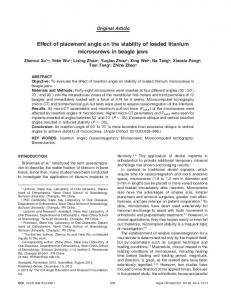
Three dimensional simplified model of matured end-to-side radiocephalic arteriovenous fistula (RCAVF) was modelled using EFD.Lab software consisting of a radial artery with 4 mm diameter connected to a cephalic vein with 6 mm diameter. The connection between radial artery and cephalic vein was made by an anastomosis, assuming an ellipsoidal cross section with a given anastomosis width (Wanas) and length (Lanas) as shown in FIGURE 1. In the design, the anastomosis width (Wanas) was fixed to 4 mm while the value of the anastomosis length (Lanas) was varied over 5 mm, 6 mm, obtained according to Galego et al. (2000), and 7 mm resulting in cross sectional area (CSA) of 15.7 mm2, 18.8 mm2 and 22.0 mm2 respectively. For each anastomosis length, the anastomosis angle was varied over 20o, 30o, 45o, 60o, 75o and 90o producing a total numbers of eighteen (18) models.
(a)
(b)
FIGURE 1. (a) Isometric view of simplified model of radiocephalic arteriovenous fistula (RCAVF). (b) Front view of simplified model.
Pressure drops over the anastomosis were calculated for six different anastomosis angles under a fixed flow rate of 900 ml/min @ 1.5x10⁻⁵ m3/s corresponding to Reynolds number 950. The outlets flow distribution was maintained at 92% at venous outlet and 8% at arterial outlet [2]. All vessel walls were assumed to be rigid and the blood flow was considered 3-D, non-pulsatile, laminar, Newtonian, incompressible and homogenous [2][5]. Fluid properties were obtained from Cole et al. (2002) and Bessa and Ortiz (2007): density, ρ = 1050 kg/m3 and dynamic viscosity, µ = 0.0035 Pa.s [5] [10].
III.
RESULTS AND DISCUSSIONS
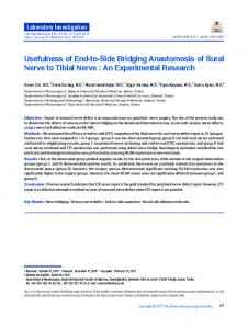
FIGURE 2. Pressure drop over anastomosis for different anastomosis angle.
Figure 2 shows the pressure drop over anastomosis for different anastomosis angle of 20o, 30o, 45o, 60o, 75o and 90o under fixed flow rate of 900 ml/ min. It can be observed that the pressure drop decreased with the increasing of anastomosis angle. The pressure drop decreased significantly for increasing anastomosis angle from 20o to 45o. For cases with anastomosis angle larger than 45o, pressure drop was shown became relatively constant. As expected, highest pressure drop was shown over smallest anastomosis angle = 20o, while lowest pressure drop was shown over largest anastomosis angle = 90o. The pressure drop decreased from 10528.9 Pa (20o angle) to 1406.3 Pa (90o angle) for anastomosis length = 5 mm, from 8251.3 Pa (20o angle) to 1085.4 Pa (90o angle) for anastomosis length = 6 mm and from 6110.8 Pa (20o angle) to 857.9 Pa (90o angle) for anastomosis length = 7 mm resulting in maximal pressure drop difference of 9122.6 Pa, 7165.9 Pa and 5252.9 Pa respectively.
(a)
(b)
(c)
(d)
(e)
(f)
FIGURE 3. Velocity vector (on the same scale) for different anastomosis angles: (a) 20o; (b) 30o; (c) 45o; (d) 60o; (e) 75o and (f) 90o for anastomosis length, Lanas = 6 mm.
(a)
(b)
(c)
(d)
(e)
(f)
FIGURE 4. Velocity vector (on the same scale) for different anastomosis angles: (a) 20o; (b) 30o; (c) 45o; (d) 60o; (e) 75o and (f) 90o for anastomosis length, Lanas = 7 mm.
Velocity vectors for different anastomosis angles were shown in FIGURE 3 and FIGURE 4. As the blood from the arterial inlet entered the anastomosis, it was separated at the heel (inner wall) where a vortex with clockwise rotation was formed. The large vortices can be seen appeared most for anastomosis angle 20o, 30o and 45o and were smaller in case of anastomosis angle 75o and 90o. Stagnation zone can be seen on the floor of the artery, on the outer wall of the vein close to the toe and also on the inner wall close to the heel as shown in FIGURE 3 and FIGURE 4. For larger angle than 60o, a large low flow zone was appeared at the inner wall as can be seen in FIGURE 3(d-f) and FIGURE 4(d-f). This stagnation and low flow zone on the floor of the artery, on the outer wall and also on the inner wall of the vein may lead to regions with increasing intimal hyperplasia and formation of stenosis. Finding an optimal anastomosis angle for a RCAVF, usable for all patients, was unrealistic due to varying, patientspecific anatomic and physiological conditions. Based on our result on the value of pressure drop over anastomosis, angle larger than 45o should be considered for construction of fistula. Stagnation zones which were known associated with the development of intimal hyperplasia and formation of stenosis were observed in all models. However, for anastomosis angle larger than 60o, a large low flow zones was observed at the inner wall compared to smaller angle than 60o. Taking into consideration, for average flowrate, it is recommended that anastomosis angle should be maintained between 45o and 60o to minimize adverse effects. IV.
CONCLUSION
In this investigation, pressure drop were shown change significantly with the variation of anastomosis angles. The geometry of the anastomosis also associated with the promotion of intimal hyperplasia and formation of stenosis. The results proved that anastomosis segment should be considered as an important component and should be chosen with care during the construction of RCAVF.
ACKNOWLEDGMENTS Support from Thermofluid Department, Faculty of Mechanical Engineering, Universiti Teknologi Malaysia is greatly acknowledged.
REFERENCES 1. Sivanesan, S., T.V. How, A. Bakran, Sites of stenosis in AV Fistulae for haemodialysis access, Neprology Dialysis Transplantation, 1999, 14: 118-120. 2. Canneyt, V., Pourchez, T., Eloot, S., Guillame, C., Bonnet, A., Segers, P., Verdonck, P., Hemodynamic impact of anastomosis size and angle in side-to-end arteriovenous fistulae: a computer analysis, Journal of Vascular Access, 2010, 11(1): 52 – 58. 3. Sivanesan, S., T.V. How, A. Bakran, Characterizing flow distributions in AV fistulae for hemodialysis access, Neprology Dialysis Transplantation, 1998, 13: 3108-3110. 4. Kumar, A., Jha M.S., Singla M., Gupta N., Raina P., Dubey D., Srivastava A., Radio-median cubital / radiocephalic arteriovenous fistula at elbow to prevent vascular steal syndrome associated with brachiocephalic fistula: Review of 320 cases, Indian Journal of Urology, 2007, 23(3): 261–264. 5. Bessa, K.L., Ortiz, J.P., Flow Visualization in Arteriovenous Fistula and Aneurysm Using Computational Fluid Dynamics, Journal of Visualization, 2009, 12(2): 95-107. 6. Ridriguez, J.A, Armadans, L., Ferrer, E., Olmos, A., Codina, S., Bartolome, J., Borrellas, J., Piera, L., The function of permanent vascular access, Neprology Dialysis Transplantation, 2000, 15: 402-408. 7. Konner, K., Nonnast-Daniel, B., Ritz, E., The Arteriovenous Fistula, Journal American Society of Neprology, 2003, 14: 1669-1680. 8. Van Tricht, I., De Wachter, D., Tordoir, J., Verdonck, P., Hemodynamics andComplications Encountered with Arteriovenous Fistulas and Grafts Access for Hemodialysis: A Review, Annals of Biomedical Engineering, 2005, 33(9): 1142-1157. 9. Hofer, M., Rappitsch, G., Perktold, K., Trubel, W., Schima, H., Numerical study of wall mechanics and fluid dynamics in end-to-side anastomoses and correlation to intimal hyperplasia, Journal of Biomechanics, 1996, 29(10): 1297-1308. 10. Cole, J.S., Watterson, J.K., O’Reilly, M.J.G., Is there a haemodynamic advantage associated with cuffed arterial anastomoses, Journal of Biomechanics, 2002, 35: 1337-1346. 11. Krueger, U., Zanow, J., Scholz, H., Computational Fluid Dynamics and Vascular Access, International Society of Artificial Organ, 2002, 26(7): 571-575.