This study examined how cells in the temporal cortex code orientation and size of a complex object. The study focused ... http://www.tandf.co.uk/journals/pp/02643294.html. 13. Requests for ... other hand, if the object's representation is prefer-.
COGNITIVE NEUROPSYCHOLOGY , 2000, 17 (1/2/3), 13–34
EFFECT OF IMAGE ORIENTATION AND SIZE ON OBJECT RECOGNITION : RESPONSES OF SINGLE UNITS IN THE MACAQUE MONKEY TEMPORAL CORTEX E. Ashbridge, D.I. Perrett, M.W. Oram, and T. Jellema University of St Andrews, Scotland, UK
This study examined how cells in the temporal cortex code orientation and size of a complex object. The study focused on cells selectively responsive to the sight of the head and body but unresponsive to control stimuli. The majority of cells tested (19/26, 73%) were selectively responsive to a particular orientation in the picture plane of the static whole body stimulus, 7/26 cells showed generalisation responding to all orientations (three cells with orientation tuning superimposed on a generalised response). Of all cells sensitive to orientation, the majority (15/22, 68%) were tuned to the upright image. The majority of cells tested (81 %, 13/16) were selective for stimulus size. The remaining cells (3/16) showed generalisation across four-fold decrease in size from life-sized. All size-sensitive cells were tuned to life-sized stimuli with decreasing responses to stimuli reduced from life-size. These results do not support previous suggestions that cells responsive to the head and body are selective to view but generalise across orientation and size. Here, extensive selectivity for size and orientation is reported. It is suggested that object orientation and size-specific responses might be pooled to obtain cell responses that generalise across size and orientation. The results suggest that experience affects neuronal coding of objects in that cells become tuned to views, orientation, and image sizes that are commonly experienced. Models of object recognition are discussed.
INTRODUCTION The visual system allows us to discriminate between objects of different orientation and size. The system also allows us to generalise across image transformations and identify an object as the same despite changes in view, orientation, and image size. How the visual system represents the appearance of
objects to enable these recognition capacities is not resolved. The study reported here investigates the effects of image transformation in orientation or size on object processing in the cortex of the anterior part of the superior temporal cortex (STSa) of the macaque monkey. This area was studied because it contains cells that are both selectively responsive to
Requests for reprints should be addressed to D.I. Perrett, School of Psychology, University of St Andrews, Scotland, KY16 9JU, UK. E. Ashbridge is currently at the Division of Psychology, South Bank University, 103 Borough Road, London SE1 0AA, UK. This research was funded by project grants from the UK MRC, BBSRC, the US ONR, and the HFSP. E. Ashbridge (née Wachsmuth) was supported by a UK SERC studentship. We acknowledge the contribution of N. J. Emery, L. K. Harrison, and J. K. Hietanen, who participated in some of the experiments. We are grateful for the support given by University technical and photographic staff and to Dr Walsh for comments. Ó 2000 Psychology Press Ltd http://www.tandf.co.uk/journals/pp/02643294.html
13
ASHBRIDGE ET AL.
the sight of one complex object—the face or head— and sensitive to viewing conditions (e.g. Bruce, Desimone, & Gross, 1981; Perrett, Rolls, & Caan, 1982; Perrett et al., 1985, 1991). The focus of studies on the face or head of previous studies (and the use of terms such as “face responsive” or even “face cells”) is perhaps misleading as to the selectivity of the cells. A substantial proportion of cells that respond to the sight of the face are also sensitive to visual information arising from body regions other than the face (e.g. Wachsmuth, Oram, & Perrett, 1994). Cells may respond to the face when this is presented in isolation, but such cells often respond to the rest of the body when the head is occluded from sight. Not surprisingly, when both the head and body are visible the response is greater than that seen to the face alone. We therefore examined the effects of image transformations on whole body stimuli. If an object’s representation can be activated independent of the orientation or size of the object’s image on the retina, then such a representation will here be referred to as “object-centred.” On the other hand, if the object’s representation is preferentially activated by a specific image orientation or size, then the representation will be referred to as viewer-centred” since activation depends on orientation and size relative to the viewer. Scalp-recorded evoked potentials to the sight of faces show orientation and size specificity (Jeffreys, 1989, 1993; Jeffreys, Tukmachi, & Rockley, 1992), implying that faces are coded at a particular stage of the human visual system in a viewer-centred manner. Neurophysiological studies of the ventral route of cortical processing have previously suggested that cells in early visual areas (V1, V2, V4) exhibit orientation -specific coding of elementary features of objects (Henry, Dreher, & Bishop, 1974; Kobatake & Tanaka, 1994). These cells project to inferotemporal (IT) cortex, where cells are selectively responsive to progressively more complex features but still exhibit orientation-specific responses (Kobatake & Tanaka, 1994; Tanaka, Saito, Fukada, & Moriya, 1991). IT cortex in turn projects to the cortex of the STSa (Seltzer & Pandya, 1978). This area contains cells selective for
14
COGNITIVE NEUROPSY CHOLOGY , 2000, 17 (1/2/3)
complex objects and which have previously been reported to respond irrespective of the stimulus orientation (Ashbridge, & Perrett, 1998; Perrett et al., 1982, 1985, 1988). Studies of temporal cortex cell responses to faces have so far tested few orientations (often restricted to upright and inverted). The first aim of the present study is to determine the extent to which cells in the STSa show object-centred orientation invariance, or viewer-centred orientation specificity in their responses to whole bodies presented in multiple orientations. This allows us to address the question of how orientation in the picture plane is processed for one biologically important object and whether it is processed in a similar way to view (see Logothetis, Pauls, & Poggio, 1995; Perrett et al., 1991; Wachsmuth et al., 1994). Furthermore, previous studies suggest mainly size-specific coding in V4 and IT and suggest a possible greater degree of size generalisation within STSa (Dobbin, Jeo, Fiser, & Allman, 1998; Ito, Tamura, Fujita, & Tanaka, 1995; Rolls & Baylis, 1986). The second aim of the study reported here is to measure the extent to which cells selective for complex objects in the cortex of the anterior STS generalise across different image sizes.
METHODS Recordings of responses of single cells from five macaque monkeys (Macaca mulatta, two females wt. 4–8kg and three males wt. 5–8kg) were carried out. The techniques applied (including surgical and recording procedures) and results from previous cellular studies of these subjects have been previously described (Perrett et al., 1991; Wachsmuth et al., 1994).
Training and Fixation Task Pre-surgical training: While in a primate chair, the subjects were trained to fixate on one of five LED lights presented on a white wall at eye level at a distance of 4 metres. For a block of 50 trials the position of the fixation LED light was constant. The
IMAGE ORIENTATION AND SIZE
monkey’s task was to discriminate the colour of the LED that followed a short signal tone to obtain the monkey’s attention. Licking resulted in fruit juice reward for the green LED. The monkey was to withhold licking in order to avoid delivery of a weak saline solution to the red LED. The LED stimuli were presented in pseudorandom order under computer control.
Visual Stimuli Pictures of eight different views (at 45° intervals rotating from the front view, see Wachsmuth et al., 1994) of the whole body were taken. For each view, eight orientations in the picture plane were constructed (resulting in 64 different stimuli); 0° (upright), 45°, 90° (horizontal), 135°, 180° (inverted), 225°, 270° (horizontal), and 315°. The images were then projected onto a white wall at a viewing distance of 4m, resulting in an image size of 24.4° (1.73m) head to toe. For some cells testing was additionally performed with different views of the head without the body in view. Testing was normally performed with the fixation light at the centre of the test image, subsidiary testing to examine effects of receptive fields was performed with the centre of the image presented 10° above, below, or to the side of the LED fixation spot. For size stimuli the same range of eight views were presented at four magnifications ranging from 100° (1.73m, head to toe height, subtending 24.4°), 75% (1.3m, 18.5°), 50% (0.87m, 12.3°) to 25% (0.43m, 6.2°) at the viewing distance of 4m. During daily life the monkey subjects saw humans at distances between 0.5 and 5m. The head to toe size of humans encountered by the subjects, at the projection distance of 4m, ranged between 1.5m and 1.8m. Control stimuli included complex 2D and 3D objects of different sizes, shapes, textures and orientations (broom, lab coats, chairs, pictures of different animals, etc.), simple 2D geometrical shapes (bars, spots and gratings), and simple 3D forms (balls, cylinders, boxes, etc.).
Testing Methods Every cell from which neuronal activity was recorded was first tested in an exploratory way by presenting a series of static and moving 3D objects (including bodies), and tactile and auditory stimuli. Where cells were found responsive to the face or body, they were tested for (a) selectivity between objects and (b) selectivity between views. Selectivity Between Objects. This was studied by comparing responses to slide images of faces, bodies, and a minimum of five control objects of approximately equivalent shape, size, and complexity (e.g. a fire extinguisher, lab coat). Test comparisons were also made using videodisk images of heads, bodies, and a variety of laboratory objects. Sensitivity to species of primate was tested (less systematically) by comparing responses to photographs of monkeys and humans. Selectivity Between Views. Each cell was tested with four views of the head and body (face, left and right profile, and back view) or with eight views (the same four views and four intermediate views). Cells found to be responsive to static views of the whole body but not to control objects were then further investigated for sensitivity to stimulus orientation and size. Size and orientation testing was performed with stimuli presented using the cell’s preferred view (front, back, etc.) once this had been established. Stimuli were presented in blocks of trials, with five trials for each stimulus condition in a computer-controlled pseudorandom order.
Data Analysis Since most cells in the anterior STS respond with a latency of 100msec (± 30msec), the magnitude of cell activity on individual trials was assessed over the 250msec time period occurring 100–350msec after stimulus onset. For some cells [with late response onset (> 200msec) or inhibitory responses (i.e. below S/A)] a 500msec time period (100–600msec post-stimulus) was used to assess cell activity. Cell responses to the whole body presented in different orientations and sizes, control objects, and COGNITIVE NEUROPSY CHOLOGY, 2000, 17 (1/2/3)
15
ASHBRIDGE ET AL.
S/A were compared on-line using a one-way ANOVA and post hoc tests [protected least significant difference (PLSD), Snedecor & Cochran, 1980] with a significance level of P < .05. Orientation Tuning A multiple linear regression analysis was performed to estimate the best relationship between response and second-order cardioid function of orientation of the stimulus (see Perrett et al., 1991). This regression analysis calculates the values of the coefficients b1–5 of the following equation for producing the highest correlation between cell responses and the angle of orientation. R = b1 + b2 cos (q) + b3 sin (q) + b4 cos (2q) + b5 sin (2q) where R is the response, b1–5 are coefficients, and q is the angle of the body orientation. This equation was used to define the optimal angle of orientation (qmax), the maximum response at this orientation (Rmax), and the sharpness of tuning. Population Response Analysis All cells, independent of their response pattern, were included in a population analysis. For each cell, the neuronal responses to different test conditions (averaged from five trials) were normalised by applying the following formula: (R-S/A)/(RmaxS/A) where R = response to test condition, Rmax = maximum response to any orientation or size (depending on the analysis), and S/A = spontaneous activity. The population response to each test condition was then computed from the average of the normalised single cell responses and was displayed after renormalising such that the maximum population response = 1, and population S/A = 0.
markers were made by injection of RRP and the fluorescent dyes, true blue and diamadino yellow. Once the last recording session had been completed, the monkey was given a sedating dose of ketamine followed by a lethal dose of barbiturate anaesthetic. After transcardial perfusion with phosphate buffered saline and 4% gluteraldehyde/ paraformaldehyde fixative, the brain was removed and put into a series of sucrose solutions with increasing concentration (10, 20, and 30%), or alternatively 2% dimethylsulphoxide and 20% glycerol. Standard histological procedures followed.
RESULTS From five subjects, 23% of anterior STS cells (1692 out of 7288 cells tested) were found to be visually responsive. These included cells selectively responsive to moving or static visual stimuli (see, e.g., Bruce et al., 1981; Oram, Perrett, & Hietanen, 1993). Of the visually responsive cells, a total of 26 cells were found to be selectively responsive to the whole body (i.e. with response to the whole human body significantly greater than that to control objects and S/A). These 26 cells were tested for selectivity to different whole body orientations in the picture plane. A total of 16 cells selectively responsive to the whole body were tested for size sensitivity. All of the cells included in this study were found unresponsive to the variety of control objects tested. Although sensitivity to identity was not tested systematically, no differences were noted in the response to the different experimenters. Moreover, the tested effects of image orientation and size were found to be comparable for images of humans and monkeys.
Histological Reconstruction After each recording session, frontal and lateral Xradiographs were taken to localise the electrode. Micro-lesions (10 microamp DC for 30sec), made at the end of some electrode tracks and subsequently identified using standard histological techniques, allowed reconstruction of the electrode position within the brain. In addition, reference
16
COGNITIVE NEUROPSY CHOLOGY , 2000, 17 (1/2/3)
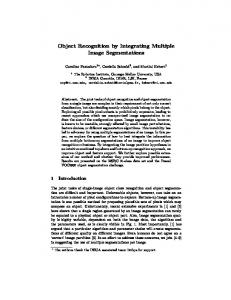
Generalisation Across Orientations Seven cells (of 26 tested) responded to all orientations at a rate significantly above S/A and control stimuli. Four of these cells showed complete generalisation in that they responded without statistical difference to all orientations (see Fig. 1).
IMAGE ORIENTATION AND SIZE
Fig. 1. The responses of a cell displaying generalisation over body orientation. The mean responses (± 1SE) to the front view of the whole body presented in eight different orientations and spontaneous activity (S/A) are illustrated for one cell (E83_38.31). Orientation is defined as the angle of anti-clockwise rotation from upright (0° or 360°). The cell response showed no significant difference between different orientations tested (P > .5 PLSD each comparison), but the cell responded to all orientations at a rate greater than control stimuli (P < .05 each comparison) and S/A (P < .05 each comparison, except 45°, P < .06 and 135°, P < .067). Overall effect of conditions ANOVA: F(9,40) = 2.28, P