seen in luxury perfusion (Raichle et aI., 1976). The luxury perfusion, an overabundant CBF, seen in the border zone around the cerebral infarct during the.
Journal of Cerebral Blood Flow and Metabolism 14:324-331 © 1 4 The International Society of Cerebral
99
Blood Flow and Metabolism
Published by Raven Press, Ltd., New York
Effect of Metabolic Alterations on the Accumulation of Technetium-99m-Labeled d,l-HMPAO in Slices of Rat Cerebral Cortex
Chang-Soo Ahn, Donald E. Tow, Chih-Chou Yu, and *Robert W. Greene Nuclear Medicine and *Psychiatry Services, VA Medical Center, BrocktonlWest Roxbury, and Harvard Medical School, Boston, Massachusetts, U.S.A.
Summary: It is widely recognized that the distribution of technetium-99m-Iabeled d,l-hexamethylpropylene amine oxime e9mTc-HMPAO) in the brain is determined by the regional blood flow. However, other factors may affect this process including the metabolism of the brain tissue. To examine this possibility we studied the effects of met abolic alterations on 99mTc-HMPAO uptake in rat brain cortex slices, with concurrent measurement of oxygen consumption (Q02)' 99mTc-HMPAO uptake was deter mined by incubating slices of rat cerebral cortex at 37°C in Krebs-Ringer phosphate glucose medium containing 99mTc-HMPAO with and without test substances. Differ ential gradients for 99mTc activity between the tissue and the suspending medium (TIM ratio) were derived from the equation TIM[99mTc] counts per gram of tissuelcounts per milliliter of medium. The Q02 of the brain slices was
measured using a biological oxygen monitor equipped with a polarographic oxygen probe. Inhibitors affecting oxidative phosphorylation caused parallel suppression of the TIM ratio and Q02' Agents that uncouple oxidation from phosphorylation increased the Q02 and decreased the TIM ratio. Incubation of slices at 22°C depressed the TIM ratio and Q02' The presence of inhibitors of oxida tive phosphorylation in the incubation medium increased the release of 99mTc activity from slices that had been prelabeled with 99mTc-HMPAO. These findings suggest that the altered metabolic status of the brain tissue mod ulates the kinetics and net accumulation of 99mTc_ HMPAO at the cellular level by either depressing uptake, increasing back-diffusion, or both. Key Words: Cerebral cortex slices-Energy metabolism of brain tissue Oxygen consumption-99mTc-Labeled HMPAO uptake.
Technetium-99m-Iabeled d,l-hexamethylpropyl ene amine oxime e9mTc-HMPAO) is used as a tracer to determine the regional cerebral blood flow (rCBF) distribution in humans utilizing the single photon emission computed tomography (SPECT) technique (Neirinckx et aI., 1987). Intracellular glu-
tathione (GSH) is considered the predominant fac tor in the retention of 99mTc-HMPAO in the brain, as it converts lipophilic compounds to a nondiffus ible hydrophilic form (Neirinckx et aI., 1988). Cur rent debates on the mechanism demand further studies to gain a better understanding of the trap ping mechanism of 99mTc-HMPAO in various pathologic situations (Babich, 1991). We investi gated the role of metabolic factors involved in the retention of 99mTc-HMPAO in the brain, indepen dent of blood flow, by measuring 99mTc-HMPAO uptake in slices of rat brain cortex incubated under various metabolic interventions, with concurrent assessment of respiratory activity. Dissociation from blood flow necessitates an in vitro approach, and according to Siesjo (1978), of all in vitro prep arations considered, brain slices represent the most physiological preparation since the majority of cells is undamaged and synaptic connections are partly intact.
=
Received February 10, 1993; final revision received August 25, 1993; accepted September 21, 1993. Address correspondence and reprint requests to Dr. C. S. Ahn at Nuclear Medicine Service, V A Medical Center, Brockton, MA 02401, U.S.A. Abbreviations used: BBB, blood-brain barrier; DCCD, dicy clohexylcarbodiimide; DNP, 2,4-dinitrophenol; OSH, glutathi one; ossa, oxidized glutathione; KRP, Krebs-Ringer phos phate buffer; KRP-O, Krebs-Ringer phosphate glucose medium; Q02, oxygen consumption; rCBF, regional cerebral blood flow; rCMR02, regional cerebral metabolic rate for oxygen; SPECT, single-photon emission computed tomography; 99mTc-HMPAO, technetium-99m-labeled d,l-hexamethylpropylene amine oxime; TIM ratio, differential gradients for 99mTc activity between the tissue and the surrounding medium.
324
ENERGY METABOLISM OF 99mTc-HMPAO UPTAKE
MATERIALS AND METHODS Preparation of cerebral cortex slices Long-Evans hooded rats weighing 150--250 g were lightly anesthetized with methoxyflurane in a close� jar and decapitated using a guillotine. The brain was qUIckly removed and cortex slices approximately 0.3 mm thick were cut from the lateral surface of each hemisphere us ing a Stadie-Riggs microtome. No more than three slices were obtained from a hemisphere to avoid the inclusion of white matter. Slices of approximately 25-40 mg were picked up from the cutting blade surface with a sharp spatula and weighed with a torsion balance, then sus pended in small individual vials containing 1 ml of chilled Krebs-Ringer phosphate glucose medium (KRP-G) of the following composition (mM): 122 NaCl, 5 KCl, 2 CaCI2, 1. 2 MgS04, 16 Na2HP04, and 5 glucose (adjusted to pH 7. 4 with 1 N NaOH). Slices from a single brain were used in experiments in which the effects of test materials were assessed by comparison with controls.
Preparation of 99mTc-HMPAO solutions A 99mTc-HMPAO solution was prepared by injecting approximately 10 mCi of 99mTc-pertechnetate in 5 D?-l of saline into a vial containing HMPAO (exametazlme, Ceretec; Amersham). The solution was eluted from a gen erator (DuPont) 3 to 5 h after the last elution and used within 30 min of elution. After shaking of the vial for 10 s, the solution was diluted with KRP-G to approximately 1.0 j.LCifml. One milliliter of this diluted 99mTc-HMPAO was added to 2 ml of KRP-G with and without test compounds in incubation vials (2. 5-cm-diameter and 5. 5-cm-long scintillation vials). Since the final 3 ml of incubation me dium contained 1 j.LCi of activity, given that the molecular weight of HMPAO is 272 and one vial of Ceretec con tained 0.5 mg of HMPAO, the concentration of HMPAO in the incubation medium was 61 nM. Radiochemical purity was determined by the three strip method according to the manufacturer's instructions with minor modifications. We used an 11 x 2-cm ITLC/ SG strip (Gelman Sciences, catalog No. 61886) for devel opment in the MEK (2-butanone) solution and a 9 x 2-cm ITLC/SG strip for the saline tube. We achieved better chromatographic separation using these strips than with the recommended 6 x 0.7-cm strips. The lipophilic com pounds ranged from 85 to 91% i� the ��eshly p�epared 99mTc-HMPAO solutions. The hpophlhc fractlOn de creased by 4. 8 ± 2. 5% (SD) at the end of 20 min of incu bation at 37°C and by 11.8 ± 4.2% at 40 min in the ab sence of brain slices.
Determination of 99mTc-HMPAO uptake by brain slices Brain cortex slices were transferred to the incubation vials containing 3 ml of 99mTc-HMPAO solution with and without test substances. After the addition of brain slices, the vials were incubated without covers for 20 min, unless otherwise stated, in a Dubnoff metabolic water bath at 37°C with shaking at 100 cycles/min. At the end of the incub ation period, the slices were removed from the vials using a platinum hook and placed on a watch glass, ad hering medium was removed using filter paper, and the slices were placed in counting tubes containing 1 ml of 2 N NaOH solution to dissolve the tissue and achieve uni-
325
formity for counting. A 0.2-ml aliquot of medium was pipetted from each incubation vial and added to 0.8 ml of 2 N NaOH solution in counting tubes. The radioactivity was determined in a 2oo-tube automatic well-type scintil lation counter. Differential gradients for 99mTc activity between the tissue and the suspending medium (TIM ra tio) were derived from the relationship T/M[99mTc]
=
countslg of tissue .
counts/ml of medlUm
Each experiment consisted of three control vials and three vials containing one test material. The same exper iment was repeated three times on different days, result ing in pooled data for nine control and nine test samples. Statistical comparison between control and test samples was done by stratified analysis using the t test (Fleiss, 1986), stratifying data from three experiments.
Determination of oxygen consumption (Q02) by brain cortex slices The Q02 by brain cortex slices was determined using a biological oxygen monitor system from Yellow Spring In strument Co. (Yellow Spring, CO) as reported previously (Robinson and Cooper, 1970; Ahn, 1982) with slight mod ifications. The apparatus consists of a Clark-type polaro graphic oxygen probe covered with a thin Teflon mem brane and housed in a Lucite plunger. This probe is in serted into a glass reaction chamber. The contents of the chamber, 3 ml of KRP-G and brain slices, were agitated continuously by magnetic stirring and maintained at 37"C by means of the surrounding water bath and a Haake thermostat. Changes in the oxygen content of the medium in the reaction chamber were monitored with a Model 53 biological oxygen monitor as changes in the anode cur rent from the probe and continuously recorded using a Perkin-Elmer 56 recorder. The medium, 3 ml of KRP-G, was first added to the water-jacketed reaction chamber and stirred for 3 min to allow temperature equilibrium and saturation with air. A preweighed brain cortex slice was then dropped into the medium. The probe was inserted, care being taken to exclude all air bubbles. The Q02 was recorded as a percentage of the decrease in the oxygen content in the medium. After obtaining the basal rate of Q02 by brain cortex slices, test materials were introduced to the medium in a volume of 30 j.Ll through the access groove of the plunger and changes in the rate of Q02 were recorded continuously (Fig. 1). Since the oxygen concen tration of 0. 23 j.Lmollml of Krebs-Ringer phosphate buffer (KRP) saturated with air at 37°C and ambient barometric pressure is known, the Q02 of brain slices can be ob tained as micromoles per gram wet weight per hour. We used KRP saturated with air for both 99mTc_ HMPAO uptake and Q02 studies because it was much simpler to handle, and use of the more physiologic Krebs-Ringer bicarbonate buffer saturated with 95% O2 and 5% CO2 did not cause significant differences in either the Q02 or the T/M ratio values. This is consistent with a previous report on Q02 (Elliott and Wolfe, 1962).
Determination of release of 99mTc activity from prelabeled slices Brain slices were placed, one each, in six vials contain ing 99mTc-HMPAO in 3 ml of KRP-G, which was preJ Cereb Blood Flow Metab. Vol.
14.
No.2.
1994
C.-S. AHN ET AL.
326
pared by the same procedure as for determination of the TIM ratio. After 20 min of incubation in the Dubnoff wa ter bath at 37°C, slices were transferred to three vials each of two groups of vials containing 3 ml of KRP-G, one group without and the other group with test materials for the second incubation. Following 20 min of the second incubation, the activity of the slices and medium was as sessed to determine the percentage released from the pre labeled slices. The experiment was repeated three times, and the pooled results were analyzed using the t test by stratifying each group of data.
Selection of metabolic inhibitors The following four compounds were utilized to sup press energy metabolism of brain slices. Azide is known to block electron flow from NADH to O2 in the respiratory chain at the stage of cytochrome oxidase, resulting in depression of both respiration and ATP synthesis (Stryer, 1988). Dicyclohexylcarbodiimide (DCCD) is an inhibitor of oxidative phosphorylation. DCCD blocks coupled phos phorylation by forming covalent bonds with an interme diary of oxidative phosphorylation at the electron trans port chain (Beechey et aI., 1967). An uncoupler of oxidative phosphorylation, 2,4dinitrophenol (DNP), blocks ATP synthesis, while sub strate oxidation and Q02 proceed maximally (Stryer, 1988). A high concentration of potassium (30-100 mM) is known to uncouple oxidative phosphorylation in brain tissue (reviewed by Elliott and Wolfe, 1962). These chemicals were obtained from Sigma (St. Louis, MO, U.S.A.): sodium azide (S-2002), DCCD (D-3128), DNP (D-7004), and potassium chloride (P-4504).
RESULTS 99mTc-HMPAO uptake and Q02 by brain cortex slices The accumulation of 99mTc-HMPAO in brain slices as expressed by the TIM ratio ranged between 9 and 14 for all experiments with an incubation time of 20 min. However, the variability within each ex periment was low, as shown in Table 1. The TIM ratio remained unchanged at a 100 x higher concen tration (6.1 fJ.M) of HMPAO in the incubation me dium. The TIM ratio was proportional to its lipophilic fraction in the medium and is shown in Fig. 2. As noted in the figure legend, the TIM ratio was 1.6 in the absence of the lipophilic fraction. This most likely reflects a residual fraction of medium pre sent even after removing adhering medium from slices with filter paper. A similar fraction of me dium was detected as a TIM ratio ranging from 1.4 to 1.8 following incubation of brain slices in the 99mTc-pertechnetate solution (which is not lipo philic). The TIM ratio increased with time from 5 to 40 min, with a gradual slowing of the rate of increase (Fig. 3). J Cereb Blood Flow
Metab, Vol. 14, No.2, 1994
100
... OJ
""-;: -
.£:l E
'"
""
u c:
90
'" �IArid
0> c:
.-
c:
'"E
OJ
a::
.. 0 ....0 � c: OJ u
...QJ
c..
=-
�
80
-70
- r--
0
10 Time (min)
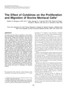
FIG. 1. Recording of the 002 by brain slices. The oxygen content of the medium, 3 ml of KRP-G, is monitored contin uously as it is consumed by brain slices. After stabilization of the control period, 30 fJ.1 of 500 mM azide was infused at the arrow, resulting in a final azide concentration of 5 mM. Prompt depression of the 002 rate is apparent. The 002 was 72 fJ.mol/g/h during the control period and 32 fJ.mol/g/h after the addition of azide.
The Q02 by untreated rat brain cortex slices ranged from 70 to 80 fJ.mol/g/h. The rate of Q02 remained constant for at least 30 min. An incubation time of 20 min was chosen for de termination of the TIM ratio in the following exper iments to minimize the effect of degradation of the lipophilic fraction of 99mTc_HMPAO (as detailed un der Preparation of 99mTc-HMPAO solutions) and because the Q02 rate of brain slices begins a gradual decline after 30 min of incubation. Effect of metabolic inhibitors and low temperatures on the TIM ratio
TABLE 1.
Addition or incubation temperature
TIM ratioa
pb
None 5 mMazide
12.1 ± 2.2 8.3 ± 2.3
u
�
10
r50
r
-
I-
�
I-
;-
r5
o
5
10
20
40
Incubation Time (min) FIG. 3. TIM ratio as a function of incubation time. Each point for the TIM ratio is the average of duplicate incubations. The incubation time ranged from 5 to 40 min.
Control
Azide 5mM
DCCD 1 mM
22· C
DNP 0.1 mM
KCI 30 mM
FIG. 4. Correlation, as a percentage of controls, of the ef fects of metabolic inhibitors and incubation conditions on the TIM ratio and 002, The bars for the TIM ratio and 002 are derived from Tables 1 and 2, respectively.
J
Cereb Blood Flow Metab, Vol. 14, No.2, 1994
328
C.-S. AHN ET AL.
3, azide and DCCD increased the release of activity from the prelabeled slices. DISCUSSION Brain slice preparations The viability of our brain cortex slice preparation was optimal since the Q02 of 70 to 80 fJ-mol/g/h was comparable to data reported in the literature (Hertz and Schou, 1962). Brain slices are an appropriate system for studies at the cellular level, eliminating flow factors. When suitably prepared, slices of mammalian brain show an electrophysiological and anatomical integrity sufficient to merit their description as cerebral sub systems (McIlwain and Bachelard, 1985). In cutting a brain tissue specimen 0.35 mm thick and weighing 100 mg, the surfaces disturbed (cut and resealed) are about 30 cm2/g, or only 0.06% of the estimated outer cell surfaces of the sample. The external area of tissue samples prepared from mammalian brain by slicing approaches that of the blood capillaries that the samples carry. From the reported length and diameter of the capillaries in the rat brain, a value of 10 mm2/mm3 is obtained for the capillary area per unit tissue volume; the excised tissue sam ple has an external area of about 6 mm2/mm3. Thus, the external areas that must form the route of sup ply of materials to the tissue sections after isolation are quite similar to the area of the capillaries that constitute the route of supply to the tissue as it exists in the brain (McIlwain and Bachelard, 1985). The maximal diffusion distance of a 0.3-mm brain slice is 0.15 mm, which far surpasses the intercap illary distance of 0.06 mm (Siesjo, 1978). In our ex perimental system the TIM ratio for 99mTc-HMPAO continued to increase at 40 min as shown in Fig. 3. In a separate experiment not reported under Re sults, the TIM ratio was still increasing at 120 min of incubation. This indicated that the 20-min incuba tion period was not long enough to reach the satu ration point, at which the differences between con-
TABLE 3.
Addition
Release of99mTc activity from pre labeled slices Released in medium
(%)a
p
b
None 5 mM azide
13.7 ± 1.6 21.2 ± 3.2