(PPBs) of microtubules (MTs) and on prophase spindle MTs in root ..... able to see the multipolar stage in onion cells using ... These multipolar spindles are.
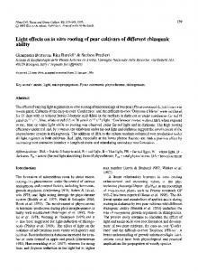
Protoplasma (1996) 192:109-121
0ROT 9 Springer-Verlag 1996 Printed in Austria
Effects of cycloheximide on preprophase bands and prophase spindles in onion (AUium cepa L.) root tip cells A. Nogami ~, T. Suzuki 2, Y. Shigenaka 2, Y. Nagahama 3, and Y. Mineyuki t' * Department of Biological Science, Faculty of Science, and 2 Laboratory of Cell Physiology, Faculty of Integrated Arts and Sciences, Hiroshima University, Higashi-Hiroshima, and 3Division of Reproductive Biology, National Institute for Basic Biology, Okazaki Received April 8, 1995 Accepted Janumy 28, 1996
Summary. Effects of cycloheximide (CHM) on preprophase bands (PPBs) of microtubules (MTs) and on prophase spindle MTs in root tip cells of onion (Allium cepa L.) were examined. When root tip cells were treated with 36 gM CHM for 0.5-4 h, the population of cells with a PPB did not decrease markedly although the population of mitotic cells and that of prophase cells with a PPB gradually decreased to half of the control root tips. In prophase cells treated with 11 and 36 gM CHM for 2 h, the width of the PPB was 1.4 times broader than that in the prophase PPB without CHM. Electron microscopic observation on the cross section of the PPB showed that the number of MTs and the distance between adjacent MTs in prophase PPBs treated with CHM were similar to those in the early developmental stage of PPBs without CHM. The bipolar spindle, that appeared in late prophase was not seen in prophase cells treated with I 1 gM or higher concentrations of CHM for 2 h. In order to examine differences of perinuclear MT arrangement between CHM treated and non-treated prophase cells, arrangement of perinuclear MTs was examined by confocal laser scanning microscopy. In control cells without CHM, MTs appeared on the nuclear surface with several "branched" or "cross over" type MT loci in the cytoplasm when broad PPB formation started. These MT loci were replaced by the "aster" type MT foci, from which several MTs radiated along the nuclear surface. The "aster" type MT loci gradually gathered to form a bipolar spindle. MTs connecting the spindle pole region and the PPB were seen in late prophase. In CHM-treated cells (11-360 ~tM for 2 h), "branched" and "cross over" type MT foci were prominent, even in prophase cells with well condensed chromosomes. Neither linkages of MTs between the spindle pole region and the PPB nor "aster" type MT foci were seen. These observations showed that CHM prevents the bundling of MTs in the PPB and also inhibits the formation of "aster" type MT foci that is essential for bipolar spindle development.
* Correspondence and reprints: Department of Biological Science, Faculty of Science, Hiroshima University, Kagamiyama 1-3-1, Higashi-Hiroshima739, Japan.
Keywords: Allium cepa L.; Cell cycle; Cycloheximide;Preprophase band; Root meristems; Spindle microtubules. Introduction A protein synthesis inhibitor, c y c l o h e x i m i d e (CHM), has b e e n reported to b l o c k the p r o g r e s s i o n of cell cycle at various points, such as interphase, prophase, prometaphase, metaphase, telophase in higher plants (Rose 1970, Webster 1973, G a r c f a - H e r d u g o e t a l . 1974, O l s z e w s k a et al. 1990). S o m e essential proteins are thought to be synthesized at these points although such m o l e c u l e s have not yet b e e n identified. R e c e n t l y we reported that C H M affects the m i c r o t u b u l e (MT) array in interphase cells ( M i n e y u k i et al. 1994). H o w ever, other effects of C H M on M T arrays have not yet b e e n well studied. Prophase is an interesting stage for the study of p l a n t M T arrays, b e c a u s e two different types of M T arrays are co-existent in prophase. One is a preprophase b a n d (PPB) of MTs that is thought to play an i m p o r tant role in the e s t a b l i s h m e n t of the d i v i s i o n plane. The other is the spindle MTs that play an essential role for c h r o m o s o m e m o v e m e n t in later stages of mitosis (Cyr 1994). The b r o a d PPB that appears in G2 phase as a broad cortical M T array a l i g n e d parallel to the future d i v i s i o n p l a n e ( M i n e y u k i et al. 1988b, G u n n i n g a n d S a m m u t 1990), b e c o m e s a narrow PPB in prophase to d e t e r m i n e the site where cell plate will fuse to the parental cell walls at the e n d of cytokinesis (Wick et at. 1981; W i c k and D u n i e c 1983, 1984; M i n e y u k i et al. 1989; M i n e y u k i and Palevitz 1990).
110
A. Nogami et al.: Effects of cycloheximide on preprophase band and spindle
The perinuclear MTs, that appear concomitantly with the broad PPB, gradually gathered to form a bipolar spindle at the end of prophase. After PPBs disappear in late prophase or prometaphase, all the MTs are incorporated into spindles in metaphase (Wick and Duniec 1983, 1984). Although the PPB is reported to exist in the CHM-treated cells (Benbadis et al. 1974, Olszewska et at. 1990, Mineyuki et al. 1994), our preliminary observation showed that there were few PPBs of mature type in the CHM-treated cells. In the present study, we examined effects of CHM on MT arrays of PPBs and prophase spindles in onion root tip cells and showed that PPBs were broad and the bipolar spindle was not formed in the presence of CHM. Based on observations by confocal laser scanning microscopy (CLSM) and electron microscopy, mechanisms of the inhibition of MT bundling in PPB s and that of bipolar spindle formation by CHM will be discussed. Material and methods Onion (Allium cepa L. cv. Highgold Nigou, Sakata Seed Co., Yokohama, Japan) seeds were sown on a filter paper moistened with distilled water and grown in a dark box at 25 ~ 2 mm long root tips of 4-day-old seedling were used for the experiments. Procedures of CHM treatment and MT observation using immunofluorescence microscopy and CLSM were done according to the methods described previously (Mineyuki et al. 1994). Shortly, 2 mm long root tips were fixed with 4% paraformaldehyde in PME buffer (50 mM Pipes, 5 mM EGTA, 1 mM MgSO4 97HzO, pH 6.8) overnight, rinsed in the buffer, and treated with a mixture of cellulase solution (Mineyuki et al. 1991 b) for 15 min. After a rinse in the buffer, root tips were squashed and air dried on the slide glass, then extracted with-20 ~ methanol for 10 min, rinsed in phosphatebuffered saline (PBS), and treated with a mouse monoclonal anti-~ tubulin antibody (Amershan Japan Co., Tokyo, Japan) for 15 min. Following a 15 min rinse in PBS, cells were exposed to fluorescein isothiocianate (FITC)-linked sheep anti-mouse immunoglobulin G (F (ab')2 fragment; Sigma Chemical Co., St. Louis, MO, U.S.A.) for 45 min, rinsed again and covered with a solution containing 50% glycerol, 50 mM Tris buffer, pH 9.0 and 1 mg/ml p-phenylenediamine. Hoechst 33258 (10 rag/l) was included in the mounting medium to identify the stage of chromosome condensation. The width of a PPB was determined by an ocular micrometer in a Nikon microscope (X2 microscope equipped for epifluorescent illumination; Nikon Co., Tokyo, Japan). As the CLSM used in this study (MRC-500; Japan BioRad Co., Tokyo, Japan) was not equipped with a UV system, we observed nuclei stained with Hoechst 33258 with a conventional fluorescence microscope mode before CLSM observations to determine the nuclear stage. The nuclear stage determined by this method is shown in the figure legends. In order to examine more detail of the MT array around the nucleus, a series of images obtained by CLSM was reconstructed as a 3-dimensional stereo-pair image using software equipped in MRC-500. For electron microscopy, 2 mm long root tips of 4-day-old seedlings which were sliced in two pieces were fixed with 3.5% glutaraldehyde
(TAAB Laboratories Equipment Ltd., Reading, U.K.) in PNIE buffer at room temperature for 1-2 h, rinsed in the PME buffer twice, postfixed with 2% osmium tetraoxide, rinsed in distilled water twice, dehydrated in acetone series, embedded in Spurr's resin (Spurr 1969) and sectioned for etectron microscopy. The sections were stained in aqueous uranyl acetate (3%) followed by Reynolds's lead citrate (Reynolds 1963). Negative films of the cross section images of PPB MTs at the magnification of 20,000 were used for the analysis of the distance between adjacent MTs. The positional information of the center of each MT was interfaced to a personal computer (PC 9801FA; NEC, Tokyo, Japan) through a video camera (KY-F30; Victor Co. Ltd., Tokyo, Japan) and the nearest-neighbor distances for the centers of MTs were measured.
Results
Kinetics of the frequency of PPBs Root tip cells treated with 3.6-360 gM CHM for 0-4 h were examined by fluorescence microscopy. Prophase cells were detectable in all cases examined, although the population of prophase cells or other mitotic cells (prometaphase, metaphase, anaphase, and telophase) in root tips changed over different concentrations and durations of treatment with CHM. The lowest value of the population of prophase (or mitotic) cells was about half of that in root tips without CHM treatment. Figure 1 shows the change in the population of prophase cells and of other mitotic cells in onion root tips treated with 36 gM CHM for various durations. The population of prophase cells or mitotic cells gradual-
A
g O
00.51
2
3
4
~
~
i
57 B
000'5 ~i
Time (h) Fig. 1. Effects of duration of treatment without (9 or with (0) solutions of 36 gM CHM on the population of prophase cells (A) and that of other mitotic cells (prometaphase, metaphase, anaphase and telophase) (B) in onion root tip cells. Each point shows the mean + S.E.M. obtained from three different samples
A. Nogami et al.: Effecl:sof cycloheximideon preprophaseband anctspindte 12- A 10,
864200
Q e5
~:
o:5 "i
g
-~
1210"
86-
4-
/
2 ~
W
o o 0:s i 12
l
-
o
5.-
A
C
1~ 1
% 0:s i
_ _ -~
U
~
Time (h) Fig. 2 A-C. Effectof duration of treatmentwith solution of CHM on the population of cells with a PPB in onion root tip cells. A Cells with a PPB, B interphasecells with a PPB, C prophase ceils with a PPB. 4-day-otd seedlings were treated without (9 or with (0) 36 gM CHM. Each point shows the mean + S.E.M. obtained from three differentsamples
ly decreased and in root tips treated with CHM for 4 h their percentage was almost half of those not treated with CHM. In contrast, the population of cells with a PPB did not decrease remarkably during 4 h treatment (Fig. 2 A). While the population of prophase cells with a PPB decreased in consequence of the decrease in prophase cell population (Fig. 2 C), the population of interphase cells with a PPB increased (Fig. 2 B), however. Thus, the total PPB population remained constant. In root tips without CHM treatment, almost all of the prophase cells had a PPB. However, 14% of prophase cells treated with 36 pM CHM for 2 h had neither spindle MTs nor PPB MTs, but had short MTs in the cytoplasm (data not shown). Double PPBs (Wick and Duniec 1983) were seen both in control cells and in the CHM-treated cells. However, the distance between these MT bands was apparently wider in CHM-treated double PPBs than in non-treated double
111
PPBs. Frequencies of the appearance of these irregular MT arrays increased when root tips were treated with high concentration (360 gM) of CHM for 2-4 h. As was reported by several researchers (Wilson 1950, Rose 1970), the well condensed or "super contracted" chromosomes were often seen in prophase cells treated with CHM for 2 or 4 h (Fig. 3 D, F). Dumbbellshaped nuclei with a PPB were also seen in CHMtreated prophase cells. The cleavage site of the dumbbell-shaped nucleus matched the plane where PPB MTs were on the cell cortex.
Width of PPBs Figure 4 A shows a histogram of the width of PPBs in interphase, early prophase (beginning of the chromatin condensation is detectable by Hoechst staining but chromatins have not yet become well defined chromosomes in this stage), and prophase cells. The width of PPB gradually narrowed when the celt cycle progressed from interphase to prophase, and most prophase cells had narrow (3 gm) PPBs (Figs. 3 A and 4 A). However, in cells treated with 36 gM CHM for 2 h, prophase PPBs with 4-5 pm width were frequent (Figs. 3 C and 4B). The widths of PPBs were compared in prophase cells of 4-day-old onion root tips that were treated with various concentrations of CHM for 2 h (Table 1). The mean width of non-treated prophase PPBs was 3.2 gin, but that in cells treated with CHM at concentrations of 11 pM or 36 gM was 4.5 gm, 1.4 times that of control PPBs. When root tips were treated with 360 gM CHM for 2 h, PPB MTs in some prophase cells dispersed in the cytoplasm and MT bands were not seen (Fig. 3 E). Because of the occurrence of prophase cells with this type of MT array, the mean width of the prophase PPBs treated with 360/aM CHM was narrow again.
Number of MTs and the distance between adjacent MTs As the immunofluorescent microscopical observation clearly showed that CHM affected the width of a PPB, we further examined the effect of CHM on a PPB by electron microscopy. As the treatment of 36 g M CHM for 2 h was the most effective on the width of prophase PPBs, the distance between adjacent MTs and the total number of MTs in a cross section of prophase PPBs were compared between 36 pM CHM-treated and non-treated PPBs (Fig. 5). Without CHM-treatment, in the early stage of PPB development in prophase only 50 MTs were seen in a
112
A. Nogami et al.: Effects of cycloheximide on preprophase band and spindle
Fig. 3. Tubulin immunofluorescence (A, C, and E) and nuclei (B, D, and F) stained with Hoechst 33258 in prophase cells of onion root tips treated or non-treated with CHM for 2 h; C and D 36 gM CHM, E and F 360 ~tM CHM. Note that the width of a PPB in the CHM-treated cell (C) is larger than that of the control cell (A). Arrowheads indicate spindle poles, asterisks PPBs. Bar: 10 gm
cross s e c t i o n o f a P P B , and f e w M T l a y e r s w e r e d e t e c t a b l e in a PPB (Fig. 6 A). H o w e v e r , the n u m b e r o f M T s and also that o f M T l a y e r s i n c r e a s e d (Fig. 6 B), and in the late PPB stage m o r e than 250 MTs w e r e seen in a P P B cross s e c t i o n a n d a b o u t 10 M T l a y e r s w e r e d e t e c t a b l e (Fig. 6 C). In C H M treated p r o p h a s e P P B s (Figs. 5 B a n d 6 D), o n l y few
M T l a y e r s w e r e seen a n d the n u m b e r o f MTs w a s 69 + 2.6 ( m e a n + S.E.M. o b t a i n e d f r o m 10 p r o p h a s e P P B s ) . W h i l e the d i s t a n c e b e t w e e n centers o f a d j a cent MTs in e a r l y P P B s was ca. 40 n m m o s t frequently, it w a s less than 3 0 n m in the late PPB stage (Fig. 7). F i g u r e 8 s h o w s the c o r r e l a t i o n b e t w e e n the m e a n n u m b e r o f total MTs and the m e a n d i s t a n c e o f
A. Nogami et al.: Effects of cycloheximide on preprophase band and spindle
Table 1. Effect of 2 h ueatment with various concentrations of CHM on the width of prophase PPBs
25
Concentration (gM)
15105r 0
O-
"6
113
L
0 3.6 11
36 360
Width of PPB (~m)
Number of PPBs
3.2 _+0.2 3.1 + 0.2 4.5 + 0.3 4.5 + 0.3 3.6 + 0.2
13 20 36 32 25
1 2 3 4 5 6 7 8 9 1011 1213 14
All values show means _+S.E.M. from 13-36 prophase PPBs
B
E -m z
202 15. 10.
0
.
~
1 2 3 4 5 6 7 8 9 1011121314
Width (• m) Fig. 4. Width of PPB MTs in interphase (open bars), earl), prophase (hatched bars), and prophase (solid bars) cells of onion root tips treated without (A) or with (B) 36 gM CHM for 2 h
adjacent MTs in non-treated or CHM-treated prophase PPBs. CHM-treated prophase PPBs appear similar to early stage control PPBs in terms of MT number and distance between adjacent MTs.
Bipolar spindle The prophase cells of 4-day-old onion root tips were treated with various concentrations of CHM for 2 h to see whether the percentage of prophase cells with a bipolar spindle decreased. The bipolar spindle is formed in late prophase of non-treated cells (Fig. 3 A, B). In the CHM-treated cells, bipolar spindles may not even be detectable in those prophase cells whose chromosomes are well condensed (Fig. 3 C, D). Without CHM, about 20% of prophase cells had a bipolar spindle. The percentage of prophase cells with a bipolar spindle decreased with increasing CHM concentration, reaching zero at a concentration of 11 gM (Fig. 9).
CLSM observations of spindle MT development In order to see differences of the perinuclear MT arrays between CHM-treated and non-treated cells,
MT arrays in the perinuclear region were observed using CLSM. In the broad PPB stage, MTs on the nuclear surface were randomly oriented and many MTs radiated from points over the entire surface of the nucleus in all directions into the cell cortex (Fig. 10 A). At this stage, several MT foci (Fig. 10 A) were seen. Most of them were in the cytoplasm apart from the nuclear surface. MT bundles were "cross over" or "branched" at these MT loci. (Here "bundle" refers to a single MT as well as to an MT bundle with several MTs, as we cannot distinguish between the two possibilities.) The population of MTs in the cytoplasm thinned out slightly when the PPB became a mature narrow band (Fig. 10 B). MTs that emanated in no particular direction were replaced by MT bundles that extended outward to the vicinity of the PPB (compare Fig. 10 A with B). The number of MT foci that were apart from the nuclear surface decreased, and most of MT loci that were on or in the vicinity of the nuclear surface. The "cross over" and "branched" type MT foci were replaced by the "aster" type MT loci, from which many MT bundles initiated on the nuclear surface (Fig. 10 C). The number of MT bundles radiating from the "aster" type MT foci increased, and the MT bundles became much more appressed to the nuclear surface after the PPB became a mature, packed MT band (Fig. 11 A). These "aster" type MT foci were not restricted to the future pole region. Some of them appeared to be near the equatorial region of the nuclear surface (Fig. 11 A). However, the more developed "aster" type MT foci gradually gathered to the nuclear polar region and the MT bundles gradually became oriented parallel to the spindle axis (Fig. 11 B). Finally, the MT foci gathered at a point of polar cap region, often slightly apart from the nuclear surface (Fig. 11 C). Although some MT bundles extended from the
114
A. Nogami et al.: Effects of cycloheximide on preprophase band and spindle
Fig. 5. Cross section of PPBs in prophase cells of onion root tips treated without (A) or with (B) 36 gM CHM for 2 h. Bar: 0.5 gm
"aster" type MT foci toward the vicinity of the PPB in mid prophase (Fig. 11 A), we could not obtain a clear image of the MT linkages between spindle MTs and the PPB. However, when prophase spindles were well developed in late prophase, MTs connecting the PPB and the spindle pole region were clearly visible
A
9
-