Chirurgia (2015) 110: 518-524 No. 6, November - December Copyright© Celsius
Elective Laparoscopic Cholangiography in Lithiasic Pathology. Intraoperative Selection Criteria O.L. Madge, Cl. Daha, C. Cirimbei, E. Brãtucu, N. D. Straja 1st Surgical Clinic, “Prof. Dr. Al. Trestioreanu” Institute of Oncology, Bucharest, Romania
“Carol Davila” University of Medicine and Pharmacy, Bucharest, Romania
Rezumat Colangiografia laparoscopicã de elecåie în patologia litiazicã. Criterii de selecåie intraoperatorii Introducere: Introducerea tehnicilor laparoscopice a fãcut ca disputa dintre adepåii efectuãrii colangiografiei intraoperatorii de rutinã sau de elecåie sã continue, dar în prezent cei mai mulåi autori recomandã practicarea ei de elecåie pe baza unor indicaåii preoperatorii şi/sau intraoperatorii bine stabilite, evitându-se astfel efectuarea unui numãr mare de proceduri inutile, cu dezavantajele şi riscurile lor inerente. Metode: S-a efectuat un studiu retrospectiv, pe parcursul a 20 de ani, ce cuprinde 100 de colangiografii laparoscopice elective. Rezultate: Cel mai fidel parametru pentru indicaåia intraoperatorie de colangiografie laparoscopicã în suspiciunea de litiazã coledocianã s-a dovedit a fi ductul cistic dilatat peste 3 mm însoåit opåional de litiazã cisticã şi/sau calea biliarã principalã dilatatã peste 10 mm. Concluzii: Conform datelor analizate colangiografia laparoscopicã s-a dovedit a fi o metodã de explorare intraoperatorie sigurã, cu mortalitate şi morbiditate specifice metodei nule, dar şi cu o fiabilitate excelentã în evidenåierea litiazei CBP.
Abstract Introduction: The introduction of laparoscopic techniques has caused the dispute between supporters of routine or elective intraoperative cholangiography to continue, but at present most authors recommend its elective practice on the basis of well-established preoperative and / or intraoperative indications, thus avoiding to carry out a large number of unnecessary procedures, with their inherent disadvantages and risks. Method: A retrospective study was conducted over 20 years, comprising 100 elective laparoscopic cholangiographies. Results: The most representative parameter for the indication of intraoperative laparoscopic cholangiography in choledochal lithiasis suspicion has proved to be common bile duct dilation of over 3 mm, optionally accompanied by cystic calculi and / or main bile duct dilation over 10 mm. Conclusions: According to the analysed data, laparoscopic cholangiography proved to be a safe method of intraoperative exploration, with mortality and morbidity specific to the null method, but also with an excellent reliability in highlighting CBD lithiasis. Key words: laparoscopic cholangiography, biliary lithiasis, ultrasound, dilated cystic duct
Cuvinte cheie: colangiografie laparoscopicã, litiazã biliarã, echografie, duct cistic dilatat
Corresponding author:
Claudiu Daha, MD, PhD 1st Surgical Clinic, “Prof. Dr. Al. Trestioreanu” Institute of Oncology 252 Fundeni Road, 2nd District, 022328, Bucharest, Romania E-mail:
[email protected]
Introduction The past 25 years have been a period of continuous change in surgery due to technical progress, the most important being the widespread of minimally invasive techniques, of
519
which general surgery benefited most. Biliary surgery is the branch where change had the greatest impact by generalizing laparoscopic cholecystectomy, with a well-codified and widely recognized technique, as the "gold standard". The laparoscopic approach of the gallbladder benefited in parallel from the development of other minimally invasive and imaging techniques, among which we mention endoscopic retrograde cholangiopancreatography (ERCP), laparoscopic cholangiography and choledochoscopy, that together collaborate for the safety of this type of approach with maximum satisfaction for both patient and surgeon. This way, complex situations of biliary pathology, including complications, can be solved by exclusive minimally invasive approach, with minimal trauma and short hospitalization (1, 2, 3). If in the era of open surgery intraoperative cholangiography was considered ananodyne gesture, easy from a technical point of view and with broad indications, in the laparoscopic era (4, 5, 6) there was an initial reluctance, quickly overcome with the recognition of the value and safety of the method. After the introduction of laparoscopic cholangiography in surgical practice, the dispute between the supporters of its routine or elective performance was rekindled. The economic factor has complicated the equation, which has remained incompletely resolved to the day. However, most authors now recommend its elective practice on the basis of well-established preoperative and/or intraoperative indications. A large number of unnecessary procedures is thus avoided, with their inherent disadvantages and risks. Laparoscopic cholangiography is a true map of the intraand extrahepatic biliary tract, with the following objectives: to identify the cystic-hepatic confluence even without dissecting it, to prevent iatrogenic lesions; to detect biliary variants or abnormalities, to avoid accidental severing of structures; to appreciate the vacuity of the main bile duct (CBD) or the existence of previously undetected or unsuspected calculi before the intervention; Oddi permeability assessment (passage of contrast agent into the duodenum) and highlighting of intrahepatic bile ducts; to identify possible iatrogenic lesions, paving the way to resolve them in the same operative session (7).
Material and Method The present study is a retrospective one, single-centre, nonrandomized, multioperatively conducted in a period of 20 years, comprising 100 consecutive laparoscopic cholangiographies relative to a number of 4,000 laparoscopic cholecystectomies (LC) - 2.5%. The study is highly informative by including all cases from the chosen period, excluding any statistical distortion secondary to the randomization process. The objective of the current paper is a retrospective analysis of the results of laparoscopic cholangiography and of the significance of the intraoperative indications of this method. Data were extracted from case report forms, including readmissions, from surgical protocols, and endoscopic retrograde cholangiopancreatography protocols. All existing records in video format or as prints on sensitive paper were observed. A record of clinical research with 30 parameters that have
been digitized and processed using a spreadsheet program EXCEL was compiled for every patient. The study group comprises 60 women and 40 men with a mean age of 53.36 years, with extremes of 26 and 77 years. Regarding laparoscopic cholangiography indications, they were highly selective, there being two distinct categories of criteria: 1. intraoperative indications (the decision to perform cholangiography was taken after laparoscopic exploration during the surgery, based on: - suspicion of main bile duct lithiasis (the presence of gallbladder microlithiasis) due to the presence of a dilated cystic duct (over 3 mm) or cystic lithiasis detected by "probing" the cystic duct with the clamp during its dissection or by presence of a CBD dilated over 10 mm; - clarifying the local anatomy in the presence of biliary anomalies or anatomical variations or due to anatomical remodelling induced by the presence of a local pathological process (most often scleroinflammatory); - suspicion of iatrogenic lesions (bile leakage or accidentally opened ducts)). 2. preoperative indications where the decision to perform cholangiography taken before surgery is based on clinical criteria (jaundice, pain, fever etc.), bio-humoral laboratory explorations (cholestasis, cytolysis, leucocytosis, evidence of acute inflammation) and imaging findings (ultrasound, ERCP, CT, MRI). A significant part of patients (56%) presented concomitant preoperative and intraoperative criteria. Preoperative indication of laparoscopic cholangiography has changed over time due to the introduction into current clinical practice of ERCP as a diagnostic and treatment method. Initially, patients with clinical or subclinical jaundice with indication of cholecystectomy, whose aetiology could not be clarified by preoperative complementary imaging explorations underwent laparoscopic cholangiography. Subsequent to the introduction of ERCP into clinical practice, the main preoperative selection criteria were attributed to patients with moderate suspicion (with indirect signs of main bile duct obstruction or episodes of jaundice in their personal history) of main bile duct lithiasis (no jaundice present at the time of the operation) and patients with migrated lithiasis previously submitted to endoscopic clearance - with the aim of intraoperatively assessing the emptiness of the main bile duct (to identify and resolve any potential migration over the period elapsing between ERCP and laparoscopic cholecystectomy according to protocol). In terms of the technique of performing cholangiography we benefited from the Olympus laparoscopic surgery standard kit and from a "C-arm" mobile X-ray machine type Siremobil Compact Siemens. As specific instruments we used the Olympus cholangiography forceps, by which it was possible to introduce a cholangiography catheter with a diameter of 6 Fr. Distribution of cases by type of cholangiography is illustrated in the chart in Fig. 1.
520
Figure 1. Types of cholangiography
Of the advantages of transvesicular cholangiography we mention: simplicity, speed, requiring no specialized surgical instruments, ability to diagnose a bilio-biliary fistula, allows diagnosis of gallbladder cancer in the early stage. The elective indication is represented by the Calot triangle, difficult to dissect due to local sclero-inflammatory remodelling. Contraindications include: gallbladder hydrops, gallbladder empyema with viscous content, sclera-atrophic chronic cholecystitis, gallbladder microlithiasis due to the danger of calculi migration. Transcystic cholangiography steps are: cystic pedicle dissection, clipping the cystic duct just below the infundibulum, cystic duct incision, then easy advancement of the cholangiocatheter (6 Fr) with the help of the cholangiography forceps, obtaining more speed through the void effect created by closing the forceps' branches. Contrast agent is then injected (although accidents due to allergy to contrast medium administration are very rare it is however recommended to use non-ionic substances with low osmolarity), followed by radiological exposure, withdrawal of the cholangiocatheter, ligation or clipping of the cystic stump.
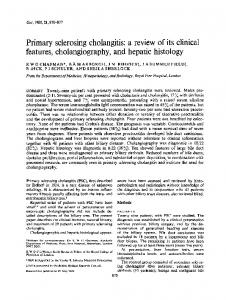
Figure 2. Transcystic laparoscopic cholangiography - migrated CBD lithiasis
Results We present some representative cases in the study group for the present study. Case 1: D. E., male., 50 years old, admitted with upper abdominal pain, nausea, scleral subclinical jaundice. In terms of paraclinical tests, he presented BT 0.9 mg / dl, BD 0.1 mg / dl, AST 33 U / l, ALT 66U / L, glucose 330 mg / dl. Ultrasound detected a 9 mm CBD with the presence of several calculi, highly distended gallbladder, with a thick wall and a double contour, with the presence of microcalculi images. CT showed normal relations between elements. Operative protocol: Gallbladder hydrops. Dilated cystic duct. Transcystic cholangiography was performed (Fig. 2), showing dilated CBD, with a faceted calculus in the lower third, with a diameter of 5 mm and good oddian passage. Cholecystectomy is finalized, with subsequent resolution of calculi by endoscopic. ERCP. A catheter is placed and ERCP is performed - CBD dilated to 14 mm, 2 faceted calculi present. PSE is performed and multiple calculi are extracted using the Dormia probe, abundant biliary magma being also discharged. The evolution has been favourable, with discharge 4 days post-ERCP. Case 2: T. E. L., female., 65 years old, admitted with right upper quadrant pain, nausea, vomiting, subclinical jaundice. In
Figure 3. Post-ERCP transcystic cholangiography
terms of paraclinical tests, she presented ESR 61 mm / h, BT 1.85 mg%, BD 0.85 mg%, AST 98 U / l, ALT 245 U / L, glucose 127 mg%. Ultrasound: Gallbladder with many intraluminal hyperechogenic images of 0.4 to 1.6 cm. CBD 1.6 cm wide at the hepatic hilum, 1.3 cm distally, with echogenic intraluminal material in the proximal segment. A catheter is place via the papillary pore and ERCP is performed: dilated CBD, presenting one voluminous calculus and multiple calculi upstream; modal distribution of intrahepatic bile ducts (IHBD), dilated cystic duct draining into the retropancreatic segment of the CBD. Wide PSE is performed, extracting multiple migrated and local calculi. Operative protocol (3 days postERCP): gallbladder with thickened wall, oedematous, with dilated cystic duct of 5 mm and CBD of about 15 mm. The cystic pedicle is dissected, the calculi is distally clipped, transcystic cholangiography is performed showing dilated biliary duct, narrowing at oddian level, but almost immediate passage of contrast agent. (Fig. 3) Cholecystectomy is finalized. The
521
postoperative evolution was favourable with discharge on the third postoperative day. Case 3: S. A., male., - 58 years old,admitted with colicky right upper quadrant and upper abdomen pain, nausea, fever, symptoms remitted under outpatient treatment. Bio-humoral explorations were within normal limits. Ultrasound detected a gallbladder moulded on calculi, with multiple calculi 0.5 to 0.7 mm with intense adhesive pericholecystic process. Normal sized CBD. Operative protocol: Small gallbladder, scleraatrophic with thick wall, oedematous. Thickened cystic duct with increased diameter, inhabited by lithiasic conglomerate. The cystic duct is incised and lithiasic material, 0.3 cm in diameter, is discharged. Transcystic cholangiography is performed, revealing the presence of calculi in the CBD and the common hepatic duct. (Fig. 4) Transcystic choledochoscopy is performed, extracting lithiasic material and revealing an inflamed, thinned papilla. Cholecystectomy is finalized. The postoperative evolution was favourable. Case 4: C. A., female, 40 years old, another case of intraoperative indication (dilated cystic duct) in which a lower duct calculus is revealed. (Fig. 5) In case of transcystic laparoscopic cholangiography there were no accidents, complications specific to cholangiography or mortality attributable to the method. Duration varied depending on the technique and instrumentation used: that of transcystic cholangiography using cholangiography forceps decreased to 8 minutes, and the average length of a transvesicular cholangiography was around 6 minutes. There were only 11 failures of the 100 cholangiographies performed. (Table 1) A number of 89 successful laparoscopic cholangiographies was recorded. 64 showed changes suggestive of various pathologies: malignancy (11 cases), benign alithiasic (7 cases), benign lithiasic (46 cases) and 25 offered normal relations between elements. The Pietrafitta classification,spanning five degrees, was used to assess the quality of results (Fig. 6): I. Contrast agent opacifies the cystic duct only. II. Contrast agent highlights the cystic duct up to its junction to the common hepatic duct. III. Contrast agent opacifies the choledochus to the Oddi notch. IV. Distally the contrast agent passes into the duodenum and proximally it reaches the right and left hepatic ducts. V. The entire biliary tree is opacified.
Table 1. Causes of failure in laparoscopic cholangiography Type of cholangiography Transvesicular Transparietohepatovesicular Transcystic
Cause of failure Image impossible to interpret (only the gallbladder is opacified) Image impossible to interpret (only the gallbladder is opacified) Catheter could not be placed in the cystic duct
No. of cases 4 1 6
Figure 4. Transcystic laparoscopic cholangiography – migrated CBD lithiasis
Figure 5. Transcystic laparoscopic cholangiography – migrated CBD lithiasis
Figure 6. Distribution of cholangiography result depending on quality
To verify the relevance of laparoscopic cholangiography indications in lithiasic pathology we removed from the initial group (100 patients) a number of 13 patients with malignancy, 7 patients with benign alithiasic pathology, 11 patients whose cholangiography images could not be interpreted, patients with CBD calculi who were submitted to preoperative ERCP followed by calculus extraction. We thus obtained a lot of 54 patients. We excluded cases of preoperative ERCP, usually
522
jaundiced patients with high suspicion of CBD calculi, subject to a standard protocol of minimally invasive treatment, on the one hand because the preoperative diagnosis of migrated lithiasis is highly probable, and on the other hand because endoscopic calculus extraction greatly alters the results of cholangiography by determining choledochal clearance. The authors chose the dilated cystic duct as the criterion for verifying the intraoperative indication. CBD dilation as a criterion was also verified, but no statistical correlation with the intraoperative indication could be found. The diameter of the cystic duct was assessed intraoperatively by comparing it to the diameter of the cholangiography catheter during cholangiography or to the right dissection forceps. Another evaluation criterion was cholangiography catheter employment facility, the cystic duct lumen having to overcome by at least one millimetre its diameter. To verify the relevance of the selection criteria we performed a statistical analysis of the group with the help of χ2 significance test. (Table 2) Sensitivity of the dilated cystic duct parameter for lithiasis is 84%, indicating it as a good method to detect choledochal calculi.Specificityhowever is unsatisfactory, with false negative cases, its value rising only to 62%. The reason behind this, we believe, is mute choledochal lithiasis, with migration at a distance from the time of surgery. Thepositive predictive value of the “dilated cystic duct“ parameter for CBD lithiasis is 55%. The negative predictive valueis 88%. In terms of performance evaluation of cholangiography in CBD lithiasis (Table 3), in 2 patients with negative results (false negative perhaps by "drowning" the microcalculi in contrast agent) presence of choledochal lithiasis was subsequently discovered (jaundice +/- angiocholitis), requiring ERCP. Sensitivity of laparoscopic cholangiography in the detection of choledochal lithiasis reaches up to 91%, specificityto 100%, positive predictive value to 100%, and negative predictive value to 92%.
Table 2.
Testing the relevance of the intraoperative indication (of the criteria cystic duct dilated over 3 mm)
Non-dilated cystic duct (D+) Non-dilated cystic duct (D-) Total χ2 =10. 972 with p=0.001
Table 3.
Cholangio+ CholangioTotal
Cholangio+(C+) Cholangio-(C-) Total 16 13 29 3 22 25 19 35 54
Assessment of cholangiography exploration performance in CBD lithiasis MBD+ lithiasis 21 2 23
MBD- lithiasis 0 23 23
Total 21 25 46
Discussions At the moment there is no consensus on the indications, advantages, role and value of laparoscopic cholangiography, but many approaches, sometimes extreme, each based however on statistical studies (8, 9, 10). Based on the study, and the literature review we performed (11, 12, 13), we consider it appropriate to address several issues, such as the place and role of laparoscopic cholangiography in the exploration of the biliary tree, performing it electively or routinely for lithiasic pathology, patient selection criteria for this method, problems related to the technique itself, and interpretation of results and subsequent therapeutic attitude. As with other laparoscopic procedures, the existence of a learning curve has been demonstrated for laparoscopic cholangiography as well. Depending on the volume of such procedures performed in that centre, the key figure of this curve is around 30-40 cases. Some authors (14) indicated a figure of 16 to 25 cases as necessary to acquire the technique, from 46 cases in performing cholangiography the success rate approaching 96%. A significant decrease in performing this procedure was registered since the introduction of ERCP and the change in preoperative indications for performing laparoscopic cholangiography. The standard protocol by which our clinic abides for minimally invasive treatment of migrated CBD lithiasis accompanied by jaundice is initial endoscopic approach, by ERCP, endoscopic papillosphincterotomy (PSE) and calculus extraction with Dormia instrument, followed by laparoscopic cholecystectomy after 48-72 hours. A relative indication of laparoscopic cholangiography in these patients was verifying the emptiness of the CBD to detect any migration of microcalculi post-ERCP or even during cholecystectomy, a situation that we encountered in 3 of 16 cases (18.75%) that presented this criterion. The high percentage discovered prompts to evaluation by laparoscopic cholangiography in all patients in this category. Only part, namely 16 of 42 (38%) patients with cholecysto-choledochal lithiasis undergoing minimally invasive therapeutic protocol were submitted to control laparoscopic cholangiography, but the trend is growing due to accumulation of experience by the surgical teams and by improvement of facilities, including a flexible choledochoscope, making the laparoscopic approach of CBD lithiasis possible. Currently there is a preference for other high-fidelity and less invasive imaging methods, which are performed before surgery, and less use of laparoscopic cholangiography as a method of first choice in exploring jaundiced patients (15). Malignant biliary pathology is not an indication for laparoscopic cholangiography. Surgical departments where this type of pathology are addressed must have access at least to ERCP, transhepatic percutaneous cholangiography and computer tomography. There is one category of information specialists largely agree on, namely intraoperative indications (intraoperative suspicion of migrated lithiasis - including cystic lithiasis, unclear anatomy due to presence of abnormal bile or local remodelling secondary to the pathological process, but also suspicion of iatrogenic injuries). In the present case the results of the study
523
are consistent with those from other centres, the authors identifying high statistical correlation between intraoperative and positive results for CBD lithiasis at laparoscopic cholangiography. ERCP maintains the elective exploratory role in jaundiced patients, but being invasive and having its own morbidity and mortality it cannot be suggested routinely to all patients with suspected CBD lithiasis. It has, however, the therapeutic advantage, allowing resolution of choledochal lithiasis in the majority of cases diagnosed. Preoperative detection of choledochal lithiasis is important because it can relieve the patient from additional interventions and allows adaptation of the therapeutic strategy. Choledocholithiasis, regarded as a contraindication for laparoscopic cholecystectomy prior to the introduction of minimally invasive manoeuvres for the CBD, today can be resolved before, during or after the laparoscopic intervention. Laparoscopic cholangiography is very useful not only to detect choledochal lithiasis, but also to highlight anatomical variants, creating a true "map" of the biliary tree, preventing thus iatrogenic biliary lesions.(16) Most surgeons recommend it, but in selected cases: cases suspected, especially intraoperatively, of choledochal lithiasis; cases with unclear anatomy of the CBD (prevents accidents during surgery); when cystic duct dissection is difficult, cholangiography provide details about the length, calibre, angle of draining into the choledochus and the possible presence of gallstones buried in the cystic duct; in the presence of a sclera-atrophic gallbladder, with creases at the level of the hilum, when presence of a bilio-biliary or biliary-digestive fistula is suspected. Once the learning curve overcome, the success rate of laparoscopic cholangiography is similar to that of open cholangiography, approaching 90% (17). The causes of failure can be: narrow cystic duct, cystic duct rupture, obstructive Heister valves, calculus impacted in the cystic duct, cystic perforation with extravasation of the contrast agent. Alternative methods were sought for intraoperative diagnosis of CBD lithiasis and for viewing local anatomy. Of these, the most used today for intraoperative exploration are intraoperative ultrasound and choledochoscopy, with proven efficiency. Statistics published in the literature, highlight an alarming increase in biliary lesions, bleeding injuries, peritonitis, with an increasing number of laparoscopic cholecystectomies performed. Prospective studies (18), reveal however a decreased incidence of ductal iatrogenic lesions using routine intraoperative cholangiography. The Canadian Society of Laparoscopic Surgery reported that only 2% of surgeons perform routine laparoscopic cholangiography, and 33% never do. (19) Supporters of routine cholangiography believe that CBD lesions can be minimized using adequate techniques for performing the cholangiography. This reveals the importance of recognizing immediately CBD lesions as a way to decrease morbidity and mortality by addressing their immediate resolution. Proponents of selective cholangiography consider that the
opportunity of performing laparoscopic cholangiography is determined mainly by intraoperative conditions. The algorithm that seems the most agreed on today divides patients into patients with high suspicion of CBD lithiasis, patients with small and no suspicion of calculi, cholangiographic exploration being reserved for those with small suspicion, those with high suspicion being preoperatively explored by ERCP. Preoperative indications of cholangiography are restricted due to the development of new exploration methods, among which we mention cholangio-MRI and EUS, thus being it rarely necessary to resort to cholangiography exploration. Arguments for selective cholangiography: • results and image qulity obtained appear to be lower than those provided by open technique; in addition, some authors have communicated false negative and false positive results, although in a minority of cases, with inadequate therapeutic consequences • in over 90% of cases exploration is unnecessary in case of practitioners of routine cholangiography • improving the means of preoperative diagnostic imaging determines the percentage of residual duct calculi in those submitted to selective cholangiography be, paradoxically, almost identical (very low) with those who practice routine cholangiography (20) • in terms of avoiding iatrogenic lesions, we mention that they mostly occur during or after the preparation of the cystic duct, so that for those who practice routine transcystic cholangiography, its prophylactic role turns into a diagnostic one; this can be avoided by using the transvesicular techniquede • although very low as a percentage, accident and complications specific of they method exist • last, but not least, the surgical act is extended up to 20 minutes in some cases and the costs involved increase
Conclusions Laparoscopic cholangiography must be performed electively on the basis of well-established intraoperative and / or preoperative indications, thus avoiding performing a large number of unnecessary procedures, with their inherent disadvantages and risks. In units provided with advanced equipment (ERCP, CT, cholangio-MRI), the main indications for performing laparoscopic cholangiography are intraoperative ones: intraoperative suspicion of common bile duct lithiasis, unclear local anatomy, biliary malformations, suspected iatrogenic injury. The most reliable intraoperative parameter indicating laparoscopic cholangiography in case of suspicion of common bile duct lithiasis proved to be cystic duct dilatation over 3 mm. Laparoscopic cholangiography proved to be a sure method of intraoperative exploration, with morbidity and mortality specific to the null method, but also with excellent reliability in highlighting CBD calculi (specificity 100%, sensitivity 91%, positive predictive value 100% and negative predictive value 92%).
524
The relationship between laparoscopic cholangiography and ERCP, although initially competitive, has later become complementary by exact delineation of indications. Although endoscopic exploration has significantly higher risks than laparoscopic cholangiography, it is preferred in jaundiced patients, because, in addition to diagnostic data, it brings forth an easy minimally invasive therapeutic option with excellent results.
Conflicts of interest. Source of Funding None declared.
References 1. 2. 3. 4. 5. 6. 7.
8.
9.
Blumgart L, editor. Surgery of the Liver and Biliary Tract. New York: Churchill Livingstone; 1994. Lefkowitch JH. Hepatobiliary pathology. Curr Opin Gastroenterol. 2006;22(3):198-208. Kersjes W, Thelen M. The importance of imaging tehniques in gallstone disease. Aktuelle Radiol. 1993;3(3):167-171. Soper NJ. Laparoscopic general surgery: past, present and future.Surgery. 1993;113(1):1-3. Dragomirescu C. Chirurgia laparoscopicã. Actualitãåi şi perspective. București: Editura Tehnicã; 1996. Duca S. Chirurgia laparoscopicã. Ed a II-a. Cluj-Napoca: Editura Paralela 45; 2001. Juvara I, Setlacec D, Rãdulescu D, Gavrilescu S, editors. Chirurgia cãilor biliare extrahepatice. București: Editura Medicalã; 1989. Brãtucu E, Ulmeanu D, Ungureanu D, Mavru M, Daha Cl, Marincaş M, et al. Colangiografia laparoscopicã - experienåa Clinicii de Chirurgie Caritas. In:Volumul Congresului de Chirurgie Laparoscopicã, Sinaia; 1996. p. 35. Lezoche E, Paganini A, Guerrieri M, Carlei F, Lomanto D, Sottili M, et al. Tehnique and results of routine dynamic cholangiography during 528 consecutive laparoscopic cholecystectomies. Surg Endosc. 1994;8(12):1443-7.
10. Willekes CL, Edoga JK, Castronuovo JJ, Widmann WD, McLean ER Jr., Chevinsky AH.Technical elements of successful laparoscopic cholangiography as defined by radiographic criteria. Arch Surg. 1995;130(4):398-400. 11. Tetik C, Thompson DM, Arregui ME. Preoperative, intraoperative and postoperative imaging techniques for diagnosis leading to the treatment of common bile duct stones. Semin Laparosc Surg. 1997;4(1):9-17. 12. Cuschieri A, Berci G. Laparoscopic Biliary Surgery. 2nd ed. Oxford: Blackwell Scientific Publication; 1992. 13. Curet M, Zucker K. Laparoscopic surgery of the biliary tract and liver. In Zuidema G, editor. Shackelford’s Surgery of the Alimentary Tract. 3rd ed. Philadelphia: WB Saunders;1996. p. 257–278. 14. Molloy M, Bower RH, Hasselgren PO, Dalton BJ. Cholangio-graphy during laparoscopic cholecystectomy – cumulative sum analysis of an institutional learning curve. J Gastrointest Surg. 1999;3(2):185-188. 15. Holzman MD, Sharp K, Holcomb GW, Frexes-Steed M, Richards WO. An alternative technique for laparoscopic cholangiography. Surg Endosc.1994;8:927-930. 16. Brãtucu E, Straja D, Ulmeanu D. Anomaliile cãilor biliare extrahepatice. Implicaåii în patologia hepatobiliarã. Chirurgia (Bucur). 1994;43(2):28-36. 17. Voyles CR, Sanders DL, Hogan R. Common bile duct evaluation in the era of laparoscopic cholecystectomy. 1050 cases later. Ann Surg. 1994;219(6):774-750. 18. Pietrabissa A, Di Candio G, Giulianotti PC, Shimi SM, Cuschieri A, Mosca F. Comparative evaluation of contact ultrasonography and transcystic cholangiography during laparoscopic cholecystectomy: a prospective study.Arch Surg. 1995;130(10): 1110-1114. 19. Soper NJ, Dunnegan DL. Routine versus selective intra-operative cholangiography during laparoscopic cholecystectomy. World J Surg. 1992; 16(6):1133-40. 20. Balogh A, Vattay P. The value of routine operative cholangiography in laparoscopic cholecystectomy (change in attitude towards the primary diagnostic and surgical treatment of bile duct lithiasis). Rom J Gastroenterol. 1996;5(2):77-83.