College of Medicine,. Bronx,. New. York. t Presently at Coney. Island. Hospital,. Brooklyn,. New. York. Supported in part by U. S. Public. Health Service Research.
FEBRUARY,
TRANSHEPATIC CHOLANGIOGRAPHY WITH SPLENOPORTOGRAPHY IN PANCREATIC CARCINOMA* By
HARRY
MILLER, JOSEPH and
M.D.,f LOUIS R. M. DEL D. COHN, M.D., NEIL R. RAMA P. COOMARASWAMY, NEW
BRONX,
P ERCUTANEOUS
this
has
procedure
been
with
percutaneous
order the extent At the Bronx
portography
delineate process.
spleno-
to more of the Municipal
in
completely obstructing Hospital
From
t Presently Supported
the
Departments at Coney
of Radiology Island
Hospital,
in part by U. S. Public
and
Surgery
Brooklyn,
Health
Service
New
of the
GUERCIO, M.D., FEINS, M.D., M.D.
Becton,
Dickinson
and
Co.,
Ruther-
ford, N. J.) were used. During performance of the splenoportography, a radiopaque skin marker was placed over the proposed site of skin puncture for the transhepatic cholangiography. This point was usually cm. below the right costal margin in the mid-clavicular line. When the anteroposterior splenoportograms were examined, the relation of the needle site to the portal vein bifurcation could be seen. This made it possible to direct the 12 cm. 20 gauge spinal needle accurately towards the hilus of the liver where the larger bile ducts are more likely to be found. Following removal of the stylet and attachment of saline filled flexible tubing, the needle was withdrawn from the liver, millimeter by millimeter, until bile was aspirated. As much bile as possible was removed and the biliary tree was filled with 50 per cent diatrizoate sodium solution (hypaque). After sufficient contrast material had been injected during visualization with image intensification, an anteropostenor roentgenogram was taken and the needle was removed. The patient was then maneuvered into various positions to ensure that the ductal system had been completely filled. A roentgenogram is also taken in the 3
Albert
Einstein
College
of Medicine,
York.
Research
F.A.C.S.,
YORK
8o-S,
Center, I 5 such combined examinations were carried out and they aided in the practical clinical management of these patients. All of the examinations were elective procedures usually performed just before the exploratory laparotomy for obstructive jaundice. They were done immediately before surgery in order to lessen the danger from bile leakage or hemorrhage. Splenoportography was attempted first and transhepatic cholangiography was undertaken immediately after these serial films had been seen and interpreted. Our technique for performing splenoportography has been previously described.3’5 Since there is seldom significant splenomegaly in these patients, autogenous, heat-denatured chromium 5 I tagged red blood cells were injected intravenously.7 A scintillation counter was placed in different positions over the left upper quadrant of the abdomen and over the left lower lateral costal margin. Scanning was done and the needle was later directed towards the point of maximum radioactivity.4 When this technique was used, it was always possible to find the B
COMBINED BILIARY
splenic pulp with the first puncture. A volume of 40 ml. of 66.8 per cent sodium iothalamate (conray_4oo) was injected in 10 seconds through an i 8 gauge spinal needle. A Stirling, manual, lever operated, pressure injector (Number AD-474o, Charles Thackray, Ltd., Leeds, England) and a 50 ml. plastic syringe (Number
transhepatic chodescribed in flumerous recent articles. The diagnostic value of this examination for patients with extraparenchymal obstructive j aundice is well established. However, no reference has been made regarding the combination of langiography
1966
Grant
468
No.
FR-66 OG i
RISI.
Bronx,
New
York.
96,
\OL.
erect
No.
iranshepatic
2
position
so
gallbladder lilese
two
getlier
edge,
an(.1
also
tile
prior
surgeon
to
operative
to
ment,
veins
to
(_)f this
Illa\’
Ilot
possible
metastatic
superior
ulesenteric
VC1lI
cava,
tOgrapil
.
during
pres-
invariably
found
since
man\’
areas
disease, vein
visualized
b’
cases v
however,
of
splenopor-
posterior
to
defects
in which performed,
w as
assess
biplane tii e
to
the
hepatic
the
pan-
nature
of
the
stone.
demonstrating ampulla
lile
nlajoritv
noma
of
of
the
complete
cases
Iii our
of
series
1)arium
bined
of
transii
ilelptul
epatic
altiiough,
in
creas,
the seen
011
tile
or
110
correlation
impression
year
old
M.R. male
with
to tile
comand
y
par
was
but
the
portal
patient gallbladder
us
to diagnose
and
pa1creatic
I1()
the
portal
region,
free of pathology. found to have
was
with evidence
duct of
or splenic
CASE
C.l).
11.
of
we were At operation,
and
with
extension
correct
presence
a carcinoma
stones
hepatic
VZt5
the
the
cause the
al)le of of
invasion
to
the the the
obstruction. ‘I’here to the vicinity’ of’
veins.
(B.\1.H.C.
18165S),
an
elderly marked
also
came
tile advanced
obstructi
ye
j au ndice. The splenoportogranl B) showed complete obstruction vein with collateral veins drain-
tile in
upper any of
( Fig. 3, Il atid
T5R905),
hospital
with
varices. The transhepatic (Fig. 4, A and B) demonstrated complete obstruction of the common bile duct with dilatation of the ductal system. The characteristic convex contour at the point of oh-
cholangiogram
suggested head of the
the diagnosis of carcinoma pancreas and the spleno-
portographic findings suggested that was far advanced and unresectahie.
an
with
to the
of the splenic ing into gastric
struction of the
CASES
presented
tumor
woman,
mentioned.
patient,
by
ticularl’
with noted
(B.M.H.C.
The wa
common
system.
gastrointes-
not of
card-
tile
series
I.
tile
roost of
ILLUSTRATIVE CASE
biliar\’
prior
invaded
not.
enabled obstruction
state,
ilead of the panof an extrinsic mass loOn of tile duodenum.
carcinonla
lesions
of
tile
were
of
otiler
those
cholangiograph
v
gastrointestinal
stric-
presented.
pancreas
made
cases
Little
benign are
the
of patients, studies
splenoportograph
the
were
head
obstruction
tinal
was
a of Vater
was
in a patient ‘I’he common
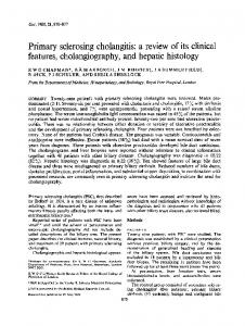
standing obstructive jaundice. Splenoportography tOlk)Wed iflinlediately by transhepatic cholangiography was perlormed before surgical exploration. ‘I’he splenoportogranl (Ii’ig. i showed a normal splenic and portal vein but the transhepatic cholangiogram (Fig. 2, 1 and B) demonstrated a mass invading the common hepatic duct, with evidence of a stone in the distal end of the common bile duct. Dilatation of the ductal system itself was also seen. These
genograns tile
splenoportogram of the gallbladder.
duct
was
studies of the
of
Normal
i.
carcinoma
vein
tumor and its extent.5 Our illustrative cases include carcinomas of the hepatic ducts, gallbladder, ampulla of Vater and pancreas. In addition, roenttore
Case
I.
with
splenopor-
filling
as
phase
those
l’IG.
of
as tile inferior
Illetastases,
veiograii
us
such the
or
revealed
be
helped
the
be true,
of collaterals
creas
in
rnalignanc’, corollary
splenoportograph
absence
displace-
Tile
Ilot
In
an’
cure.6
C
tile
tography.
tile
was
Hepati
iisuallv
C1fl
If
for
are
\‘
the
advance.
pancreatic
lesion at operation be unresectal)le
tile
enables
occlusion
or
of
knowland
showed
or
I)iliarv
of
appraisal
This
approach
in
deformity
ence
to-
exploration,
tile
plan
portal
or
tile
diagno-
an
lesion.
procedure
splenic
of
anatomic
permit
of
gaitled
the
filling performed
accurate
an
resectal)ility
tile
possible
place.
exanlinations
Illake
possible
sis
that
takes
469
Cholangiographv
long-
tion,
this
impression
was
confirmed.
the lesion At opera-
l\’Iiller
470
et (ii.
IEBRlJR\’,
J
1966
(
In
common
duct
0
____
I. 11G.
2.
Case i. (‘4) Transhepatic with i!lVItSiOn of the
male and
III. A.E. underwent transhepatic
tion
of the
hepatic
duct.
(R)
cause
of severe
unremitting
splenoportogram
(Fig.
metastases operation
of the advance the lesion and
venous
knowledge of the uninvolved
the
na-
portal
system.
)
was
entirely
CASE
or tumor extension was was quickly performed
year
old
E.W.
Negro
woman, the second
of
denum. portogram hold-up
Jaundice
(Fig.
7,
ofcontrast
junction.
This
posterior
and
trinsic of the
(B.M.H.C.
IV.
struction
interpreted
lateral due axis
263268),
was found portion
was also 1 and material
was
pressure celiac
-.l5.
a
to have of the
29
obduo-
present. A splenoB) demonstrated at the splenoportal from
the
antero-
roentgenogranls to pathologic lymph nodes.
a
as
ex-
enlargement The trans-
1
carcln:m:to:s
l1G.
of the gallbladder of Vater.
jaundice.
but the transhepatic cholangiogram (Fig. 6, 1 and B) showed complete obstruction of the distal common duct. On the basis of these findings we believed that we would find a resectable tumor of the head of the pancreas. At surgery, it was possible to perform a pancreaticoduodenectomy and no evidence of node The
demonstrating carcinoma found in the ampulla
was
because ture of
negative
lymph found.
sketch
A stone
(B.\I.H.C. 198234), an elderly combined splenoportography cholangiography for delinea-
CASE
The
and
cholangiogram
common
3. Case
pancreas. of contrast portal
II.
There vein.
(A)
Splenoportogram
is complete
material
is seen
and
obstruction in the
spleen.
(B)
ofthe
obstruction
0
sketch showing far advanced carcinoma of#{149} the head of the splenic vein with gastric varices. Subcapsular extravasation
Some
of the
contrast
material
can
be seen
to have
reached
the
96,
\o1.,
No.
Transhepati
2
lI(;. 4. Case
II.
hilc
COi11i1U)n
hepatic showed
(A) duct
Transhepatic cholangiogram with a characteristic convex
cholangiogranis obstruction ‘I’he
of
diagnosis
the of
and
bile
bile
C)
duct
ducts
carcinoma
(B)
and of
the
471
sketch showing due to carcinoma
contour
B and
( Iig. 8, 1, the common
of
dilatation
proximal
Witil
gallbladder.
c Cholangiographv
‘Ehe ance
fusit’orm of the
rather than obstructed
may
material
diagnosis
the complete of the head
have
in this
of \ater was confirmed at surgery and although the lesion WaS technically resectable, the tumor had metastasized to the regional lymph nodes including those surrounding the celiac axis. Several months following the
exploration
pancreat
dice
obstruction of
the
of
the
pancreas.
convex appearof contrast a clue to the correct blunt
column
been
case.
aflll)ullIt
icoduodenectom
tography
performed.
was
currence
of the intrahepatic
plus
metastatic
v,
splenopor-
This
obstruction filling
deposits.
repeat
showed
of defects
This
the
was
a re-
splenic suggestive
later
vein of
confirmed
at autopsy.
old
CASE male
V.
R.l). had
progressive
(B.i\l.H.C. 2 month
a
was
a
technical
cholangiography
the
bile
constriction.
Because
make
portal
attempt
at
contrast material (l’ig. 9, /1 and
a distal evidence
Our
denlonstrated duct was
system
benign
that
in
was
stricture
was excised Operative
rest
normal.
of the
an
colic. B) was
branch
of
was suggestive transhepatic
elderly
and to surgical
3atm-
obstructive
The splenoportograrn normal except for the
right
of an intrahepatic cholangiograrn
portal
( [‘ig.
an
vein. lesion. I I , .I
and B) showed choledocholithiasis and cholecvstolithiasis. In addition, irregularities of the gallbladder lumen and partial obstruction of the common hepatic duct were interpreted its malignant
disease
of the
gallbladder.
of
any to
a normal
upon the
diagnosis
the
This The
long-standing
biliary .1 and
obstructed
indicating
tion
of decided
we
revealed
carcinoma of the distal common out metastases or extension. At of Oddi WLS found.
10,
290787),
fusiform
superimposed
remaining B).
for
with
( 1’ig.
(B.M.l-I.C.
combined splenoportograph’ cholangiographv prior
splenoportography.
and
system
A.C.
VI.
Transhepatic
findings.
successful
venous
sphincter tumor
splenoportogd isten
with was no
of these
a second l)r0Ved
first
failure.
duct
There
stones.
of
demonstrated
common
‘Fhis
‘I’he
jaundice.
raphv
5$ year painless
a
319910),
history
CASE
male, had transhepatic
the
hiliary
was
tree
that
bile duct operation,
locally
and
the no
pancreatograms
of The
the final
ampulla
-‘
of a with-
pancreatic diagnosis
of Vater.
11G.
5.
patient
Case with
the pancreas volvement.
m.
Normal
resectable
without
splenoportogram carcinoma of the
evidence
of lymph
in
a
head
of
node
in-
Miller
472
et al.
I
i It RI
I RI
I (11)1)
0 l’l(;.
Case
.
At
surgical and chvma were CASE
(l’ig.
its
cholangiogram in carcinoma
tree
nature the liver
into
this paren-
of
confirmed.
VII.
but
12,
the
extension
(B.M.H.C. partial ol)structive
,‘.\\‘.
male, had splenoportogram
spects
(A) Transhepatic of the hiliary
exploration,
lesion
elderly The
iii.
was
the
‘I
and
of’ the
gallbladder
There
was
a partial
showed
and
tion
of
amount:of
the
common contrast
massive
bile material
appearance
duct
was
and
seen
a small trickling
through the area ofstenosis into a normal distal duct. At exploration, a carcinoma of the common bile duct was found and it was assessed to be surgically resectable.
re-
cholangiogram
intrahepatic
obstruction
an
jaundice. in all
normal
transhepatic
B)
297738),
and (B) sketch showing typical of the head of the pancreas.
DI S C U S S I 0 N
distention
bile of the
Ill
ducts.
all
cases
portographv
midpor-
in and
wilicil transhepatic
conibined
splenocholangiog-
..
partial spI.no
Fi;.
7. Case IV. (A) Splenoportogram and (B) sketch in a young woman. The lesion was resectable but the the obstruction at the splenoportaljunction, precluded
obstruction of portal junction
demonstrating
carcinoma of the ampulla of Vater to the celiac lymph nodes, shown here as the possibility ofa surgical cure,
metastases
96,
\on.
No.
Transhepatic
2
Cholangiograph
z_
gall
I’m.
8. Case
showing
the head
iv. the
(A
dilated
of the
-
bladder
and
B)
gallbladder
pancreas.
473
obstructed bile duct
Different and
views
total
and (C) obstruction
composite of the
sketch common
common
of the transhepatic bile duct secondary
cholangiogram to carcinoma
of
lIC. 9. Case demonstrating was normal
v.
raph\’
used,
(A) Splenoportogram what later has proven in all respects.
was
assessment
of
provided
an the
accurate of
this approach. are elderly and, risks. FFi erefore,
operative technique
which consuming
a time
the head of Splenoportographv
tile
superimposed to be a l)enign
diagnosis
extent
b’
patients
tree.’
By not
can help the surgeon but futile mobilization pancreas
is
itself,
and
is worth a particularly
however,
delineate
tile
portal hepatic, tases.2
of
and
use
of
the
our
nor
inflamof tile
Neither alone or
celiac however,
in the be gained the
from
series
sense from
simple each
of
sum
when
cases,
can reveal
all
transin tra-
axis
metastwo other and these
t each
complemen
than
tamed
primary
obstruction
periportal combination,
are synergistic mation can
a splenoportogram site
In
examinations
biliary
1)iliary
and (B) sketch splenoportogram
malignant from or obstruction
tile
venous pathways. cholangiograph’
hepatic
pursuing. sensiand
causing
cholangiogram of Tater. The
distinguish displacement
it
matorv
avoid
growth
transhepatic of the ampulla
tumor can
1966
IIIIIRIARY,
on the stricture
the lesion were Most of these in general, poor a roen tgenologi c
tive procedure for detecting the spread of tumors of the pancreas can
et cii.
1\liller
474
that more infortheir combined of
tile
data
oh-
used separately. In degrees of involve-
‘I
.--
N
obstruct.d of right
portol
branch v#{149}ln /-_
FIG.
10.
Case
VI.
(A)
Splenoportogram and (B) sketch, and obstructing a branch
the liver
showing carcinoma of the intrahepatic
L)
of the gallbladder portal vein.
extending
into
96,
\oi..
Transhepatic
No.
Cholangiography
475
7
.
I,
mon with
bile
duct
stones
tsl7o l’ic. I I. Case cholelithiasis
vi. (A) Transhepatic and choledocholithiasis,
cholangiognam there are
and
(B)
sketch
irregularities
of
demonstrating the gallbladder
that, in addition to the contour suggestive of
ncoplasm. of the pancreaticobi1iar’
nlent
Ill
all of
the
portal cases
system carcinomas where
an-
associated were abnormality
with noted.
the portal system external compression of
obstruction
(Fig.
was
3/f),
from sligilt to complete
seen,
(Fig. the
7/f)
tunlor
was
found
0 FIG.
12.
Case
VII.
(A)
Transhepatic cholangiogram and (B) sketch showing common bile duct causing a partial ol)struction.
a resectable
carcinoma
of tile
Miller
476
to be tion.
too
far
advanced
for
curative
resec-
et al. 2.
0.,
ARNER,
of dilated
under
Louis
R. M.
Department
Del
Guercio,
M.D.
of Surgery
Albert
Einstein
Bronx
6:, New
College
of Medicine
York REFERENCES
I.
ABEATICI,
S., and
CAMPI,
spl#{233}noportographiques
de l’abdomen 59, 803-834.
L. des
Les caract#{233}nistiques tumeurs
sup#{233}nieur. Acta
chir.
du foie et beig., 1960,
roentgen
H.,
MILLER,
STATE,
oportography
and
with
patients
4. JOHNSON,
and
nondilated
television
R. P.,
COOMARASWAMY,
Obst.,
cholangiography:
bile
ducts
Surgery,
control.
561-571.
1962,52,
3.
S. I.
SELDINGER,
transhepatic
puncture
Splenoportography combined wi th transhepatic cholangiography has been found to be of definite clinical value in determining the cause of obstructive jaundice and in estimating the resectability of biliary pancreatic carcinomas. A series of7 cases illustrating the value of the combined studies is presented.
S., and
HAGBERG,
Percutaneous CONCLUSIONS
1966
FEBRUARY,
cirrhosis
1964, zz8, P. M., and
considerations
in
DEL
portal
M. Splen-
ELKIN,
vein
of liver. HERION,
L. R. M.,
GUERCIO,
and
D.,
thrombosis Surg.,
Gynec.
in
&
J. C. Technical
scintillation
scanning
of
spleen. Radiology, 1961, 76, 438-443. 5. MILLER, H., COOMARASWAMY, R. P., DEL GUERCIO, L. R. M., ELKIN, M., and STATE, D. Value of biplane splenoportography. Radiology, 1963, 8z, 53-57. 6. R#{246}SCH, J., and HERFORT, K. Contribution of splenoportography to diagnosis of diseases of pancreas. I. Tumorous diseases. Acta med. scandinav., 1962, 171, 251-261. 7. WINKELMAN, J. W., WAGNER, H. N., JR., MC AFEE, J. G., and MOZLEY, J. M. Visualization human
of
spleen
Radiology,
in
man
1960, 75,
by radioisotope 465-466.
scanning.