articles
Encoding social signals in the mouse main olfactory bulb Da Yu Lin1, Shao-Zhong Zhang2, Eric Block2 & Lawrence C. Katz1 1
HHMI and Department of Neurobiology, Box 3209, Duke University Medical Center, Durham, North Carolina 27710, USA Department of Chemistry, University at Albany, State University of New York, Albany, New York 12222, USA
2
...........................................................................................................................................................................................................................
Mammalian urine releases complex mixtures of volatile compounds that are used in reproduction, territoriality and conspecific recognition. To understand how such complex mixtures are represented in the main olfactory bulb, we analysed the electrophysiological responses of individual mitral cells to volatile compounds in mouse urine. In both males and females, urine volatile compounds evoke robust responses in a small subset of mitral cells. Fractionation of the volatile compounds using gas chromatography showed that out of the hundreds of compounds present, mitral cells are activated by single compounds. One cohort of mitral cells responded exclusively to male urine; these neurons were activated by (methylthio)methanethiol, a potent, previously unknown semiochemical present only in male urine. When added to urine, synthetic (methylthio)methanethiol significantly enhances urine attractiveness to female mice. We conclude that mitral cells represent natural odorant stimuli by acting as selective feature detectors, and that their activation is largely independent of the presence of other components in the olfactory stimulus. Much of the social signalling between mammals involves the secretion, detection and interpretation of chemical cues. In addition to non-volatile, species-specific signals (which require physical contact between an animal and an odour source), mammals use numerous other chemical signals that are secreted in urine or other bodily fluids and can be detected at some distance from their source. Ewes, for example, recognize their offspring based on volatiles1, newborn rabbit pups locate their mothers’ mammillary glands using a volatile compound found in milk2, and mice can recognize one another based on diffusible signals originating from urine3. Whereas non-volatile cues are detected by the accessory olfactory system, volatile social cues are processed by the anatomically separate main olfactory system, which relies on an entirely distinct set of peripheral receptor molecules (see refs 4, 5 for recent reviews). Mouse urine, like most natural olfactory stimuli, consists of a plethora of distinct volatile compounds, of which only a fraction are known. The presence and level of many urine components vary according to the sex, strain, social status and oestrous cycle of individual mice6–9. Although this suggests that urine contains information that could be used to assign identity to conspecific mice, the chemical identity of the salient cues, together with the process by which individual neurons encode this complex mixture, remain unknown. To investigate how the stimulus features of urine volatile compounds are encoded by individual mitral cells, we used a combination of extracellular single-unit recordings and gas chromatographic separations of urine components.
we recorded from 2,733 mitral/tufted cells in the main olfactory bulbs of 102 sexually mature BALB/c female mice. Outside the responsive clusters, urine-responsive cells (neurons for which firing rates were elevated by .30% during stimulus presentation) were rare (Supplementary Fig. 2). A total of 208 neurons (almost all recorded in the responsive clusters) responded to volatiles derived from diluted urine of at least one mouse strain and sex (Fig. 1). This number substantially overestimates the overall frequency of urineresponsive mitral cells (see Supplementary Fig. 1c), as responsive regions were sampled with many more electrode penetrations. Subsequent recordings from an additional 536 neurons in male mice (11 BALB/c and 7 C57Bl/6 mice), focusing on the ventral– lateral regions, yielded a similar proportion (10%) of urine-responsive neurons (n ¼ 53). Post-hoc analyses of a random sample of neurons (n ¼ 113) indicated that ,5% of neurons (n ¼ 6) classified as unresponsive had changes of .2 standard deviations in their firing rates during stimulus presentations (as expected from a random distribution), whereas all neurons classified as responsive (n ¼ 50) had changes of .2 standard deviations (we cannot exclude the possibility that very weakly responsive neurons might be missed using our screening procedure). Thus, despite the chemical diversity of the dilute urine stimulus, the mitral cells responsive to urine in both male and female mice comprise a small proportion of neurons, confined to restricted regions of the main olfactory bulbs.
Mitral cell responses to fractionated urine Urine-responsive mitral cells in the olfactory bulb Urine activates immediate early gene expression in glomeruli located in the ventral–lateral region of the main olfactory bulb10,11. We began searching for urine-responsive mitral cells in this region in female mice and located two clusters, on the ventral and lateral aspects of the bulb (each extending about 300 mm along the anterior–posterior axis) in which urine-responsive mitral/tufted cells were present at moderate densities (Supplementary Fig. 1). The relative positions of these two clusters potentially reflects the activation of mirror-selective glomerular maps12,13. To assess whether urine-responsive neurons were found elsewhere, we systematically recorded from neurons at 100-mm intervals along the anterior–posterior, medial–lateral, and dorsal–ventral axes, sampling roughly two-thirds of the bulb (see Methods). In total, 470
Responses to urine began shortly after the onset of the first inhalation following stimulus presentation, and continued for the duration of the 2-s stimulus. Almost all responses were excitatory (241/261, 92%), and were frequently synchronized to the animal’s breathing cycle (Fig. 1a, b). A small fraction of neurons (15/261, 6%) were inhibited by urine volatiles (Fig. 1c), and an even smaller number (5/261, 2%) were excited by some types of urine and inhibited by others. To distinguish whether the activation of a mitral cell in response to urine reflects the integration of responses to several distinct volatile components, or a selective response to an individual component, we combined solid-phase microextraction (SPME) of urine volatiles with gas chromatography (GC) and single-unit electrophysiological recording (gas chromatography-electrophysiology, or GC-E14,15).
© 2005 Nature Publishing Group
NATURE | VOL 434 | 24 MARCH 2005 | www.nature.com/nature
articles SPME is used to concentrate volatiles in the headspace above a volume of urine by dissolving them in a series of coatings that adsorb compounds of low molecular mass on the basis of size and polarity. The total amount of a volatile absorbed by a SPME fibre is linearly proportional to its initial concentration in urine16,17. To minimize the bias of SPME fibres towards chemicals with certain molecular weights and polarities, we used a fibre consisting of three phases: Carboxen, divinylbenzene and polymethylsiloxane. Although these are currently the most ‘unbiased’ fibres available18–20 they nonetheless concentrate some molecules to a greater extent than others, and might not concentrate some volatile compounds effectively (see Discussion). Despite these potential caveats, this approach allowed us to record the activity of urine-
responsive mitral cells to at least 100 sequentially presented volatile components of urine (Supplementary Fig. 3). We used GC-E to assess the responses of 104 urine-responsive mitral cells (from 57 BALB/c female mice, 10 BALB/c male mice and 4 C57Bl/6 male mice) to separated volatile components. For 83 neurons, responses were confirmed by at least two independent GC runs. After isolating neurons that responded to urine volatiles (Fig. 2a, d), we continuously recorded action potentials for 20 min while eluting volatiles that had been concentrated by SPME (see Methods). Responses to eluted compounds were unambiguous: an abrupt increase in firing rate occurred at the same elution times on successive GC runs (Fig. 2b, c, e, f). The size of eluted peaks and the magnitude of neural responses were uncorrelated (Fig. 3c), and minor peaks elicited responses as robustly as large peaks (Fig. 2g, h). Most urine-responsive neurons (80%, 83/104) were activated at a single time point, corresponding to a single eluted volatile urine component (Fig. 3a, b); 11% (11/104) responded at two time points and the remainder (10/104) responded at 3–5 time points (Supplementary Fig. 4). Averaging multiple runs to the same urine sample increased the signal-to-noise ratio of the responses, but did not reveal additional weak responses (Supplementary Fig. 5). The specific times of the responses were widely distributed over the course of the GC run. We identified at least 25 distinct response times, ranging from 2.5 min to 16 min after the start of the run (Fig. 3a). Several neurons responded to volatiles eluting at a specific time point, suggesting that we recorded from several mitral cells with similar or identical chemical selectivity across different animals. One particular subpopulation responded to a volatile eluting at ,508 s. As detailed below, these cells, observed in both males and females, responded to a component present exclusively in male urine (Fig. 3a, boxed region; cells that responded at this elution time but were not male-selective are not included in the box). These findings demonstrate that mitral cells are highly selective when confronted by a wide range of odorants at near-natural concentrations. This is not due to a weakened stimulus. Using a similar combination of SPME and GC-separation of urine volatiles, expert human olfactory analysts (Microanalytics) detected over 30 distinct odours eluting from the column (Supplementary Fig. 6). Mice probably detect a considerably larger repertoire of components.
Sex-selectivity of mitral cell responses
Figure 1 Urine-responsive mitral cells in the mouse main olfactory bulb. a–c, Responses of three single units in the mitral cell layer to stimulation by urine volatiles (2 s, indicated by black bar). An extracellular recording trace is shown for each cell, with the animal’s breathing rhythm shown directly underneath. Raster plots and post-stimulus time histograms show responses to multiple stimulus presentations. Most responsive cells were rapidly excited by urine volatiles (a, b) with responses synchronized to the breathing rhythm, but a few cells were inhibited by urine volatiles (c). In the raster plots, responses to different types of urine are indicated by different colours: green, C57Bl/6 male; yellow, C57Bl/6 female; blue, BALB/c male; red: BALB/c female. NATURE | VOL 434 | 24 MARCH 2005 | www.nature.com/nature
To assess the sex- and strain-selectivity of the urine-selective neurons in the main olfactory bulb, we calculated a sex-selectivity index (SexSI) and a strain-selectivity index (StrSI) for 98 of the 104 neurons analysed using GC-E (six neurons were excluded owing to incomplete data). Both indices range from 21 (activated exclusively by female or C57Bl/6 urine) to þ1 (activated exclusively by male or BALB/c urine) (Fig. 4g, h). Most neurons responded to urine from both strains, but 14% (14/98) responded much more robustly to BALB/c urine (from both sexes) than to C57Bl/6 urine (StrSI . 0.5). Neurons recorded in female mice showed an overall preference for male urine, with a median SexSI of 0.46; this bias was not observed among urine-responsive cells in male mice (median SexSI ¼ 0.03). We found a single female-selective neuron in one male mouse (SexSI , 20.5), but no female-selective neurons in female mice, even though we recorded from almost three times as many urine-responsive neurons in females compared with males. Within the population showing male preference (SexSI . 0.5), a subset of neurons responded exclusively to male urine (SexSI ¼ 1.0, n ¼ 14) (Fig. 4a, b). Three of these neurons were also tested with mouse urine from castrated males, and each failed to respond. Interestingly, 10/14 cells in this group were activated at virtually identical elution times (normalized time, 508 ^ 1.8 s). Almost all (9/10) were recorded at similar stereotaxic coordinates in several different animals. These neurons showed an unusual firing pattern
© 2005 Nature Publishing Group
471
articles in response to the eluted stimulus, consisting of a sustained discharge that persisted well beyond the putative elution peak. The activation onset of these neurons (at 508 s) closely matched the elution time of a known mouse pheromone, 6-hydroxy-6methyl-3-heptanone (HMH, ref. 21), which we identified by mass spectroscopy as a large peak in male urine eluting at 500 ^ 6.8 s. However, closer inspection of the GC profile revealed a small peak (partially embedded in the falling phase of the HMH peak) that reached maximum amplitude at ,508 s. This peak was present in male urine GC profiles but absent in female and castrated male urine profiles (Fig. 4c–f). To establish that these neurons were responding to this second component, and not to HMH or a similar volatile, we sequentially tested responses to volatiles eluted on two different GC columns (n ¼ 5). When neurons were presented with volatiles eluted from our standard column (BP-5), which separates molecules according to boiling point, they responded at the expected 508 s. When we switched to a column that separates volatiles according to polarity (BP-20), the responses of the same neurons invariably shifted to 705 s, coincident with a small GC peak. This peak was completely distinct from the HMH peak, which, on this column, eluted at 375 s (Supplementary Fig. 7). The similarity of the neuronal responses to the elution of volatiles using two
Figure 2 Urine-responsive cells are activated by single components. a, A neuron’s response to complete mouse urine volatiles. b, c, Firing rate (Hz) of the cell in a, as individual urine components are separated and presented using GC-E. Lower panels show gas chromatograms of C57Bl/6 male (b) and C57Bl/6 female (c) mouse urine. Larger peaks are truncated to enhance the visibility of smaller peaks. The cell responded at virtually the identical, single time point (433 and 434 s), in two independent GC-E recordings. Upper panel: vertical bar, 10 Hz; horizontal bar, 1 min. Lower panel: vertical 472
columns that substantially change the presentation order of the stimuli also demonstrates that the responses of these neurons were independent of antecedent odours.
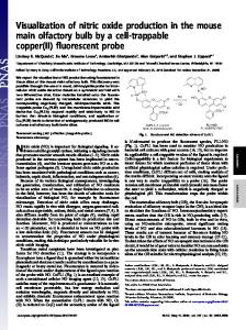
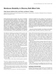
A novel sex-specific compound in male urine Data from high-resolution mass spectrometry indicated that the active compound had an exact mass corresponding to the formula C2H6S2 (molecular mass 93.9911). Analysis of sulphur isotope ratios also indicated that the compound contained two sulphur atoms. This peak showed fragment ions at m/q ratios of 61 (loss of SH), 47 (CH3S or CH2SH) and 45 (base peak; HCS). Taken together, the data suggest three candidate structures: dimethyl disulphide, 1,2-ethanedithiol and (methylthio)methanethiol (MTMT). As the retention time of this unknown compound on both GC columns was clearly different from that of dimethyl disulphide or 1,2ethanedithiol, MTMT was synthesized22 (see Supplementary Methods). The retention times on both the BP-5 and BP-20 columns, and the mass spectral fragmentation patterns for synthetic MTMT precisely matched those for the unknown compound with m/q of 94 (Fig. 5c). Some structurally similar sulphur-containing compounds were also found in urine, including dimethyl disulphide (CH3SSCH3), bis(methylthio)methane(CH3SCH2SCH3) and
bar, 100 mV; horizontal bar, 1 min. d–f, Another urine-responsive mitral cell activated by a specific component of BALB/c male (e) or C57Bl/6 male (f) urine eluting at a different time (240 s) from that seen in b, c. Scale bars as above. g, h, Expanded representations of the regions indicated by dashed lines in e and f. The maximal cell response coincides with a small peak in the GC profiles (black arrows). Upper panel: vertical bar, 10 Hz; horizontal bar, 10 s. Lower panel: vertical bar, 20 mV; horizontal bar, 10 s.
© 2005 Nature Publishing Group
NATURE | VOL 434 | 24 MARCH 2005 | www.nature.com/nature
articles methyl (methylthio)methyl disulphide (CH3SSCH2SCH3), but none of these elicited neuronal responses during GC runs. To humans, both synthetic MTMT and the native compound eluting at 508 s have a strong garlic or savory odour. We next tested four neurons (three in females and one in males) that responded to eluted urine volatiles at 508 s, this time using synthetic MTMT. When presented either as a volatile or by elution from the GC, the cellular responses to MTMT precisely matched those of the endogenous compound (Fig. 5a, b). Using synthetic MTMT to construct a dose–response curve, we established the response threshold as 10 p.p.b. (parts per 109, v/v) in water (Fig. 5d); male and female neurons had similar thresholds. By adding various amounts of MTMT to mouse urine, then performing SPME, we estimated that this molecule is naturally present in male mouse urine at a concentration of approximately 20 p.p.b. (v/v), but is almost undetectable in female urine. This value is similar to the behavioural threshold value reported for responses to another known volatile semiochemical that acts via the main olfactory system2. Native MTMT was detected in bladder urine, indicating that its presence in urine is not a consequence of bacterial metabolism, fecal contamination or compounds added to urine via glandular secretions. Thus we conclude that the male-specific responses in the main olfactory bulb primarily reflect activation of neurons by a previously unreported sulphur-containing compound secreted in male mouse urine.
Behavioural responses to MTMT Other sulphur-containing compounds, such as dimethyl disulphide and carbon disulphide, play important roles in rodent social behaviours23,24. To examine whether the presence of MTMT correlated with a behavioural role in sex discrimination, we took advantage of the fact that female mice strongly prefer to smell male (as opposed to female) urine, and that they largely ignore the urine from castrated males25, which has greatly reduced
Figure 3 Activation of urine-responsive neurons by single components. a, GC retention times at which individual neurons were activated. Normalized time points (squares) at which each of 66 urine-responsive cells were activated are aligned with a reference GC profile of male mouse urine. Red squares indicate female mice, black squares indicate male mice. Box indicates putative MTMT-responsive cells (see text and Fig. 5). b, Most NATURE | VOL 434 | 24 MARCH 2005 | www.nature.com/nature
concentrations of many volatiles including known pheromones and MTMT (we identified 112 peaks in the GC profile of intact C57Bl/6 male mouse urine but only 57 peaks in the urine of castrated C57Bl/6 male mice). Using a ‘Y’ maze assay, we found that all sexually experienced female mice (BALB/c, n ¼ 14) clearly preferred smelling urine from intact male mice to that from castrated male mice (P , 0.01, Fig. 6); most females (86%) also visited the arm of the maze containing the intact male urine first. To see whether MTMT could restore the attractiveness of urine from castrated males, we next presented female mice with a choice between ‘castrated’ urine and castrated urine to which 20 p.p.b. (v/v) MTMT was added. All but two females (n ¼ 16 out of 18) spent significantly more time investigating the castrated urine containing MTMT (P , 0.01, Fig. 6 and Supplementary Fig. 8). Female mice also showed a strong initial preference for the urine sample containing MTMT: 78% chose to sniff first at the arm with MTMT-containing urine. MTMT alone was ineffective in this behavioural paradigm, although female mice showed a slight preference for water containing 50 p.p.b. MTMT versus pure water. This implies the behavioural effects of MTMT are elicited in the context of other volatiles present in urine. The preference for castrated urine with added MTMT was not simply due to the presence of a novel odour. Acetophenone (another compound present in urine) added to castrated urine to a final concentration of 10 p.p.m. (at least tenfold higher than its endogenous concentration in urine) had no effect on female preference (Fig. 6). Although replenishing a single component—MTMT—does not fully restore female mouse preference for intact male mouse urine, it substantially increases the behavioural attractiveness of an otherwise uninteresting stimulus. Given that female mice respond differently to MTMT depending on whether it is presented in water or castrated urine, we tested whether the presence of castrated urine volatiles modulated the responses of MTMT-responsive mitral cells (Supplementary
mitral cells recorded from both sexes respond at single elution times; a small fraction respond at multiple times (colour conventions as in a). c, The amplitude of the eluted peaks in the GC record are unrelated to their ability to activate urine-responsive neurons (386 responses, compared with the GC signal, in mV, from the FID detector)
© 2005 Nature Publishing Group
473
articles Fig. 9). After isolating male-selective cells responsive to MTMT, we quantified the neuronal responses to 20 p.p.b. MTMT diluted in water, compared with 20 p.p.b. MTMT diluted in castrated urine (n ¼ 3). In all cases the responses were remarkably similar. Thus, the contextual dependency of female interest in MTMT is unlikely to arise from modulating the responses of MTMT-responsive cells themselves, but is probably a result of integration somewhere outside the olfactory bulb. Volatiles from body secretions signal the presence of potential rivals and mates and motivate investigational behaviour in many mammalian species4. Even humans can reportedly distinguish sex on the basis of olfactory cues26,27. However, few of these sexual attractants are known. Here we show that a male-specific compound, MTMT, isolated by its ability to activate single neurons, considerably enhances the attractiveness of urine to female mice. Unlike previously described pheromones, MTMT is highly volatile, with a calculated vapour pressure at room temperature of ,15 torr. This is about 20-fold higher than exo-brevicomin and 2,000-fold higher than farnesene, two well-described pheromones that act via the accessory olfactory system28–30. MTMT seems likely to advertise the presence of a male from a distance, either as a signal to other males or to attract females. Levels of MTMT could serve as an indicator for the freshness of urine, especially since other sulphurcontaining compounds in mouse urine, methanethiol (CH3SH) and dimethyl disulphide (CH3SSCH3), can react with MTMT
to form methyl(methylthio)methyl disulphide and thereby gradually decrease MTMT concentration over time. The finding that almost a third of urine-responsive neurons in both the male and female main olfactory bulb were activated by MTMT but that only a few cells were activated by any other single urine component (Fig. 3a) suggests either that multiple glomeruli are activated by MTMT, or that the glomerulus activated by MTMT is innervated by an unusually large number of mitral cells. These neurons could be associated with the 15–20 sexually dimorphic, ‘atypical glomeruli’ that have been implicated in the detection of biologically significant odours31–33. In other sensory systems, central representations of stimuli of particular significance have enlarged central representations, and the central representation of MTMT may represent an example of such an enhanced representation in the mammalian olfactory system, perhaps analogous to the enlarged representation of sex pheromones in the macroglomerular complex in insects34. A persistent issue in the field of olfactory coding has been the odorant specificity of mitral cell activity35–39. The idea of ‘specialist’ and ‘generalist’ responsive neurons34 has been invoked to explain the variability in molecular response ranges. However, the response range of mitral cells in mammals has historically been defined using relatively high odorant concentrations and restricted stimulus sets35,37. This does not necessarily relate to the discriminatory ability of mitral cells in biologically relevant contexts, where they are unlikely to encounter stimuli containing high, nearly identical concentrations of nearly identical stimuli. A more relevant question
Figure 4 Sex- and strain-specific responses to urine volatiles. a–f, A mitral cell activated specifically by male urine volatiles (a) and not by female urine (b). Bar indicates time of odour delivery. c, d, Individual components of male BALB/c urine (c) elicit a response at 508 s (normalized response, conventions as in Fig. 3a), female C57BL/6 urine (d) elicits no response. Scale bars as in Fig. 2b–f. e, f, Enlarged portions of the GC profiles (boxed regions in c and d) show a small peak in male urine (e, black arrow) as the putative peak
activating the cell response; this peak is absent in female urine (f). Upper panel: vertical bar, 10 Hz; horizontal bar, 10 s. Lower panel: vertical bar, 100 mV; horizontal bar, 10 s. g, Sex-selectivity index shows a substantial subpopulation of neurons with an index score close to 1, indicating responses exclusively to male urine; such cells were less common in males (black bars). h, Most neurons responded to urine from both strains tested (colour conventions as previously).
Discussion
474
© 2005 Nature Publishing Group
NATURE | VOL 434 | 24 MARCH 2005 | www.nature.com/nature
articles
Figure 5 Male-specific neurons respond to a novel semiochemical. a, Activation of a mitral cell by a single component of BALB/c male mouse urine eluting at 506 s (normalized response). b, Synthetic MTMT (structure shown at upper left) elutes at the same time (507 s) and evokes an identical response (scales as in Fig. 2b–f). The small apparent size of the MTMT peak results from the low concentration used. c, Mass spectra of
endogenous MTMT from mouse urine (upper panel) and synthetic MTMT (lower panel) obtained at the retention time corresponding to activation of the neuron in a and b. d, Dose–response curve to synthetic MTMT (averaged from four responsive cells, bars indicate mean ^ s.e.m.). Response threshold is approximately 10 p.p.b.
is the selectivity of mitral cells when confronted with arbitrarily large sets of distinct chemicals. In the case of projection neurons in the Drosophila antennal lobe (the fly analogue of mitral cells), imaging data suggest that most individual projection neurons respond robustly to only a small subset of stimuli40, but electrophysiological recordings of the same neurons indicate that most respond to a considerably broader range41. Broad tuning of mitral cells has also been reported in zebrafish36,39. Mice, however, have a far larger repertoire of olfactory receptors than either flies or fish42–44, and we see little evidence for such broad tuning; out of perhaps hundreds of possible volatile stimuli in urine, mouse mitral cells generally respond to just a single one. Although this apparent selectivity could reflect a limitation of the SPME approach (such as a systematic failure to adsorb certain urine volatiles) we consider this unlikely. We found no neurons that responded to presentations of complete urine volatiles but subsequently failed to respond to at least one component during the GC run, as would be expected if we failed to capture the relevant molecular species by SPME. In addition, urine volatile profiles isolated by a theoretically unbiased technique (cryogenic trapping) identified a total of 98 chemicals45. Using SPME, we detected between 100 (in BALB/c female urine) and 131 volatile compounds (in BALB/c male urine) and identified several components (in addition to MTMT) that were not observed by cryogenic trapping. Moreover, the chemical families isolated by the two approaches were the same. Other studies comparing cryogenic trapping with SPME have concluded that the sensitivity, linearity and reliability of SPME are comparable to or superior than cryogenic trapping46–48. Although it remains possible that when only a single activity peak is observed by GC-E, a small number of additional compounds might also induce responses, mitral cells clearly respond to only a very small fraction of the volatile stimuli present in urine. This specificity could reflect a case of ‘specialist neurons’, but preliminary results suggest the same degree of specificity in response to components in a range of other natural stimuli as well as to very large libraries of
pure odorants (I. Davison, D. Y. L. and L. C. K., unpublished observations). Although hundreds of compounds are present in natural stimuli, a much more restricted set of compounds may be responsible for constructing the olfactory percept. The olfactory image of a particular urine might be formed by the immediate, coordinate activation of a specific cohort of narrowly-tuned feature detectors49, rather than a time-dependent, emergent property of more moderately selective detectors50. Understanding how such a complex signal is represented provides insight not only into neural
NATURE | VOL 434 | 24 MARCH 2005 | www.nature.com/nature
Figure 6 Synthetic MTMT enhances the attractiveness of urine to female mice. In a Y-maze preference test, females strongly prefer urine from intact males compared with castrated male mice, as indicated by investigation time of the different samples (shown as mean ^ s.e.m.). Addition of 20 p.p.b. synthetic MTMT to castrated urine roughly doubles investigation time. Much higher concentrations of acetophenone (ACE, 1 p.p.m. or 10 p.p.m.) have no effect. Although 50 p.p.b. MTMT in water slightly increased investigation time compared with water alone, the absolute investigation time is much lower than that of MTMT presented in a castrated urine background. Asterisks indicate statistically significant differences (Wilcoxon signed-ranks test, P , 0.01).
© 2005 Nature Publishing Group
475
articles strategies for the coding of social signals, but also suggests a paradigm for understanding more generally how natural olfactory scenes are represented in the olfactory bulb and beyond. A
Methods All animal experiments were performed according to a protocol approved by the Duke University Institutional Animal Care and Usage Committee.
Electrophysiology After initial anaesthesia (10:1 ketamine:xylazine, intraperitoneal injection), sexually mature (3–9-month-old) BALB/c or C57Bl/6 mice (Charles River Laboratory) were maintained on isofluorane (1–3% in 100% O2). The intersection of the sagittal suture and the sinus separating the olfactory bulbs and cortex was used as a reproducible zero point for mapping. Isolated single units were recorded with tungsten microelectrodes (1.5–3 MQ, Microprobe Inc.), digitized (20 kHz) and analysed using Spike2 software (Cambridge Electronic Design Limited). Small electrolytic lesions (4 trains of 7 mA for 5 s, at 5 s intervals) were occasionally made and verified histologically.
Data analysis A cell was classified as urine-responsive if its firing rate during urine presentation changed by more than 30% from its spontaneous firing rate in the 2-s pre-stimulus interval. For each urine stimulus, responses to 2–5 stimulus presentations were averaged. We calculated a SexSI and StrSI as follows: SexSI ¼ (male urine response) 2 (female urine response)/ (male urine response þ female urine response) and StrSI ¼ (BALB/c urine response 2 C57Bl/6 urine response)/(BALB/c urine response þ C57Bl/6 urine response). Male and female urine responses are the sum of cell responses to BALB/c and C57Bl/6 animals of the respective sexes, and strain-selective responses are the sum of responses of both sexes to urines from different strains.
Odour stimuli Using custom-built metabolic cages, urine was collected from 5–10 animals into vials immersed in dry ice, and stored at 280 8C. Oestrous cycle was not controlled. Urine vapours (10% urine in dH2O) were presented in charcoal filtered room air (1.2 l min21) using a TTL-triggered, 6-channel olfactometer. Later experiments used a robotic 64channel olfactometer in which 100% urine vapour (0.2 l min21) was diluted into a 2 l min21 flow stream. To concentrate volatiles from urine, a SPME fibre (2 cm–50/30 mm DVD/Carboxen/PDMS StableFlex, Supelco) was inserted for 3–6 h into the headspace of a 1.5-ml vial with a Teflon septum (Agilent) containing 150 ml of mouse urine (saturated with NaCl, mildly heated to 37 8C–40 8C and constantly stirred). Extracted volatiles were desorbed at the GC injection port (Agilent model 6980, 260 8C, 3 min) and separated on either a nonpolar BP-5 or a polar BP-20 column (SGE, 30 m, inside diameter 0.25 mm) using helium (1.2 ml min21) as the carrier gas. Oven temperature was maintained at 50 8C for 2 min, ramped at 10 8C min21 to 200 8C, and at a further 30 8C min21 to a final temperature of 260 8C, maintained for 1 min. The column effluent was split equally and each half was routed to either a flame-ionization detector (FID, operating at 280 8C) or a mass spectrometry detector (Agilent model 5973, electron impact mode, 70 eV electron energy, 34–400 AMU at 3.93 scans s21) or the animal’s nose via a heated transfer line. To compensate for slight shifts in retention times, the retention times for an unknown compound (UC) from different GC runs were adjusted according to the retention time (RT) of HMH as follows: adjusted RT of UC ¼ (averaged RT of HMH/actual RT of HMH) £ actual RT of UC. During GC-E experiments, cells were analysed only if their firing rate changed significantly (99% confidence level) from their mean firing rate, and their activity lasted longer than one breathing cycle. If multiple GC-E runs were performed, a given response had to be repeated at least twice.
Behavioural analysis Sexually experienced, light-cycle adapted (light on from 1800 to 0600 h) BALB/c female mice (n ¼ 37) were used (during their subjective night, between 1200 and 1800 h) to examine the behaviour effects of synthetic MTMT in a urine preference test. Preference tests were conducted in a custom-made Y-maze (32 £ 12 £ 30 cm) with a sliding door regulating access to each arm. The test urine (50 ml, applied to a 1-cm2 piece of filter paper) was placed at the bottom of a clean 1.5-ml eppendorf tube, which was in turn fitted into a circular port (1.3 cm inside diameter) located at the end of each arm. The mouse could sniff at the opening of the tube but was unable to contact its contents. Tests were performed in darkness (videotaped under infrared illumination). After habituation to the test apparatus (5 min), the animal was restricted to one arm, and two tubes containing filter paper with water only were inserted into the sniffing access ports. The animal was released and allowed to freely investigate for 5 min. The procedure was then repeated with urine samples placed on the filter paper (2-min trials). Videotapes were scored for the time spent sniffing each urine stimulus (snout oriented towards the opening and held within 1 cm of it) by an individual blind to the conditions being tested. Received 23 September 2004; accepted 2 February 2005; doi:10.1038/nature03414. Published online 20 February 2005. 1. Baldwin, B. A. & Shillito, E. E. The effects of ablation of the olfactory bulbs on parturition and maternal behaviour in Soay sheep. Anim. Behav. 22, 220–223 (1974). 2. Schaal, B. et al. Chemical and behavioural characterization of the rabbit mammary pheromone. Nature 424, 68–72 (2003). 3. Yamaguchi, M. et al. Distinctive urinary odors governed by the major histocompatibility locus of the mouse. Proc. Natl Acad. Sci. USA 78, 5817–5820 (1981). 4. Brennan, P. A. & Keverne, E. B. Something in the air? New insights into mammalian pheromones.
476
Curr. Biol. 14, R81–R89 (2004). 5. Dulac, C. & Torello, A. T. Molecular detection of pheromone signals in mammals: from genes to behaviour. Nature Rev. Neurosci. 4, 551–562 (2003). 6. Andreolini, F., Jemiolo, B. & Novotny, M. V. Dynamics of excretion of urinary chemosignals in the house mouse (Mus musulus) during the natural estrous cycle. Experientia 43, 998–1002 (1987). 7. Schwende, F. J., Wiesler, D., Jorgenson, J. W., Carmack, M. & Novotny, M. Urinary volatile constituents of the house mouse, Mus musculus, and their endocrine dependency. J Chem. Ecol. 12, 277–295 (1986). 8. Harvey, S., Jemiolo, B. & Novotny, M. Pattern of volatile compounds in dominant and subordinate male mouse urine. J. Chem. Ecol. 14, 2061–2072 (1989). 9. Jemiolo, B., Xie, T. M., Andreolini, F., Baker, A. E. M. & Novotny, M. The t complex of the mouse: chemical characterization by urinary volatile profiles. J. Chem. Ecol. 17, 353–367 (1990). 10. Schaefer, M. L., Young, D. A. & Restrepo, D. Olfactory fingerprints for major histocompatibility complex-determined body odors. J. Neurosci. 21, 2481–2487 (2001). 11. Schaefer, M. L., Yamazaki, K., Osada, K., Restrepo, D. & Beauchamp, G. K. Olfactory fingerprints for major histocompatibility complex-determined body odors II: relationship among odor maps, genetics, odor composition, and behavior. J. Neurosci. 22, 9513–9521 (2002). 12. Lodovichi, C., Belluscio, L. & Katz, L. C. Functional topography of connections linking mirrorsymmetric maps in the mouse olfactory bulb. Neuron 38, 265–276 (2003). 13. Belluscio, L., Lodovichi, C., Feinstein, P., Mombaerts, P. & Katz, L. C. Odorant receptors instruct functional circuitry in the mouse olfactory bulb. Nature 419, 296–300 (2002). 14. Wadhams, L. Coupled gas chromatography-single cell recording: a new technique for use in the analysis of insect pheromones. Z. Naturforsch. 37c, 947–952 (1982). 15. Stensmyr, M. C., Giordano, E., Balloi, A., Angioy, A. M. & Hansson, B. S. Novel natural ligands for Drosophila olfactory receptor neurones. J. Exp. Biol. 206, 715–724 (2003). 16. Kayali-Sayadi, M. N., Bautista, J. M., Polo-Diez, L. M. & Salazar, I. Identification of pheromones in mouse urine by head-space solid phase microextraction followed by gas chromatography-mass spectrometry. J. Chromatogr. B Analyt. Technol. Biomed. Life Sci. 796, 55–62 (2003). 17. Louch, D., Motlagh, S. & Pawliszyn, J. Dynamics of organic compound extraction from water using liquid-coated fused silica fibers. Anal. Chem. 64, 1187–1199 (1992). 18. Baek, H. H. & Kim, H. J. Solid phase microextraction-gas chromatography-olfactometry of soy sauce based on sample dilution analysis. Food Sci. Biotech. 13, 90–95 (2004). 19. Lee, S. N., Kim, N. S. & Lee, D. S. Comparative study of extraction techniques for determination of garlic flavor components by gas chromatography-mass spectrometry. Anal. Bioanal. Chem. 337, 749–756 (2003). 20. Torrens, J., Rui-Aumatell, M., Lopez-Tamames, E. & Buxaderas, S. Volatile compounds of red and white wines by headspace ETH solid-phase microextraction using different fibers. J. Chromatogr. Sci. 42, 310–316 (2004). 21. Novotny, M. V. et al. A unique urinary constituent, 6-hydroxy-6-methyl-3-heptanone, is a pheromone that accelerates puberty in female mice. Chem. Biol. 6, 377–383 (1999). 22. Schutte, L. One-step synthesis of dithiohemiacetals, a new class of compounds. Tetrahedr. Lett. 12, 2321–2322 (1971). 23. Singer, A. G. et al. Dimethyl disulfide: an attractant pheromone in hamster vaginal secretion. Science 191, 948–950 (1976). 24. Galef, B. G. Jr, Mason, J. R., Preti, G. & Bean, N. J. Carbon disulfide: a semiochemical mediating socially-induced diet choice in rats. Physiol. Behav. 42, 119–124 (1988). 25. Scott, J. W. & Pfaff, D. W. Behavioral and electrophysiological responses of female mice to male urine odors. Physiol. Behav. 5, 407–411 (1970). 26. Doty, R. L., Green, P. A., Ram, C. & Yankell, S. L. Communication of gender from human breath odors: relationship to perceived intensity and pleasantness. Horm. Behav. 16, 13–22 (1982). 27. Wallace, P. Individual discrimination of humans by odor. Physiol. Behav. 19, 577–579 (1977). 28. Novotny, M., Schwende, F. J., Wiesler, D., Jorgenson, J. W. & Carmack, M. Identification of a testosterone-dependent unique volatile constituent of male mouse urine: 7-exo-ethyl-5-methyl-6,8dioxabicyclo[3.2.1]-3-octene. Experientia 40, 217–219 (1984). 29. Leinders-Zufall, T. et al. Ultrasensitive pheromone detection by mammalian vomeronasal neurons. Nature 405, 792–796 (2000). 30. Novotny, M., Harvey, S. & Jemiolo, B. Chemistry of male dominance in the house mouse, Mus domesticus. Experientia 46, 109–113 (1990). 31. Shinoda, K., Shiotani, Y. & Osawa, Y. “Necklace olfactory glomeruli” form unique components of the rat primary olfactory system. J. Comp. Neurol. 284, 362–373 (1989). 32. Weruaga, E. et al. A sexually dimorphic group of atypical glomeruli in the mouse olfactory bulb. Chem. Senses 26, 7–15 (2001). 33. Lin, W., Arellano, J., Slotnick, B. & Restrepo, D. Odors detected by mice deficient in cyclic nucleotidegated channel subunit A2 stimulate the main olfactory system. J. Neurosci. 24, 3703–3710 (2004). 34. Hildebrand, J. G. & Shepherd, G. M. Mechanisms of olfactory discrimination: converging evidence for common principles across phyla. Annu. Rev. Neurosci. 20, 595–631 (1997). 35. Mori, K. Relation of chemical structure to specificity of response in olfactory glomeruli. Curr. Opin. Neurobiol. 5, 467–474 (1995). 36. Friedrich, R. W. & Laurent, G. Dynamic optimization of odor representations by slow temporal patterning of mitral cell activity. Science 291, 889–894 (2001). 37. Motokizawa, F. Odor representation and discrimination in mitral/tufted cells of the rat olfactory bulb. Exp. Brain Res. 112, 24–34 (1996). 38. Tanabe, T., Iino, M. & Takagi, S. F. Discrimination of odors in olfactory bulb, pyriform-amygdaloid areas, and orbitofrontal cortex of the monkey. J. Neurophysiol. 38, 1284–1296 (1975). 39. Friedrich, R. W., Habermann, C. J. & Laurent, G. Multiplexing using synchrony in the zebrafish olfactory bulb. Nature Neurosci. 7, 862–871 (2004). 40. Wang, J. W., Wong, A. M., Flores, J., Vosshall, L. B. & Axel, R. Two-photon calcium imaging reveals an odor-evoked map of activity in the fly brain. Cell 112, 271–282 (2003). 41. Wilson, R. I., Turner, G. C. & Laurent, G. Transformation of olfactory representations in the Drosophila antennal lobe. Science 303, 366–370 (2004). 42. Vosshall, L. B., Amrein, H., Morozov, P. S., Rzhetsky, A. & Axel, R. A spatial map of olfactory receptor expression in the Drosophila antenna. Cell 96, 725–736 (1999). 43. Clyne, P. J. et al. A novel family of divergent seven-transmembrane proteins: candidate odorant receptors in Drosophila. Neuron 22, 327–338 (1999). 44. Ngai, J., Dowling, M. M., Buck, L., Axel, R. & Chess, A. The family of genes encoding odorant receptors in the channel catfish. Cell 72, 657–666 (1993).
© 2005 Nature Publishing Group
NATURE | VOL 434 | 24 MARCH 2005 | www.nature.com/nature
articles 45. Miyashita, K. & Robinson, A. B. Identification of compounds in mouse urine vapor by gas chromatography and mass spectrometry. Mech. Ageing Dev. 13, 177–184 (1980). 46. Bocchini, P., Andalo, C., Bonfiglioli, D. & Galletti, G. C. Solid-phase microextraction gas chromatography/mass spectrometric analysis of volatile organic compounds in water. Rapid Commun. Mass Spectrom. 13, 2133–2139 (1999). 47. Contarini, G. & Povolo, M. Volatile fraction of milk: comparison between purge and trap and solid phase microextraction techniques. J. Agric. Food Chem. 50, 7350–7355 (2002). 48. Fu, S. G., Yoon, Y. & Bazemore, R. Aroma-active components in fermented bamboo shoots. J. Agric. Food Chem. 50, 549–554 (2002). 49. Brody, C. D. & Hopfield, J. J. Simple networks for spike-timing-based computation, with application to olfactory processing. Neuron 37, 843–852 (2003). 50. Laurent, G. Olfactory network dynamics and the coding of multidimensional signals. Nature Rev. Neurosci. 3, 884–895 (2002).
Supplementary Information accompanies the paper on www.nature.com/nature.
NATURE | VOL 434 | 24 MARCH 2005 | www.nature.com/nature
Acknowledgements We thank R. Axel, M. Ehlers, R. Mooney, D. Fitzpatrick and members of the Katz laboratory for critical comments on the manuscript; J. Jin, R. Irving, G. Dubay and L. Nielsen for technical assistance; and X. Han for critical assistance with chemical analysis. This work is supported by the NIH (NICDS) (L.C.K.), the Broad Foundation (D.Y.L.), NSF (E.B.), the Petroleum Research Fund, administered by the American Chemical Society (E.B.), and the Berryman Institute (E.B.). L.C.K. is an Investigator in the Howard Hughes Medical Institute.
Competing interests statement The authors declare that they have no competing financial interests. Correspondence and requests for materials should be addressed to D.Y.L. (
[email protected]).
© 2005 Nature Publishing Group
477