© 1999 Nature America Inc. • http://neurosci.nature.com
news and views
Taming time in the olfactory bulb Idan Segev
© 1999 Nature America Inc. • http://neurosci.nature.com
The biophysics of an unusual synaptic arrangement within the olfactory bulb suggests a way in which rapidly inactivating potassium channels could modulate the timing of oscillations that underlie odor recognition. Olfaction is something of a luxury for humans, allowing us to enjoy flowers, perfumes and good food; for more highly olfactory animals, however, it is critical for survival—escaping danger, hunting for food and finding a mate. To gain useful information from the barrage of molecules that reach the olfactory epithelium, the olfactory system must identify the quality and intensity of each odor as well as its spatial source. How is this done? Olfactory processing in both vertebrates and invertebrates depends critically on rhythmic neural activity, and the encoding and discrimination of odors are known to involve the synchronization of oscillatory activity in neural networks1–3. GABAergic inhibition is critical for the generation and synchronization of olfactory oscillations 2, but how exactly are these rhythms controlled? On page 1106 of this issue, Schoppa and Westbrook 4 describe a new biophysical mechanism by which rapidly inactivating potassium channels at an unusual type of synapse can filter the activity of the inhibitory neurons that are responsible for controlling oscillatory activity in the mammalian olfactory bulb. The olfactory bulb is the first site of olfactory processing within the brain (Fig. 1a). Sensory axons from the olfactory epithelium converge onto the distal dendritic tufts of mitral cells, which are the principal neurons of the bulb. Mitral cells also receive inhibitory input from local interneurons, the granule cells. These inhibitory synapses are formed on the secondary dendrites of the mitral cells, which extend laterally for long distances across the bulb. Modern understanding of olfactory bulb function is based on a groundbreaking paper that appeared over thirty years ago in a new journal5. In this study, a theoretician, a physiologist and two anatomists joined forces to provide a completely new interpretation for the field Idan Segev is in the Department of Neurobiology and Center for Neural Computation, The Hebrew University, Jerusalem, 91904, Israel. e-mail:
[email protected]
potentials that were recorded at different depths in the bulb following antidromic stimulation. They proposed that “mitral dendrites synaptically excite granule dendrites, and granule dendrites then synaptically inhibit mitral dendrites”. Their proposal was based on an unusual synaptic arrangement that they found in the bulb (which has subsequently been found in several other systems, including the retina, olivary nucleus and thalamus6). Within the same dendritic spine of granule cells, there are two synaptic contacts with opposite polarities (Fig. 1b). The authors suggested that this reciprocal dendrodendritic synaptic connection mediates mitral-to-granule excitation and granule-to-mitral inhibition, and that this negative-feedback loop is responsible for the rhythmic oscillatory behavior found in the olfactory bulb7. Subsequent work has confirmed this model, and as we now know, dendrodendritic interactions in the bulb mediate not only reciprocal inhibition but also lateral inhibition; depolarization of a single mitral cell activity leads to a prolonged (200 ms) inhibition in that same cell, as well as inhibiting other mitral cells that are connected to the same granule cells. This lateral inhibition is believed to provide the basis for odor discrimination8, and plasticity in this circuitry may underlie the formation of olfactory memories6. The inhibitory input to mitral cells is mediated by GABAA receptors, and the excitatory input from mitral to granule cells involves both AMPA- and NMDAtype glutamate receptors8. AMPA receptors, with their short open times, mediate fast excitation, whereas NMDA receptors have longer open times and therefore mediate prolonged excitation. Blocking either NMDA receptors or AMPA receptors abolishes the granule-to-mitral inhibition, but it is the long time course of the NMDA receptor-mediated input that determines the duration of the GABAA receptor-mediated output from granule dendrites, and hence the oscillatory behavior of the bulb. Schoppa and Westbrook 4 have now examined the basis of this tight
nature neuroscience • volume 2 no 12 • december 1999
NMDA–GABA receptor link, and in doing so have unraveled a new mechanism for controlling the timing of activity in the olfactory bulb. Using whole-cell and outOlfactory receptors
Detail A
Mitral cell Granule cell To piriform cortex
Detail A Mitral cell dendrite
GABAAr
+
— NMDAr AMPAr KA
Granule cell dendrite
Bob Crimi
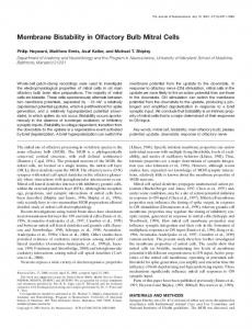
Fig. 1. Schematic drawing of the anatomy of the mammalian olfactory bulb. (a) Odor information is carried via the olfactory nerves into the mitral cell tufted dendrites. Mitral cells send axons to the piriform cortex; they also make excitatory, dendrodendritic synapses onto granule cell dendrites. The axonless granule cells have spiny dendrites; they form both reciprocal and lateral dendrodendritic inhibitory synapses onto the mitral cell dendrites. (b) Reciprocal synaptic contacts with opposite polarities. Mitral dendrites release glutamate and depolarize (+) the granule cell dendritic spine. The granule spine in turn releases GABA and inhibits (–) the mitral cell. The A-type potassium conductance (IA) in the granule cell dendrites specifically filters the brief AMPA receptor component, but not the prolonged NMDA receptor component, of the mixed AMPA/NMDA receptor mitral-togranule excitation. As a consequence, the granule-to-mitral inhibition follows the slow kinetics of the NMDA receptors. 1041
© 1999 Nature America Inc. • http://neurosci.nature.com
news and views
aA
Mitral GABA
GABA
NMDA
Granule
NMDA
Odor
© 1999 Nature America Inc. • http://neurosci.nature.com
bB
Mitral GABA
Granule
AMPA +NMDA
GABA
AMPA +NMDA
AMPA +NMDA Bob Crimi
Odor
Fig. 2. Postulated mechanism by which IA may control the frequency of odor-induced oscillations in the olfactory bulb. (a) In response to a puff of odor, mitral cells generate repeated bursts of spikes (upper trace). Each burst induces a slow NMDA receptor-mediated EPSP in the granule cells, which triggers a train of spikes in these cells (lower trace). (b) If IA current in the granule-cell dendrites is blocked, the mitral cell burst triggers an earlier and larger EPSP in the granule cells, which is now mediated by AMPA as well as NMDA receptors. This causes early spiking in the granule cells, which in turn produces earlier and stronger inhibition of the mitral cells. The mitral cell burst is therefore shorter, as is the duration of granule-to-mitral inhibition. This could cause an increase in the frequency of the odor-induced oscillations.
side-out patch recordings from granule cells in slices of rat olfactory bulb, they showed that granule cell dendrites possess a high density of A-type potassium channels. These channels mediate a current known as IA, which is activated very rapidly upon depolarization and then inactivates with a time constant of about 20 milliseconds. Because of its rapid kinetics, IA filters (repolarizes) brief depolarizations, whereas prolonged depolarizations inactivate IA and thus are not filtered. Schoppa and Westbrook found that IA in granule cell dendrites severely limits the effectiveness of the fast AMPA receptor-mediated, but not the slow NMDA receptor-mediated, mitral-to-granule excitation. When a mitral cell releases glutamate, postsynaptic AMPA receptors are activated, leading to a local depolarization at the granule cell spine head mem1042
brane that is sufficient to relieve the magnesium block from NMDA receptors. However, because the AMPA receptor-mediated response is filtered by IA, it is not sufficient to cause release of GABA from dendritic spines on the granule cells. GABA release requires the activation of the slow NMDA receptor-dependent excitatory potentials, and it is the time course of these slower NMDA receptor responses that determine the duration of GABA release, and hence of the reciprocal and lateral inhibitory inputs. The authors showed that blocking IA with the drug 4-AP unmasked a prominent AMPA receptor-mediated excitatory response in the granule cells. This enhanced AMPA receptor-mediated mitral-to-granule excitatory input reduced the onset time of the granule-tomitral inhibition. The earlier inhibition
results from both earlier and stronger local depolarization in the granule spine membrane, as well as from earlier spike firing in granule cells (Fig. 2). The A-type potassium channel is an impressive example of the precision with which nature has designed its basic biophysical tools. These channels can be directed to specific locations in both axons 9 and dendrites 10. In axons, they affect repetitive firing, enable spike firing at very low rates11, and are responsible for channeling spike trains preferentially to selected axonal subtrees9. In dendrites they can serve as ‘shock absorbers’, dampening dendritic excitability and strong excitatory inputs10,12. Because of their fast kinetics, A-type channels are also ideal for discriminating between fast and slow inputs, as Schoppa and Westbrook have shown4. Most importantly, however, the effectiveness of I A is modifiable in a dynamic way. Indeed, its degree of inactivation (and therefore its availability) is very sensitive to membrane potential near rest. IA is also sensitive to a variety of second messengers and neurotransmitters, and to changes in intracellular and extracellular ion concentration. Schoppa and Westbrook 4 found that in their slice preparations, about half of the A-type channels were inactivated at the resting membrane potential of the granule cells. It is thus possible that in the olfactory bulb, IA may undergo dynamic modulation, either through changes in the resting potential of granule cells or through the action of protein kinases. Changes in the availability of IA would alter the relative contribution of AMPA and NMDA receptor inputs to granule cells and thus the kinetics and strength of both reciprocal and lateral inhibition. This in turn would affect oscillatory rhythms as well as the spatial extent of inhibition within the bulb, which could allow modification of the bulb’s ability to discriminate and remember different odors. The big challenge now is to go beyond the mechanistic description of olfactory bulb circuitry, and to understand how its function arises from the machinery. This will mean taking information at many levels—the anatomical elements of the bulb, their biophysical properties and insights gained from theory—and integrating them into a compact model. A compact model is one that captures the essential features required to reproduce the global behavior of the system, rather than replicating all its details13–15. To be considered successful, the model should be able to discriminate between different odors and learn to rec-
nature neuroscience • volume 2 no 12 • december 1999
© 1999 Nature America Inc. • http://neurosci.nature.com
news and views
ognize new odors—in other words, it should ‘smell’. Only then will we be able to say that we really understand olfaction.
© 1999 Nature America Inc. • http://neurosci.nature.com
1. Sopfer, M., Bhagavan, S., Smith, B. H. & Laurent, G. Nature 390, 70–74 (1997).
5. Rall, W., Shepherd, G. M., Reese, T. S. & Brightman, M. W. Exp. Neurol. 14, 44–56 (1966). 6. Shepherd, G. M. & Greer, C. A. in The Synaptic Organization of the Brain 4th edn. (ed. Shepherd, G. M.) 159–203 (Oxford Univ. Press, New York, 1998).
10. Hoffman, D. A., Magee, J. C., Colbert, C. M. & Johnston, D. Nature 387, 869–875 (1997). 11. Rush M. E. & Rinzel, J. Bull. Math. Biol. 57, 899–929 (1994). 12. Yuste, R. Nature 387, 851–853 (1997). 13. Rall, W. & Shepherd, G. M. J. Neurophysiol. 31, 884–915 (1968).
2. MacLeod, C., Backer, A. & Laurent, G. Nature 395, 693–698 (1998).
7. Segev, I., Rinzel, J. & Shepherd, G. M. in The Theoretical Foundation of Dendritic Function (eds. Segev, I., Rinzel, J. & Shepherd, G. M.) 149–159 (Bradford, Cambridge, Massachusetts, 1995).
3. Tank, D. W., Gelperin, A. & Kleinfeld, D. Science 265, 1819–1820 (1994).
8. Isaacson, J. S. & Strowbridge, B. W. Neuron 20, 749–761 (1998).
14. Protopapas, A. D., Vanier, M. & Bower, J. M. in Methods in Neuronal Modeling (eds. Koch, C. & Segev, I.) 461–498 (Bradford, Cambridge, Massachusetts, 1998).
4. Schoppa, N. E. & Westbrook, G. L. Nat. Neurosci. 2, 1106–1113 (1999).
9. Debanne, D., Guerineau, N. C., Gahwiler, B. H. & Thompson, S. M. Nature 389, 286–289 (1997).
15. Hopfield, J. J. Proc. Natl. Acad. Sci. USA 88, 6462–6466 (1991).
Eyes wide shut Mark Hübener and Tobias Bonhoeffer Crowley and Katz cast doubt on the idea that correlated activity is critical for visual cortex development by showing that ocular dominance maps can emerge without any retinal input. Despite its bewildering complexity, the circuitry of the mammalian visual cortex represents information about the outside world in remarkably regular patterns. As with other vertebrates, the visual scene that is projected onto the retina is represented by a corresponding ‘retinotopic’ map on the cortical surface. However, in higher mammals, several additional functional maps are superimposed onto the retinotopic map. For instance, input from the two eyes is segregated into an intricate pattern known as an ocular dominance map (Fig. 1), in which adjacent regions of the cortex (termed ‘columns’ because they span the thickness of the cortex) respond to the same location in the visual field, but are driven mainly by input from only one of the two eyes. Further features such as contour orientation, motion direction, spatial frequency and probably others are also mapped in a systematic fashion on this cortical surface. The resulting patterns differ from animal to animal, but their general features are highly reproducible. Although the significance of these maps for neural processing remains enigmatic, much effort has been devoted to understanding how they arise during development, so far without the advent of a generally accepted theory. In this issue of Nature Neuroscience, Crowley and Katz1 describe a rather unexThe authors are at the Max Planck Institute of Neurobiology, Am Klopferspitz 18A, 82152 Munich-Martinsried, Germany. e-mail:
[email protected]
pected result that might change our thinking about how functional maps develop. The authors report that ocular dominance columns in the visual cortex of ferrets form even if both eyes are removed very early in development. To appreciate the significance of this finding, it is useful to review how our ideas of map formation in the visual cortex have evolved over the last few decades. Hubel and Wiesel showed in the early 1960s that the visual cortex is highly developed even in very young animals with no visual experience2. From the earliest stages at which recordings are possible, cortical neurons respond preferentially to contours of particular orientations, and neighboring neurons prefer similar orientations, indicating the presence of an orientation map. This suggested that the neural circuitry underlying orientation preference and its systematic mapping does not depend on experience, but must be innate. However, in another study3, the same authors also showed that the ocular dominance map can be profoundly influenced by experience. If one eye is temporarily closed during the so-called critical period in early development, then the ocular dominance columns corresponding to that eye shrink in size, with a corresponding expansion of the territory occupied by the open eye. Although this process is still not fully understood, there is now ample evidence that it depends on neural activity, and that axons conveying signals from the two eyes compete with each other for synaptic tar-
nature neuroscience • volume 2 no 12 • december 1999
gets in the cortex. Because so many experiments demonstrated that visual experience can influence the layout of cortical maps, it became widely accepted that vision might actually be instrumental in their establishment. This notion was further strengthened by theoretical models showing that plausible assumptions about Hebbian synaptic plasticity and patterns of correlated activity among neighboring axons can explain the systematic mapping of both ocular dominance and orientation. With so much evidence suggesting a role for experience, the alternative possibility, that molecular mechanisms might control the emergence of these maps, lost more and more ground. A challenge for activity-dependent models came when the initial observation by Hubel and Wiesel2 was eventually confirmed and extended by studies showing that both ocular dominance columns4 and orientation maps5,6 can develop almost normally without visual input. However, although these studies showed that visual experience was not essential, they did not exclude a role for neuronal activity, and indeed it had been discovered in the meantime that neurons in the visual system are spontaneously active long before
Fig. 1. Ocular dominance map of monkey striate cortex. The black and white areas represent cortical regions innervated by the different eyes. 1043