Department of Cell Biology, Wellcome Laboratories, Langley Court, Beckenham, Kent, UK BR3 3BS. EPR spectra of an insoluble fraction from the rodent ...
3409
J. CHEM. SOC. FARADAY TRANS., 1994, 90122), 3409-3410
EPR Spectroscopic Studies of Haemoglobin Breakdown in Malarial Parasite-infected Erythrocytes? Richard Cammack* and Daulat S. Pati11 Centre for the Study of Metals in Biology and Medicine, King’s College, London, UK W8 7AH David Linstead Department of Cell Biology, Wellcome Laboratories, Langley Court, Beckenham, Kent, UK BR3 3BS
EPR spectra of an insoluble fraction from the rodent malarial parasite Plasmodium berghei, grown in mouse erythrocytes, have been measured. Signals due to high-spin iron(r1r)haem and two low-spin h a e m species were detected. Comparison with other derivatives of haemoglobin, and the use of a simple ligand-field model, identified the low-spin species as imidazole-hydroxy and bis-imidazole derivatives of haem. These species may represent intermediate stages in t h e breakdown of haemoglobin to the insoluble form, haemozoin. The utility of EPR spectroscopy for the study of t h e breakdown pathway is discussed.
Malarial parasites in the erythrocyte derive a considerable part of their nutrients and energy of growth from proteolytic degradation of haemoglobin. The pathway of degradation of haemoglobin is a subject of current interest, because it may be a target for chemotherapy. The porphyrin from haemoglobin, together with some of the polypeptide, is finally deposited in an insoluble form known as haemozoin. The formation of this material avoids the toxicity of porphyrin to the malarial parasite, probably because it sequesters the iron and helps to prevent the formation of destructive oxygen radicak2 There is some evidence that antimalarial drugs such as chloroquine act by inhibition of the formation of haemo~oin.~ We have used EPR spectroscopy to examine insoluble, haemozoin-containing fractions from erythrocytes infected with the malarial parasite Plasmodium berghei. Distinct signals were observed from low-spin iron(@ haems.
Materials and Methods P . berghei was grown in vivo using male CD1 mice. Infected erythrocytes were separated from the blood and treated by the procedure of Fry and B e e ~ l e y After . ~ centrifugation the erythrocytes were treated with cellulose, to remove platelets and white cells. Malarial parasite cells were extracted by lysis with 0.15% (v/v) digitonin, centrifuged for 5 min at 4000 rpm in a Denley BS400 bench centrifuge, and resuspended in icecold 0.07 mol dm-3 sucrose-0.21 mol d m P 3 mannitol-1 mmol dm- ethyleneglycol tetraacetate-5 mmol dmMgCl,-5 mmol dm-3 KH,PO,-4 mmol d m P 3 HEPES, pH 7.4 (‘H medium’). After washing three times in H medium, the parasite pellet was lysed by brief sonication. A darkbrown insoluble fraction of the parasite cells was obtained by centrifugation at 10000 g for 5 min, and washed with H medium. Samples were resuspended in a minimum volume of H medium then frozen in liquid nitrogen. Some of the samples were oxidized with 0.5 mmol dm-3 2,6-dichloroindophenol for 2 min before freezing. EPR spectra were recorded on a Varian E4 spectrometer with an Oxford Instruments ESR900 liquid-helium flow cryostat.
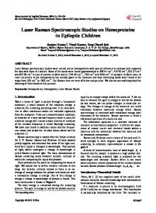
Results EPR spectra of the washed insoluble fraction from P . berghei, recorded at 24 K, showed the axial spectrum of high-spin ferrihaemoglobin, at g = 6.0, 2.0 (Fig. 1). In addition, the spectra showed distinct low-spin Fe”’ species (Fig. 2). The preparations appeared to be somewhat reduced, since the intensity of these signals was enhanced by oxidation with 2,6dichloroindophenol. In the sample shown in Fig. 2(a), the amplitude of the low-spin signal in the untreated sample (not shown) was 14% that of the oxidized form. Spectra of different samples, of which Fig. 2(a) and (b) are examples, may be interpreted in terms of varying amounts of two principal species. For one species (haem l), the g-factors were 2.77, 2.25, 1.7 (the latter being relatively broad) and for the other (haem 2) 2.59, 2.18, 1.83. These values are similar to, but different from the literature values for haemoglobin-histidine (2.70, 2.21, 1.69) and haemoglobin-hydroxide (2.56, 2.18, 1.88).5 Assuming that the two low-spin species are haem derivatives, the type of axial ligands attached to the iron may be investigated by an analysis of the rhombicity and tetragonal field. In a classic study, Blumberg and Peisach’ examined the spectra of a number of derivatives of haemoglobin and myoglobin, and compared them with data from cytochromes and other proteins. In the EPR spectrum of a distorted iron(rI1) site there is insufficient information to derive all the ligandfield parameters. Blumberg and Peisach’ used an empirical
g-factor
8.06.0 I
50
4.0
3.0
I
I
I
100
150
200
1.5
2.0
1
250
300
350
400
450
magnetic field/rnT
t This paper was presented at the 27th International ESR Conference at the University of Wales, Cardiff, 21st-25th March, 1994. $ Present address : Department of Biochemistry, University of Georgia, Athens, GA 30602, USA.
Fig. 1 EPR spectrum of a haemozoin-containing insoluble fraction from P. berghei, recorded at 23 K. Other conditions of measurement: microwave power 20 mW, frequency 9.18 GHz, modulation amplitude 1 mT.
3410
J. CHEM. SOC. FARADAY TRANS., 1994, VOL. 90
I
l
g-factor 2.2
2.6
3.0 l
I
I
I
erythrocytes in culture. P . berghei is also unusual in that reticulocytes as well as mature erythrocytes can become infected, so that some of the infected cells might be expected to contain cytochromes. However, the amount of low-spin haem detected is much greater than would be expected for cytochromes. Therefore, there may be differences in the breakdown of haemoglobin in P . berghei as compared with P . yoellii or P .falciparurn.
1.8
I
I
Discussion
200
250
300 magnetic field/mT
350
400
Fig. 2 EPR spectra of two different preparations from P. berghei, oxidized with 0.5 mmol dm- 2,6-dichloroindophenol, showing the low-spin haem region. Conditions of measurement as for Fig. 1, except that the gain setting is increased eight-fold.
approach, based on the t2* hole m0de1.~-~ It gives a classification of low-spin haem compounds based on the distortions of the haem iron from octahedral symmetry. The parameter V/A, representing the rhombicity (distortion in the x-y plane) is plotted against the tetragonal field A/A (representing the distortion along the z axis); V is the rhombic splitting, A the axial splitting and A the spin-orbit coupling constant. It was demonstrated that haem proteins with different axial ligands fell within defined areas on the plot. The advantage of this method is that it requires only a determination of the g-factors of the spectrum. The classification is effective for derivatives such as those described here, where the g-factors are less than 3. It is not applicable to highly axial cases such as bis-histidine coordination where the two histidine planes are orthogonal, and bis-methionine, both of which give g, > 3.2.8-11 The values of V/A and A/A for the two-low-spin haem compounds, were calculated in the Blumberg-Peisach coordinate frame." For haem 1, this gives V/A = 0.75, A/A = 3.9, which places it in Group H (haemoglobin-histidine). This type of spectrum is observed, for example, when haemoglobin is denatured, so that the oxygen-binding cavity on the distal side of the haem collapses, and the distal histidine becomes coordinated to the iron. For haem 2, V/A = 0.82, A/A = 5.5, which places it in Group 0. This spectrum resembles the spectrum of alkaline iron(m) haemoglobin, and certain peroxidases. In parallel studies, spectra were recorded of similar fractions from rat erythrocytes infected with P . yoellii, or in human erythrocytes infected by P . fakiparum. No low-spin haem signals were detected with these malarial parasites. One possible reason for this might be the high level of infection in the mouse cells; these cells were derived from an animal, whereas the others were from malarial parasites grown in
In the trophozoite stage of the human malarial parasite P . falciparurn, haemoglobin is ingested into a vacuole, and then hydrolysed to yield amino acids and haemozoin. Goldberg et a1.12 have presented evidence that this occurs in an ordered sequence of reactions. A specific protease first cleaves the haemoglobin a-chain at a site in a hinge region, between phenylalanine-33 and leucine-34. Further hydrolysis of the polypeptides is catalysed by other proteases. In this study we have used EPR spectroscopy to observe breakdown products of haemoglobin in their iron@) states, in an insoluble fraction from red cells infected with the malarial parasite P. berghei. A high-spin species and at least two different types of low-spin species were distinguished. In view of the extremely high concentration of haemoglobin in erythrocytes, the high-spin species might be a denatured form of methaemoglobin, or methaemoglobin adsorbed onto the haemozoin pellet. The low-spin haem compounds might represent denatured forms of haemoglobin itself, or intermediate stages in the hydrolysis, consisting of polypeptides coordinated to haem. We thank Susan Bradley for assistance with sample preparation. The work was supported by the SERC.
References 1 D. E. Goldberg and A. F. G. Slater, Parasitology Today, 1992,8, 280. 2 J. Golenser, E. Marva, R. Hare1 and M. Chevion, Free Radical Res. Commun., 1991, 12, 639. 3 A. F. G. Slater and A. Cerami, Nature (London), 1992,355,167. 4 M. Fry and J. E. Beesley, Parasitology, 1991, 102, 17. 5 W. E. Blumberg and J. Peisach, in Bioinorganic Chemistry, ed. R. Dessy, J. Willard and L. Taylor, Advances in Chemistry Series, vol. 100, American Chemical Society, Washington, 1971, pp. 27 1-291. 6 J. S. Grifith, The Theory of Transition-metal lons, Cambridge University Press, Cambridge, 1961, p. 363. 7 C. P. S. Taylor, Biochirn. Biophys. Acta, 1977,491, 137. 8 G . Palmer, in The Porphyrins, ed. D. Dolphin, Academic Press, New York, 1979, vol. 4, pp. 313-353. 9 G. Palmer, in Methods for Determining Metal Ion Environments in Proteins, ed. D. W. Darnall and R. G. Wilkins, Elseviermorth Holland, New York, 1980, pp. 153-181. 10 G. Palmer, Biochern. SOC.Trans., 1985,13,548. 11 M. R. Cheesman, A. J. Thomson, C. Greenwood, G. R. Moore and F. Kadir, Nature (London), 1990,346,771. 12 D. E. Goldberg, A. F. G. Slater, A. Cerami and G. B. Henderson, Proc. Natl. Acad. Sci. USA, 1990,87,2931.
Paper 4/02233E; Received 14th April, 1994