(Eb), the major component, and erabutoxin a (Ea) are single- ... ent in composition from Eb, and neurotoxin a, a 62-residue protein, two .... Inc., Foster City. CA).
Eur. J . Biochein 153, 521 - 577 ( I 985) ( FEBS 1985
Erabutoxin b Initial protein refinement and sequence analysis at 0.140-nm resolution Philip E. BOURNE’, Atsushi SATO’, Peter W. R. CORFIELD‘. Lawrence S. KOSEN’, Steven BlRKEN’and Rarbara W. LOW
’ Department of Biochemistry and Molecular Biophysics; and Department of Medicine, Columbia University, New York (Received May 9/September 5, 1985) - EJB 85 0506
The crystal structure of the protein postsynaptic neurotoxin, erabutoxin b, has been refined at 0.140-nm resolution ( R = 0.22) by restrained least-squares and interactive computer graphics. The study has established complete structural identity of the two sea-snake venom toxins, erabutoxin b and neurotoxin b, isolated from Laticaudi smzifusciutu snakes taken in different Pacific Ocean waters. Two chemical-sequence inversion errors in erabutoxin b have been discovered during refinement, corrected a n d subsequently confirmed in both erabutoxin b and erabutoxin a by chemical analysis. The correct sequences are Hish-Gln7,hitherto unsuspected, and SerlxPro”. The sequence correction His6-Gln7explains the anomalous results of ‘H N M R solution studies and those of early chemical modification experiments, which were in conflict with the previously published three-dimensional structure of erabutoxin b. On refinement, the five-stranded beta sheet described earlier is now shown to be discontinuous, split into a two-stranded beta loop and a three-stranded beta sheet. Unique features of the Pro5“Gly4’ peripheral segment have now been identified. 51 water moleculc positions have been located. The erabutoxins b and a, the two principal postsynaptic neurotoxins of venom from the sea-snake Luticauda .smzifusc.iatu found off‘ the Okinawas (Ryuku Islands) were isolated, purified, crystallized [ I ] and the sequences determined [2] by Tamiya and his colleagues. Both erabutoxin b (Eb), the major component, and erabutoxin a (Ea) are singlechain proteins with 62 amino acid residues and four disulphide bridges [3]; in erabutoxin a, a single residue variant of erabutoxin b, Amz6 replaces His’”. Tu and his coworkers [4] subsequcntly reported the compositions of two major postsynaptic neurotoxins froin venom of the same species of snake caught off the Philippines some 2000 kin distant from the Okinawas: these were neurotoxin b, reported as a 61 -residue protein, seven residues different in composition from Eb, and neurotoxin a, a 62-residue protein, two residues different from Ea. X-ray crystal structure determination at 0.275-nm resolution of the erabutoxin b structure in this laboratory [ 5 ] was followed by X-ray analysis at 0.22 nm of neurotoxin b by Tsernoglou and Petsko [6], who employed the complete Eb chemical sequence [2] in structure determination; they thus established neurotoxin b as a 62-residue protein, similar in both sequence and conformation to Eb. Later Tsernoglou and Petsko reported [7] a sequence inversion in neurotoxin b, corrected to Glu21-Ser2’, a correction made independently by chemical analysis [8] for erabutoxin b. An implied singleresidue difference in composition, Arg59 for Val5’, was also reported for neurotoxin b, in conflict with the later composiCorrespondence 10 B. W. Low, Department of Biochemistry, Columbia University, 630 West 168 Street, New York City, New York, USA 10032 Note. The atomic co-ordinates and B pararnetcrs for this erabutoxin b ytrueture refinement have been deposited in the Brookhaven Protein Data Bank, Chemistry Department, Brookhaven National Laboratory, Upton, NY 11973, USA (Tsernoglou, D. and Pctsko, G . A. (1980). Ahhreviutions. Erabutoxin b, Eb; erabutoxin a, Ea.
tion analyses [9] for this toxin. A least-squares refinement of neurotoxin b at 0.138-nm resolution [lo] still failed to identify rcsidue 59 unambigiously, although a second sequence inDespite the version was found and corrected to SerlX-ProL9. apparent sequence differences, we remained confident that neurotoxin b and erabutoxin b were identical and that no subspecies differences exist between these neurotoxins from L. scwzifusciatu snakes caught in different waters. X-ray crystallographic studies appeared t o confirm [I 11 the early identification of Ea as [Asn”] Eb [2] and to establish that neurotoxin a was [Asn”] neurotoxin b [12]. These conclusions, however, drawn from relatively low-resolution X-ray studies, left open the possibility of inversion sequence differences between Ea and Eb. An independent partial chemical analysis was therefore undertaken.
EXPERIMENTAL PROCEDURE X-rci.y studies
Erabutoxin b crystallizes in the space group P212121,Z = 4. Conditions used to prepare crystals at pH 9.5 for data collection in the range 0.26 -0.14 nm were described earlier [5]for the crystals employed in data collection over the range GC: -0.25 nm. Complete duplicate sets of diffraction data for Friedel pairs were collected over the range 0.26-0.20 nm from two crystals using omega scans with a Nicolet P21 diffractometer. Data in the range 0.206-0.14 nm were collected from three crystals with a 30% overlap. With one exception crystals used were discarded when standard retlection intensities dropped by 10% (20% in the unique case). Intensity data were corrected by standard methods with empirical absorption corrections made as suggested by North et al. [ 131. Scaling and merging of data sets were carried out by the Steigemann (Munich) PROTEIN system [14]. Of31 103 independent measurements, 3450 were rejected by use of the
522 criterion II),k, - < I,,,, > I/ < > is > 0.6 where < / h k [ > is the mean of lhe intensity of reflection I h k l . Remaining values for cach reflection were averaged to give 9511 independent intensities: 91% of the possible number in the range m 0.14 nm. All 9468 reflections in the range 1.00-0.14 nm were used in least-squares refinement; no weak reflections were removed. Starring model. As an exploratory model for refinement, we chose to employ the atomic co-ordinates from the 0.138nm neurotoxin b refinement [lo], rather than those of the 0.25-nm erabutoxin by structure [I 51. An initial comparison betwecn the two sets of main-chain atomic co-ordinates had shown differences small enough to suggest that they resulted simply from the independent structural interpretations of one protein at two different resolutions. Possible effects of different crystallization conditions on protein conforination appeared minimal and well within the compass of the refinement method. The conditions employed [6] in the crystallization of neurotoxin b are bracketed by the two sets of conditions used [5] for the crystallization of isomorphous preparations of Eb. Procedure. In the first refinement step the atomic coordinates [lo] of the neurotoxin b molecule (without the coordinates of side-chain 59 all water molecule oxygens and the sulfate group), were refined against erabutoxin b data by the restrained least-squares refinement procedure of Konnert and Hendrickson 116 - 181. After seven cycles of refinement (three with 0.5-0.14-nm data, followed by four with 1.0-0.14-nm data) the R factor dropped from 0.41 to 0.31 with a rms accumulated shift from initial positions of 0.049 nm. The conventional R factor is a means for the comparative evaluIn the ation of model against data, where R = llFol - lFcll/lF,l. 2(FJ - lFcl difference map, then calculated with erabutoxin b data at 0.1 5-nm resolution, the Ser”-Pro” residue sequence found earlier for neurotoxin b [lo] was clearly evident. Two further cycles of more highly constrained refinement led to an R factor of 0.34 with a rms accumulated shift of 0.019 nm. Model-building studies with a new difference map, in conjunction with iterative computer graphics using the MSX graphics system [I91 and FRODO [20], led to some side-chain shifts and the recognition of 29 water positions and one sulphate group, which were then included in the refinement. After a further nine cycles of refinement the R factor dropped to 0.26, with a rms total shift during this stage a t 0.032 nm. The Val” side-chain carbon positions were all clearly evident in the difference map calculated at this stage. After additional model rebuilding followed by five further cycles of refinement the R factor decreased to 0.24 with an accumulated rms shift of 0.021 nm during this stage. A 21FJ - IFc[ difference map then calculated indicated, at residues 6 and 7, a sequence inversion to His6-Gln7. The corrected His6-Gln7 sequence and further model shifts were employed during the final five cycles of refinement, from which the sulfate group co-ordinates were omitted. A further 24 water molecule positions had been recognized in the difference map and included as oxygen atoms with occupancies permitted to vary in the final refinement. The accumulated rms positional shift for this phase was 0.012 nm and the final R factor was 0.22 for the 9468 reflections in the range 1.O-0.14 nm. The mean B parameter for both protein and water molecules was 0.151 nm2. Of the 1334 bond lengths, 116 deviate from ideality by more than two standard deviations. Choice of the refined (0.138-nm) neurotoxin b atomic co-ordinates [lo], as starting model for refinement with Eb data, presented no problems and was a marginally better choice than the 0.25-nm erabutoxin
structure [I 51. The rnms shift from the matched neurotoxin b starting co-ordinate set to this refinement model was 0.12 nm whereas from the matched erabutoxin b set it was 0.13 nm. Shifts in position of main-chain atoms on refinement were, with few cxceptions, small ( 6 0 . 1 nm) and non-cumulative in effect along the peptide chain. The few exceptionally large shifts involve the peptide chain a t Amh2, Ser9, Ser’ and the terminal N of Arg’. They are accompanied by even larger than average side-chain shifts.
Clzemical analysis Automated Edman degradation analyses of the proteins erabutoxins a and b after performic acid oxidation 1211 were carried out initially in a Beckman 890B sequencer. In two separate runs, each using 17 nmol erabutoxin a, 12 nmol histidine were recovered from cycle 6 in both runs, which is consistent with a sequence repetitive yield of 94%. Erabutoxin b and a sequences were then subsequently redetermined, for ten and twenty cycles respectively, using the gas-phase microsequencer (model 470A, Applied Biosystems Inc., Foster City. CA). The resultant amino acid phenylthiohydantoins were analyzed on a Hewlett-Packard 1084B highperformance liquid chromatograph using either a Dupont Zorbax C-18 column by the method of Zimmerman et al. [22] or ;in IBM cyano-column (IBM Instruments, Walhgford, CT) according to the recommended procedure [23]. Results of microsequencer studies for the first twenty residues of Ea and the first 10 residues of E b are tabulated in Table 1.
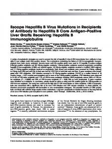
RESULTS A N D DISCUSSION X-ray crystal structure refinement has established the complete structural identity of neurotoxin b and erabutoxin b and has led to the correction for erabutoxin b of two chemical sequence errors [2]. The chemical analyses undertaken here and elsewhere [24] confirm these X-ray findings. The X-ray evidence for the two chemical sequence errors is shown in the two 21FJ - lFcl difference Fouriers shown in Fig. 1. Only C-p atoms of the side-chains were included in F, calculations for residues 6 and 7 (Fig. l A ) , and only C-a atoms for the side chains 18 and 19 (Fig. 1B). Atomic positions are shown superimposed. The original side-chain assignment Val59 121, seen at lower resolution in Eb [5, 151, is evidently correct and reconfirmed. The established sequence of erabutoxin b, the major neurotoxin component of all L. semifasciata venom, is shown as part of Fig. 2, where it serves also to identify residues in the stereodrawing.
Significance of the His-Gln sequence correction Establishment in E b of the sequence His6-Gln7 by structure refinement resolves several anomalies which arose from the assumption of the incorrect sequence ‘Gln6-His7’. These findings and interpretations of chemical 125, 261 and physicochemical solution studies [26,27] were in conflict with the three-dimensional erabutoxin b structure [5, 151 when the chemical sequence identification of residue 7 as His was employed; they are now explained. As can be seen in the stereoscopic drawing (Fig. 2 A and b) residue 7, which protrudes from the upper concave surface of the molecule (Fig. 2A), is exposed and accessible to solvent. Residue His6, on the other hand, although on the ‘outer’ surface of the
523 0
v!
0
-
wmm
v! c'!
0 0 0
524
A
B Fig. 1. Vii~icsof‘diff2,rence mupps, phases calculured after omission of’ side-chirin a t w m beyond C-B,for side chuirzs 6 and 7 arid C - x f o r ,sidtwhuiti.\ 18 rind I Y . (A) 2 ~ F o ~ - ~maps F c ~ showing the region near residuc 6 with the outline of His superposcd on the three-dimensional contour map (left) and the region ncar residue 7 with the outline of Gln superposed (right). (B) 21F01- IFcI map (left) and lFoi- lFcl map (right) showing the region ncar residue 19 with the outlinc of Pro superposcd on thc thrcc-dimensional contour maps. In the IFo/- lFcl map only those atoms left out o f the phase calculation are expected to appear
molecule is relatively inaccessible; it is surrounded and held by a total of 31 van der Waals packing interactions of less than 0.40 nm plus a further 33 interactions between 0.4 nm and 0.45 nm in length to residues 4, 8, 10, 11, 12, 30 and 39. The B parameters of the tightly packed His‘ side-chain range from 0.1 nm2 to 0.1 5 nm’ while those from the exposed Gln7 are 0.24 nm2 to 0.30 nm2. The inaccessible His’ position thus cxplains the results of iodination studies of E b and Ea [25] where the putative ‘His” residue, common to both erabutoxins, was not iodinated. Further, the observation of Inagaki et al. [26]that ethyl mercury phosphate attacks Hiszh first is explained in the same way. The assumption of a ‘His7’ identification led also to misinterpretation of the ‘H NMR solution studies of erabutoxin b [26, 271 which show a ‘His” near to Phe4. It was, therefore, concluded that the first erabutoxin loop must be twisted in solution in order to bring the side-chains 4 and 7 close to each other. This would have implied a gross conformational shift on crystallisation, requiring great intrinsic molecular flexibility. The experimental findings in solution
are explained by the correct sequence without invoking conformation changes on crystallisation. Residucs Phe4 and His6 are adjacent near-neighbor side-chains on the same side of the beta strand of the first peptide loop, which can be seen on the extreme right in Fig. 2B. There are six van der Waal’s packing interactions between the Phe4 and His6 side-chains with C.. .C and C.. .N distances in the range 0.36-0.395 nm. Moltwdrir structure
The hydrogen-bonded beta structure of erabutoxin b [15] remains well defined with five anti-parallel beta strands A, B, C, D, E and a beta bulge in the AB loop, although on refinement the sheet is seen to be disrupted between the two-stranded AB loop and the three-stranded CDE sheet. Only two hydrogen bonds are formed between the A and D strands as indicated in Fig. 3A and B; only one of these, NsH...037(C),is a main-chain linkage. At the wide part of the gap between the A and D strands a second hydrogen bond links carbonyl oxygen atom ( C ) 0 6to a terminal N H of A1-g39.
525
A
B
P B C
Fig. 2. Stereoscopic drawinx with the backbone truce q f t h e erabutoxin h structure ujter refiiiement. Both front (A)looking into the molecular concavity and back view (B) looking at the convex under surface of the molecule are shown. The first loop (1 -17) of the peptide chain is thus on the extreme left in (A) and the extreme right in (B). The backbone trace of the front view of the structure i s shown in (C). The established amino acid sequence of erabutoxin b given here serves to help identify residues in the stereo drawing: Arg Ile Cys Phe Asn His Gin Ser Ser Gln Pro Gln Thr Thr Lys Thr Cys (17) Ser Pro Gly Glu Ser Ser Cys (24) Tyr His Lys Gln Trp Ser Asp Phe Arg Gly Thr Ile lle Glu Arg Gly Cys (41) Gly Cys Pro Thr Val Lys Pro Gly Ile Lys Leu Ser Cys (54) Cys Glu Ser Glu Val Cys Asn Asn (62)
The local conformation here is further stabilized by a contact between His6(C)0 and a water molecule, which is hydrogenbonded to the peptide NH of Arg3”. Projections show some of the curves and twists in both segments of beta structure.
I D
C
Fig. 3. Projerted view of the beta structure in erabutoxin b. (A) AB loop; (B) DEC sheet. Hydrogen bonds are indicated by dotted lines between carbonyl oxygen and amide nitrogen and C-cc positions are indicaled by filled circles
There are five tight turns in this structure wherc the Eb peptide chain reverses direction. Two of these maintain conformations assigned at lower resolution [I 51: the type l/Hl beta bend 31 -34 [28] and the type 11 beta bend 47-50. The 18 -21 turn is now established as type 11, as would be expected from the 18 - 19 sequence inversion. The 7 - 10 turn is also type 11. The last bend, 57-60, with the C-terminal closed loop, is a more poorly defined type I1 bend. The turns 18 21, 31 - 34 and 47 - 50 are close to ideal. The refinement revealed unique features of one peripheral strand segment from Pro44 to Gly49. Here the atomic B parameters are exceptionally large for both main-chain and side-chain atoms, with a mean B parameter for this segment of 0.261 nm2 compared to 0.131 nm2 for the rest of the molecule.
526 Table 2. Disi/lphi& 1inkuge.c in ercihutoxin h Handedness, LM or RH and x1x2;13x4x5 Bond
C-x...C-x
xi
x2
13
K4
3-24 17-41 43-54 55-60
nm 0.61 (LH) 0.54 (LH) 0.51 ( R H ) 0.62 ( R H )
-62 152 -172 59
-70 -50 74 85
-82 -87 80 86
-
15
64 89 177 87
-56 -75 -59 62
molecules in this Eb refinement form a hydrogen-bonded network with bonds to one or more protein oxygen or nitrogen atoms, as well as, in many instances, to other water molecules. Ordered water structure is evidently a n important feature of this crystal structure.
CONCLUSION The least-squares refinement of the erabutoxin b structure at 0.14 nm has established the structure and provided a detailed description of the stereocheinistry of this the major neurotoxic component of venom from the sea snake L. smizfilsciatu found in Pacific Ocean waters. The quality of the X-1-ay data and power of the refinement procedure led to the discovery of a chemical sequence error not detected in earlier refinements nor at lower resolution. Refinement has provided more detailed information about molecular conform at'ion. The structure,/function significance of the detailed stercochemistry of invariant and conservatively substituted residues in the reactive site and elsewhere i s discussed in a following communication, where the stereochemical relationships between residues in the epitope region(s) [29, 301 will also be considered.
Such large localized differences in B parameters could indicate a region of erroneous fitting of the structure to the electron density. The electron density map in this region is, however, diffuse and ill-defined even for the main-chain atomic positions, in sharp contrast to the excellent resolution in the remainder of the map. This would suggest a region of intrinsic molecular flexibility. The segment 44- 40 curves away from the rest of the molecule, as can be seen in Fig. 2A (extreme right). With the exception of one long hydrogen bond (0.315 nm) at the (C)O4 7 . . . H N 5 0 beta turn, there are no stabilizing hydrogen bonds and few van der Wa,d 1s contacts between this scgment and the adjacent beta strand E. This contrasts sharply with the rest of the molecule, which is We are grateful to Dr Waync Hendrickson for his hospitality at extensively cross-linked by an elaborate network of intramolecular hydrogen bonds and by van der Waals interactions. the U.S. Naval Research Laboratory Washington DC, where the D h d f k l e bridges. The chiralities (sense of screw) of the computer graphics study described here was carried out. We thank four disulfide bridges in erabutoxin b were earlier [15] him and Dr Janet I,. Smith for generous help and advice during this study. We thank also Professor Nobua Tamiya for his generous gifts characterized as three right-handed and one left-handed. The of crabutoxin a and b. This work was supported by a grant NS07747 handedness of the C ~ s ~ - C linkage y s ~ ~ was shifted to left- (B.W.L.) from the National Institute of Neurological and Communihanded when the neurotoxin b starting model was adopted cative Diseases and Stroke by a federally funded Biomedical Scienccs for refinement. Reexamination of earlier Eb electron density ~Rescarch Grant (B.W.L.) from the College of Physicians and inaps [5, 151 showed this interpretation to be equally accept- Surgeons and by a grant HD15454 (S.B.) from the National Institute C-a distances are givcn in (>[Child Health and Human Development. We also thank Dr M . A. able Dihedral angles and C-a,.. Gawinowicz for his contribution to thc gas-phase scquencing bttidier. Table 2. Water molecu1e.v atid the sulfate group. 2 of the 53 water molecules (oxygen) positions moved to positions 0.12 nm apart in the last refinement phase. In the final electron density map they formed part of a broad peak of high density, at REFERENCES former sulEate sulfur and oxygen positions, and are therefore 1. Tamiya, N. & Arai, H. (1966) Biochem. J . YY, 624-630. so identified. All but two of the remaining 51 water molecular 2. Sato, S. & Tamiya, N. (1971) Biochem. -7. 122, 453-461. positions refined with occupancies in the range 1.O-0.5. 3. Endo, Y.. Sato, S.. Ishii, S. & Tamiya, N. (1971) Biochem. .J. 122. 67 water molecule positions were listed for the neurotoxin b 463-467. structure and cited as unrefined [lo]. A comparison of 4. TLI,A.. Hong, B. S . & Solie. T. N. (1971) Biochemisrrj, 10. 1295the water positions in the erabutoxin b and neurotoxin b 1304. structures was made after some 27 water positions in the 5. Low, B. W., Preston, H. S., Sato, A., Rosen, L. S.. Searl, J. E.. Rudko, A. D. & Richardson, J. S. (1976) Proc. Nail ilcuu'. Sci. neurotoxin b structure had been deleted. These were all less USA 73, 2991 - 2994. than 0.175 nm from another water and/or protein atom(s). A 6. Tsernoglou, D. & Pctsko, G. A. (1976) FEBS Lerr. 68, 1-4. similar but more rigid criterion, applied to the 51 erabutoxin 7. Tscrnoglou, D.. Petsko, G. A . & Tu, A. T. (1977) Bioclim Biob water sites, leaves 46 acceptable positions. Only nine water p/1j'.s. Ac'Iu 491, 605-608. molecule positions appeared as probable equivalents in the 8. Macda, N. & Tamiya, N . (1977) Biochern.1.167, 289-291. two structures, with equivalence here defined as binding to 9. Tamiya, N. & Takasaki, C. (1978) Biochim. Biophj.s. Actu 532, the same protein atom(s), and a difference of no more than 199-201. 0.1 nm between their positions. Neurotoxin b water molecule 10. Bernstein, F. C., Koctzle, T. F., Williams, G. J. B., Meyer. E. F., Jr, Brice, M. D., Rodgers, J. R., Kennard, O., Shimanouchi, positions were not refined and thus not in their optimal T. & Tasumi, M. (1977) J . Mol. Biol. 112, 532-543. positions. On the other hand at each stage of refinement in the Eb study water molecules were introduced only after fitting of 1 1 . Unpublishcd studies in this laboratory cited in Low. B. W. (1979) in Snake venoms. Handbook of' experimenral phurmucology the protein molecule and only when there was evidence of (Chen-Yuan Lee, ed.) vol. 52, p. 236, Springer-Verlag. Berlin. hydrogen bonding to another atom. Thus, shifts in atomic co- 12. Tscrnoglou. D. & Petsko, G. A . (1977) Proc. Nut1 Ac,trd.Sci. C'SA ordinates of erabutoxin b atoms led to associated changes in 74, 971 -974. water positions. N o conclusions can, thcrefore, be drawn 13. North, A. C . T., Phillips, D. C. & Mathews. F. S . (1968) Actci concerning any possible effects of pH, salt concentration and/ Crystallogr. A24, 351 -359. or buffer identity on the location of ordered water in these 14. Steigeniann, W. (1974) Ph. D. Thesis. Technical University. Munich. protein crystal preparations. 44 of the acceptable 46 water
527 15. Kinihall, M. R., Salo, A,, Richardson, J. S., Kosen, L. S. & Low, B. W. (1 079) Bioclievn. Biophys. Res. Commun. 88, 950 -959. 16. Konnerl, J. H. & Hendrickson, W. A. (1980) A c f u Crystallogr.
A36, 344-350. 17. Hendrickson, W. A. & Konnert, J. H. (1980) in Conzpufing in cry.~fulkographj(Diamond, R., Rameseshan, S. & Venkatesan, K., eds) pp. 1301 - 1323, Indian Academy of Sciences, Bangalore. 18. Hendrickson, W. A. & Konnert, J. H. (1981) in Biomulccular siructurc, cmforrnarion fbncfion and evolution (Srinivasan, R., ed.) pp. 43 -- 57, Pergamon Press, Oxford. 19. Barry, C. D., Molner, C. E. & Rosenherger, F. U. (1976) Twhnical Mcvno no. 22Y, Computer Science Laboratory, Washington University, St Louis, Missouri. 20. Jones, T. A . (1978) J . Appl. CrystaIl(igr. 11, 268-272. 21. Hirs, C. H. W. (1967) Method.7 Enzymol. 11, 59-62.
22. Zimmcrman, C. L., Apclla, E. & Pisano, J . J . (1977) Anul. Biu< . / I C t ~ l .77, 569 - 573. 23. Hunkapillcr, M. W. & Hood, L. E. (1983) Mef1md.s Enzyrnol. Y I , 486-493. 24. Nishida, S., Kokohun, Y. & Tamiya, N. (1985) Biochem. J . 226, 879- 880. 25. Salo, S. & Tamiya, N . (1970) J . Biochcvn. (Tokyo) 68, 867-872. 26. Inagaki, F., Miyazawa, T. & Williams, R . 1. P. (1981) Biosci. K P ~ . 7. 743 - 755. 27. Inagaki. F., Miyazawa, T., Hori, H . &Tamiya, N. (1978) E~4r.J . Biochcrn. 8Y, 433 -442. 28. Venka(achalem, C . M. (1968) Biopolymers 6 , 1425- 1436. 29. Mcnez, A. & Boulain, J . C . (1983) Toxicon suppl. 3, 295 -298. 30. Tamiya, N. & Abe, T. (1980) in Natural toxins (Eaker, D. & Wadstrom, T., eds) pp. 91 -98, Pergamon Press.