MARK R. GEIER, 1 ABDEL FATTAH M. ATTALLAH, .~D CARL R. MERRIL. The Laboratory o] General and Comparative Biochemistry [M. R. G., C. R. M.],.
TN VIT]~O
Ynl. 11. No. 1, 1975
CHARACTERIZATION OF ESCHERICHIA COL! BACTERIAL VIRUSES IN COMMERCIAL SERA MARK R. GEIER, 1 ABDEL FATTAH M. ATTALLAH, .~D CARL R. MERRIL The Laboratory o] General and Comparative Biochemistry [M. R. G., C. R. M.], The National Institute o] Mental Health, Bethesda, Maryland 2001~, and Children's Hospital [A. F. M. A.], 2125-18th Street N.W. Washington, D. C. 20009 This was done by plating phages and bacteria with a multiplicity of infection of five phages per bacterium on tryptone agar media. Viralresistant colonies were selected and colony-purified. Most of the bacterial mutants selected in this way can be expected to be resistant because they have lost the ability to absorb the phages to which they are resistant. This, however, is not the only way in which bacteria can become resistant (6). The bacterial strains that result from such selection are called indicator strains. An indicator strain of bacteria is resistant to one class of phage but susceptible to another. CS1, CS2, CS3, and CS4 phages were found, by spot testing against E. coli C mutant strains (Table 1), to fall into two classes (I and II). By subjecting bacteria that were resistant to phage class I to phage from class II, a strain of E. coli C resistant to both classes I and I I phage was prepared. This strain failed to plate any of the four calf serum viruses. Bacterial viruses from various commercial sera were tested for their ability to form plaques on the doubly resistant strain, each of the two singly resistant strains, and the nonresistant parent strain. The data from these tests are shown in Table 2. These data indicate that a significant proportion of the bacterial viruses found in commercially available sera from various sources could fit into the two classes we had defined. The percentage of phages that did not belong to either of the resistance classes I or I I ranged from 0% for serum 13, to 57% for serum 11. On the whole, for the 13 sera batches tested, 72.5 • 18.9% of the E. coli phages present belonged to resistance classes I or II.
Bacteriophages have been shown to be present in sera from a variety of commercial sources (1). These studies may have grossly underestimated the extent of bacteriophage contamination in these sera. This is due to the fact that Merril and co-workers only screened for the presence of coliphages that were capable of growing on Escherichia coli C. Since this initial study, bacteriophages that have hosts other than E. coli have also been detected in commercial fetal calf sera (2, 3). Commercially available sera also occasionally contain bovine viruses and mycoplasmas (4, 5). This contamination occurs even though commercial suppliers monitor their sera for bacteria, fungi, adventitious agents of bovine origin, and mycoplasmas. Bacteriophages have been found in serum lots that are labelled "virus screened." Investigation was undertaken of the bacteriophages that were found by plating on E. coli C, and of their distribution in various types of sera, with special emphasis on typing of the phage by use of resistant strains of bacteria. METHODS AND RESULTS
Commercially available fetal calf serum was plated against E. coli C, a restriction- and modification-deficient strain. Various clear and turbid plaque types were observed. Four plaques with clear centers were selected and designated CS1, CS2, CS3, and CS4. The plaques were purified by repeated passage on E. coli C. A sterile plate stock, containing more than 108 plaque-forming units per ml, was prepared from each plaque. E. coli C mutants were selected for their ability to resist infection by one of the four fetal calf serum viruses: CS1, CS2, CS3, and CS4.
DISCUSSION
1Send requests for reprints to Mark R. Geier, The Laboratory of General and Comparative Biochemistry, The National Institute of Mental Health, Bethesda, Md. 20014.
Our data indicate that three-fourths of the E. coli phage found in the batches of serum tested were of the same type with respect to 55
56
GEIER, ATTALLAH, AND MERRIL TABLE
1
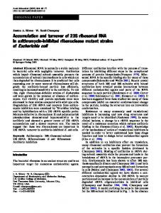
SERUM BACTERIOPHAGE RESISTANCE OF THE E . coli C STRAINS USED Serum Bacteriophages Resistance Class of Bacteria 2Sl CS2 CS3 CS4
Bacterial Strain No.
Selected Bacterial Resistance
1 2 3 4 5 6
None CS1 CS2 CS3 CS4 CS2 + CS4
+
+
+
-
+
+
+ +
I
+ +
-
_ _
+ +
II II
-
+
+
-
I
l+II
Table 1 shows the results of spot testing the four serum bacteriophage isolates on lawns of each of the bacterial strains used in this study. (+) indicates lytic action of the phage on the bacterial strain, ( - ) indicates no effect of the phage on the bacteria. The bacterial strains were isolated by subiecting E. coli C to a high multiplicity of infection of the indicated bacteriophage (CS1-4). Bacterial strain 1 is the parent E. coli C which has not undergone any selection for serum phage resistance. Strains 2 to 5 were selected for their ability to resist the indicated serum phage. Strain 6 was selected first for its resistance to serum phage CS2, and then for its resistance to phage CS4. This doubly resistant strain of E. coli C is resistant to all four of the serum phages. The lytic action of phage on the various strains of E. coli C tested are presented as Resistance Class I and:II, as indicated in the table.
their host range, as defined by the resistance classes we have described. A series of phages, all active against the original bacterial strain, can be grouped into types by examining the way a mutant bacterial strain, selected for its resistance to one phage, behaves with respect to its ability to be destroyed by the other phage isolates (7). Selection of bacterial mutants that are resistant to a particular bacteriophage is known to be specific, in that each change observed usually involves resistance to only a few of the bacteriophage strains capable of attaching to the original strain of bacteria (8). An example of the use of this technique is the separation of seven T coli-dysentery phages into six resistance classes (6). The technique is not completely reliable in determining whether or not phages are related. For example, the colidysentery phage T~ and the coli phage phi-80 are known to share the same attachment site, although they are not closely related (9). However, on the whole, this technique is superior to electron microscopy, evaluation of plaque morphology, and other methods for rapidly evaluating how closely a phage in one sample is related to a phage in another sample. The presence of bacteriophages in commercial sera indicates that the sera were contaminated
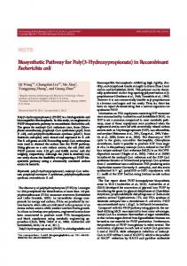
TABLE 2 ABILITY OF BACTERIOPHAGESIN COMMERCIALSERA TO FORM PLAQUES ON STRAINS OF E. coli C E. coli C Strains Comments
Lot No.
Type of Serum
Source
Fetal calf Fetal calf Calf Fetal calf Fetal calf Lamb Fetal calf
GIBCO GIBCO Flow GIBCO GIBCO Microbiological Associates Flow
A2201N 17 R7173R 12 421243 481 E014515/2 39 C41470 16 84 77336 455534 8
5 4 240 5 7 12 4
Calf Fetal calf Fetal calf Fetal calf Fetal calf Chicken Horse
Microbiological Associates GIBCO Microbiological Associates GIBCO GIBCO Microbiological Associates Microbiological Associates
80374 C3202K 79168 C6132G E0145J 80612 77928
345 45 13 14 39 4 0
105 2 4
r
8
9 10 11 12 13 14
8 287 13 12 54 2
12 10 560 42 7 59 4
Virus screened
215 13 11 6 8 3 8 0 0 0 O
225 36 4 14 18 7 0
Dialysed
1
Screened for bovine viruses and mycoplasm
Table 2 shows the number of plaques formed when 1 ml of each of 14 lots of commercial serum was plated against strains of E. coli C with various resistances to fetal calf sera viruses. The selection and phage resistance properties of the bacterial strains are summarized in Table 1.
PHAGES IN COMMERCIAL SERA with bacteria before filter sterilization. A study of fetal calf sera before filter sterilization detected bacterial contamination in nine of nine samples from two distributors (10). Inasmuch as filter sterilization of the sera failed to remove bacteriophages, other molecules of prokaryotic origin probably also pass through the filter. Thus, the sera may contain prokaryotic proteins, nucleic acids, etc. The presence of bacterial molecules in sera might well affect a wide range of tissue culture experiments. For example, studies involving antibiotics and drug metabolism in tissue culture could be confused by the presence of penicillinase or other bacterial molecules capable of breaking down antibiotics. On the other hand, certain bacterial or viral products might themselves have antibiotic activities. A specific antiviral, peptide-like substance has been isolated from lambda phage. The substance has been shown to have activity, both in vitro and in vivo, against vaccinia and Herpes simplex viruses (11). Furthermore, electron microscopic studies of low level virus production by various cell lines could possibly be confused by the presence of bacteriophages in the serum. Similarities between electron micrographs of bacteriophage particles and "C" and other virus-like particles may be seen, by comparing examples in the literature (12, 13). Furthermore, investigations which have been performed in many laboratories indicate that bacteriophage DNA can undergo transcription and translation in eukaryotic animals and plants (14-18). The presence of bacteriophages in vaccines was predicted by Merril and co-workers and confirmed by Petricciani and co-workers (19). Vaccines containing related bacteriophages and possibly other bacterial components, present a potentially very serious medical problem, in need of further investigation (20). The presence of related bacteriophages, bacterial toxins and certain other bacterial molecules might help to explain many of the adverse reactions to vaccination that are encountered. The possible importance of the presence of bacteriophages in commercially available sera has been emphasized by this study. The data (Tables 2 and 3) show that the distribution of these viruses is systematic rather than random. This means that any effects which the viruses
57
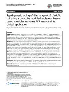
have in eukaryotic systems may be observed wherever these sera are used. Furthermore, the influence of phages in tissue culture experiments cannot be ruled out by simply repeating the study with a different serum lot. The relatedness of the serum viruses capable of plating on E. coli C may indicate that the workers who prepare the sera contaminate it with the same type of viruses; or, as appears more likely, properties of factors in serum itself are selected for the growth of certain types of phages. A more remote possibility is that the viruses are indeed intrinsic and that their relatedness is due to progenitors of the organisms from which they are derived. Attempts to destroy phages in serum without harming its quality include differential centrifugation and chemical treatment of the sera. Ultrafiltration (21) and immunoabsorbent columns (22), which have been shown capable of removing bacteriophages from sera, may prove unsatisfactory because they may not remove toxins and other undesirable bacterial molecules from the sera. A better solution is to prevent serum from being exposed to prokaryotic organisms in the first place, by handling the serum in a sterile TABLE 3 PERCENTAGE OF BACTERIOPHAGES IN COMMERCIAL SERA THAT D o NOT BELONG TO THE DEFINED RESISTANCE CLASSES
Code No.
1
2 3 4 5
6 7 8
9 10 11 12 13
Percentage of Phages Capable of Forming Plaques on Bacterial Strain
29 33 49 12 43 14 50 30 4 30 57 7 0
5
66 59 33 75 64 25 62 28 84 42 20 0
70 83 116 107 43 70 50 65 80 30 100 46 175
Table 3 shows the percentage of phage in various sera that are capable of making plaques on each of the resistance strains described above and summarized in Table 1. The code numbers designate serum lots described in Table 2.
58
GEIER, ATTALLAH, AND M E R R I L
manner. When serum has been successfully made free of prokaryotic contaminants, tissue culture systems may become cleaner and more reliable. REFERENCES 1. Merril, C. R., T. B. Friedman, A. F. M. Attallah, M. R. Geier, K. Krell, and R. Yarkin. 1972. Isolation of bacteriophages from commercial sera. In Vitro 8: 92-93. 2. Haselkorn, R. 1973. Bacterial viruses in fetal bovine sera. Proceedings o] the Workshop on the Problems o] Bacteriophage Contamination. Food and Drug Administration, Bureau of Biologics, Bethesda, Md. 3. Rosanoff, E. I. 1973. The development of a salmonella phage assay and its application to the testing of EGG biological products. Proceedings o] the Workshop on the Problems o] Bacteriophage Contamination. Food and Drug Administration, Bureau of Biologics, Bethesda, Md. 4. Boone, C. W., N. Mantel, J. D. Caruso, Jr., E. Kazam, and R. E. Stevenson. 1972. Quality control studies on fetal bovine serum used in tissue culture. In Vitro 7: 174-189. 5. Molander, C. W., A. J. Kniazeff, C. W. Boone, A. Paley, and D. T. Imagawa. 1972. Isolation and characterization of viruses from fetal calf serum. In Vitro 7 : 168-173. 6. Adams, M. H. 1959. Bacteriophages. Interscience Publishers Inc., New York. 7. Burnet, F. M. 1929. Smooth-rough variation in bacteria in its relation to bacteriophage. J. Path. Bact. 32 : 15-42. 8. Demerec, M., and U. Fano. 1945. Bacteriophage-resistant mutants in Escherichia coli. Genetics 30: 119-136. 9. Signer, E. R. 1966. Interactions of prophages at the att,o site with the chromosome of Escherichia coli. J. Mol. Biol. 15 : 243-255. 10. Morello, J. A. 1973. Isolation and characterization of bacteria in fetal bovine sera. Proceedi~gs o] the Wo~'kshop on the Problems o] Bacteriophage Contamination. Food and Drug Administration, Bureau of Biologics, Bethesda, Md. 11. Centifanto, Y. 1968. Antiviral agent from lambda-infected Escherichia coli K-12. Appl. Microbiol. 16: 827-834. 12. Simon, L. D. 1972. Infection of Escherichia coli by T2 and T4 bacteriophages as seen in the
electron microscope: T4 head morphogenesis. Proc. Natl. Acad. Sci. U.S.A. 69: 907-911. 13. Hooks, J., C. J. Gibbs, H. Chopra, M. Lewis, and D. C. Gajdusek. 1972. Spontaneous transformation of human brain cells grown in vitro and description of associated particles. Science 176: 1420-1425. 14. Merril, C. R., M. R. Geier, and J. C. Petricciani. 1971. Bacterial gene expression in human cells. Nature 233 : 398--400. 15. Geier, M. R., and C. R. Merril. 1972. Lambda phage transcription in human fibroblasts. Virology 47 : 638-643. 16. Ledoux, L. 1971. In: In]ormative Molecules in Biological Systems. North Holland and American Elsevier, New York. 17. Doy, C. H., P. M. Gresshoff, and B. G. Rolfe. 1973. Biological and molecular evidence for the transgenosis of genes from bacteria to plant cells. Proc. Natl. Acad. Sci. U.S.A. 70: 723-726. 18. Doy, C. H., P. M. Gresshoff, and B. G. Rolfe. 1973. Time course of phenotypic expression of Escherichia coli gene Z following transgenosis in haploid Lycopersicon esculentum cells. Nature New Biol. 244: 90-91. 19. Chu, F. C., J. Johnson, P. Carter, and J. Petricciani. 1973. Bacteriophage isolation from viral vaccines. Proceedings o] the Workshop on the Problems o] Bacteriophage Contamination. Food and Drug Administration, Bureau of Biologics, Bethesda, Md. 20. Geier, M. R., M. E. Trigg, and R. Merril. 1973. A model system for the evaluation of the fate of phage in contaminated vaccines: physiological disposition of bacteriophage in mice. Proceedings o] the Workshop on the Problems o] Bacteriophage Contamination. Food and Drug Administration, Bureau of Biologics, Bethesda, Md. 21. Attallah, A. F. M., J. C. I-Iouck, and J. C. Petricciani. 1973. The elimination of phages from sera by molecular ultrafiltration. Proceedings o] the Workshop on the Problems o] Bacteriophage Contamination. Food and Drug Administration, Bureau of Biologics, Bethesda, Md. 22. Orr, H. C., H. H. Westall, P. G. Probst, D. C. Littlejohn, F. C. Chu, J. B. Johnson, and J. C. Petricciani. 1974. Removal of bacteriophages from serum by immunoadsorbents. In Vitro 9 : 354.