Appl Microbiol Biotechnol (2009) 82:463–470 DOI 10.1007/s00253-008-1774-x
BIOTECHNOLOGICALLY RELEVANT ENZYMES AND PROTEINS
Improving aquaporin Z expression in Escherichia coli by fusion partners and subsequent condition optimization Jiazhang Lian & Shinghua Ding & Jin Cai & Danping Zhang & Zhinan Xu & Xiaoning Wang
Received: 27 August 2008 / Revised: 26 October 2008 / Accepted: 27 October 2008 / Published online: 13 November 2008 # Springer-Verlag 2008
Abstract Aquaporin Z (AqpZ), a typical orthodox aquaporin with six transmembrane domains, was expressed as a fusion protein with TrxA in E. coli in our previous work. In the present study, three fusion partners (DsbA, GST and MBP) were employed to improve the expression level of this channel protein in E. coli. The result showed that, compared with the expression level of TrxA-AqpZ, five- to 40-fold increase in the productivity of AqpZ with fusion proteins was achieved by employing these different fusion partners, and MBP was the most efficient fusion partner to increase the expression level. By using E. coli C43 (DE3)/ pMAL-AqpZ, the effects of different expression conditions were investigated systematically to improve the expression level of MBP-AqpZ in E. coli. The high productivity of MBP-AqpZ (200 mg/l) was achieved under optimized conditions. The present work provides a novel approach to improve the expression level of membrane proteins in E. coli.
J. Lian : J. Cai : D. Zhang : Z. Xu (*) Institute of Bioengineering, Department of Chemical and Biochemical Engineering, Zhejiang University, Hangzhou 310027, China e-mail:
[email protected] S. Ding Department of Biological Engineering, University of Missouri, Columbia, MO 65211, USA X. Wang School of Bioscience and Bioengineering, South China University of Technology, Guangzhou 510006, China
Keywords Aquaporin Z . MBP . GST . DsbA . Membrane protein
Introduction Aquaporin, a ubiquitous water channel, is characterized by high water permeability and low activation energy (Borgnia et al. 1999b). AqpZ, a water channel in Escherichia coli, has an open reading frame of 693-base and is a typical orthodox aquaporin with 6 transmembrane domains and five connecting loops (Calamita et al. 1995). NPA box (residues Asn-Pro-Ala), the most highly conserved motifs, is the centre of the so-called Hourglass Model which is believed to be directly involved in the selectivity of the channel (Calamita 2000). It was reported that, upon reconstitution into proteoliposomes, AqpZ exhibited very high water permeability (Pf =10×10−14 cm3 s−1 subunit−1) and had low Arrhenius activation energy (Ea=3.7 kcal/mol; Borgnia et al. 1999a). About 12 ng functional AqpZ is effective to recover the requisite 2.4 l water for a single person per day. Moreover, AqpZ is a specific biological water channel with high osmotic water permeability, lack of N-linked glycan, resistant to proteolysis, and not inhibited by any biological macromolecules, making AqpZ-based water recovery technology a promising tool for water filtration and water recovery for space flight (Swartz 2006). Therefore, AqpZ is not only an ideal model for the study on how to improve the expression level of channel proteins in E. coli, but also greatly promises a novel waterfilter biotechnology. Membrane proteins, considered as the bridge between cytoplasm and extracellular environment, are important drug targets for modern medicinal and pharmaceutical research (Wang et al. 2003) and play a variety of critical
464
Appl Microbiol Biotechnol (2009) 82:463–470
physiological roles, such as transduction of extracellular signals, transportation of substances through membrane, and generation of energy. High-resolution structural information is essential for understanding the function of these proteins. The structural study of these proteins is hampered by low expression level due to the highly hydrophobic nature of membrane proteins that is toxic to host itself. Furthermore, the overexpression will result in dramatic decrease in cell growth, and even immediate cell disruption (Tate 2001). In addition, overexpression of these proteins may also block the insertion machinery responsible for establishing correct topology of membrane proteins (Kiefer et al. 1996). In order to achieve high expression of AqpZ, fusion protein that can prevent the insertion of the recombinant protein into membrane might be a promising strategy. Fusion protein expression with a highly soluble partner, such as thioredoxin (TrxA; LaVallie et al. 1993), disulphide oxidoreductase (DsbA; Zhang et al. 1998), glutathione-S-transferase (GST; Nygren et al. 1994), maltose binding protein (MBP; Pryor and Leiting 1997), Protein A (Samuelsson et al. 1994), and ubiquitin (Power et al. 1990), would improve the yield as well as the solubility of recombinant protein. However, when the target protein comes to be membrane proteins, such as Gprotein coupled receptors (Kiefer et al. 1996; Kiefer et al. 2000), phospholamban (Yao et al. 1996), and bacterioopsin (Chen and Gouaux 1996), the fusion protein tends to aggregate to form inclusion bodies in E. coli. Although tremendous efforts have been invested in expressing membrane proteins with a variety of strategies, the expression level is low, about 1 mg/l culture (Borgnia et al. 1999a; Kozono et al. 2003; Maduke et al. 1999; Voges and Jap 1998). Therefore, it may be an efficient strategy to express and refold membrane proteins with these fusion tags in insoluble form with relative high yield to facilitate the subsequent purification and structure determination. In the present work, high-level expression of AqpZ fusion proteins was achieved in E. coli by employing three fusion partners (DsbA, GST and MBP), and MBP was found to be fusion partner to achieve the highest productivity of AqpZ fusion protein with least toxicity to cell growth.
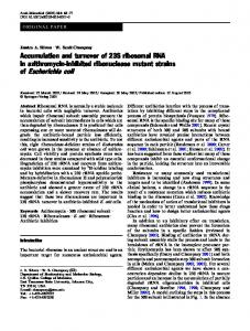
Materials and methods Host strains, vectors, and enzymes E. coli DH5α was used as the host for gene manipulation, while C43 (DE3) (Lucigen, Middleton, WI), a mutant derived from BL21 (DE3) with high efficiency expression of toxic proteins (Bruno and John 1996), was chosen as recombinant protein expression host. pMD19-T Simple Vector (Takara, Dalian, China) was used as cloning vector, and pET-39b(+) (Novagen, Madison, WI), pGEX-4T-2 (Pharmacia, Piscataway, NJ) and pMAL-P2 (New England Biolabs, Beverly, MA) were used to construct expression vectors. The restriction enzymes NcoI, HindIII, BamHI and SalI as well as LA Taq DNA polymerase and T4 DNA ligase were all purchased from Takara. Amplification of AqpZ DNA fragments Except the selection of restriction enzymes, gene manipulation was exactly the same as that for the construction of TrxA-AqpZ in our previous study (Lian et al. 2008), including the substitute of the rare codon AGA with CGC and the introduction of two consecutive stop codons (TAATAA; Table 1). Construction of cloning and expression vectors The amplified DNA fragments were cloned into the pMD19-T Simple Vector, and then transformed into E. coli DH5α. The cloning vectors were extracted from the overnight culture with a QIAprep Spin Miniprep Kit (Qiagen, GmbH, Germany). Nucleotide sequences of the target DNA fragment in the cloning vectors were confirmed by DNA sequencing. Following enzyme digestion (NcoI/ HindIII or BamHI/SalI), the target fragments from the cloning vector, pET-39b(+), pGEX-4T-2 and pMAL-P2 were recovered using a QIAquick Gel Extraction Kit (Qiagen) and ligated by T4 DNA ligase to construct the fusion expression vector pET39-AqpZ, pGEX-AqpZ and pMAL-AqpZ (Fig. 1). Then the reconstructed vectors were transformed into E. coli C43 (DE3) for following studies.
Table 1 Oligonucleotides used for the construction of expression vectors Primer
Sequence
Enzyme
F1 R1 F2 R2
5′-GGTGCGGATCCATGTTCCGCAAATTAGCAGCTGAATGTT-3′ 5′-AACGCGTCGACTTATTAATCACGCTTTTCCAGCAGGGTCCGG-3’ 5′-TACCATGGCTATGTTCCGCAAATTAGCAGCTGAATGTT-3′ 5′-GCAAGCTTTTATTAATCACGCTTTTCCAGCAGGGTC-3′
BamHI SalI NcoI HindIII
F and R represent the forward and reverse primers, respectively. F1, R1 for the construction of pGEX-AqpZ and pMAL-AqpZ, while F2, R2 for pET39-AqpZ. The restriction enzyme sites introduction in the primers are italicized, the initiating codon and the double terminating codons are shown in bold, and the substituted codon is underlined
Appl Microbiol Biotechnol (2009) 82:463–470
465
f1 origin
HindIII (6580)
or 100 μg/ml ampicillin at 37°C overnight. The culture was inoculated into 50 ml fresh SOC medium per 250 ml flask, and then cultivated and induced at certain conditions according to optimization purpose.
NcoI (5876)
DsbA-AqpZ
kan
Preparation of inclusion bodies pE T 39 -A qpZ 6772 bp
Ori
lac I
a tac Promoter
BamHI (931)
lac I
GST-AqpZ
pGEX-A qpZ 5655 bp SalI (1636)
pBR322 origin Amp
b
p B R 322 Or ig in Tac Pr o mo ter
pMAL-P2-AqpZ
SDS-PAGE analysis
74 12 bp
f1 o rig in
MB P-Aq p Z BamH I (2777)
Amp n n rb Termin ato r
The cells were harvested by centrifugation at 4°C (5,000×g, 30 min), resuspended in 3 ml cell lysis buffer containing 50 mM Tris–HCl (pH8.0), 1 mM EDTA, 100 mM NaCl, 1 mM phenylmethylsulfonyl fluoride (PMSF) and 0.5 mg/ ml lysozyme, and stirred at 4°C for 30 min. Next, 4 mg deoxycholic acid was added and the resulting suspension was incubated at 37°C with occasional stirring until the suspension turned to be viscous. Then 10 μg/ml DNase I was added and the mixture was incubated at room temperature (RT) for about 30 min until the suspension became clear. Following centrifugation at 4°C (17,000×g, 30 min), the pellet, containing inclusion bodies and cell debris, was washed three times by resuspending in wash buffer containing 50 mM Tris–HCl (pH8.0), 10 mM EDTA, 100 mM NaCl, 1 mM PMSF and 0.5% Triton X-100. Soluble fusion protein was obtained by stirring the washed inclusion bodies in 1 ml solubilization buffer I containing 50 mM Tris–HCl (pH8.0), 10 mM EDTA, 100 mM NaCl, 1 mM PMSF, and 8 M urea at RT for 1 h. The resulting solution was diluted with 9 volume of solubilization buffer II containing 50 mM KH2PO4 (pH 10.7), 1 mM EDTA, 50 mM NaCl, and 1 mM PMSF, incubated at RT for 30 min, and then adjusted to pH8.0 followed by additional 1 h incubation at RT. Insoluble materials were removed by centrifugation at RT (17,000 g, 30 min), and the resulting supernatant containing most of the inclusion bodies was analyzed by SDS-PAGE.
Sa lI (34 82)
c Fig. 1 Expression vectors for fusion proteins. a pET39-AqpZ with T7 promoter, DsbA tag and kanamycin resistance; b pGEX-AqpZ with tac promoter, GST tag and ampicillin resistance; c pMAL-AqpZ with tac promoter, MBP tag and ampicillin resistance
Expression of AqpZ fusion proteins A fresh clone of E. coli harboring the expression vector was grown in 3 ml LB medium containing 30 μg/ml kanamycin
SDS-PAGE was performed using 12% resolving gels and 5% stacking gels. Gels were initially run at 100 V and then at 200 V once the dye entered the resolving gel. The protein bands were visualized by Coomassie brilliant blue R250 and then analyzed with Quantity One (Bio-Rad, Hercules, CA) to evaluate the expression level of target fusion proteins.
Results Gene cloning and sequencing AqpZ genes were successfully amplified from E. coli DH5α genomic DNA, and ligated into pET-39b(+), pGEX-4T-2 and pMAL-P2, respectively. Using a 3730 DNA Analyzer (Applied Biosystems, CA, USA), the
466
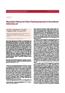
sequence of AqpZ and the introduction of DsbA, GST, or MBP fusion tag were confirmed. There were certain specific protease sites (enterokinase for pET39-AqpZ, thrombin for pGEX-AqpZ and Factor Xa for pMAL-AqpZ) between the fusion tags and the target protein to facilitate the purification of mature AqpZ by specific removal of the fusion partners. Fusion expression of AqpZ with DsbA, GST, and MBP The expression vectors (pET39-AqpZ, pGEX-AqpZ, and pMAL-AqpZ) were transformed into E. coli C43 (DE3) to get three types of recombinant E. coli, respectively. When the culture was propagated (37°C, 200 rpm) to an optical density (OD600) of 1.0, the expression was induced by 1.0 mM isopropyl-β-D-thiogalactoside (IPTG), and then followed by additional 6 h culture at 37°C. As shown in Fig. 2a, DsbA-AqpZ, GST-AqpZ and MBP-AqpZ were all
Fig. 2 Expression of AqpZ fusion proteins in E. coli. a SDS-PAGE analyses of AqpZ expression with different partners. Lane 1, 3, 5, negative control without induction; Lane 2, 4, 6, the inclusion bodies from C43 (DE3)/pET39-AqpZ, C43 (DE3)/pGEX-AqpZ, and C43 (DE3)/pMAL-AqpZ, respectively. Numbers on the left denote the
Appl Microbiol Biotechnol (2009) 82:463–470
effectively expressed as inclusion bodies in E. coli. Further analysis of the supernatant after cell lysis by SDS-PAGE and western blotting indicated that there are little soluble or membrane-targeted fusion proteins (Data not shown). With a BCA protein assay kit (Pierce, Rockford, IL), the productivity of DsbA-AqpZ, GST-AqpZ, MBP-AqpZ were determined to be about 25 mg/l, 30 mg/l, 65 mg/l, respectively. It was also seen in Fig. 2b that, after 6 h IPTG induction, the cell densities for three different cultures were 1.8, 2.5, and 4.5, respectively. Apparently, the reduced toxicity of MBP-AqpZ overexpression to cell growth contributed to the highest productivity of MBPAqpZ in E. coli. Interestingly, although with a periplasmic location signal at the N-terminus of MBP, no target fusion protein in the periplasm was detected. To explore the possibilities of improving expression levels, several onefactor experiments were conducted to investigate different expression conditions in the subsequent experiments.
position of protein markers. Arrows indicate the position of target proteins. b Biomass of recombinant E. coli after 6 h induction by 1.0 mM IPTG at 37°C and the corresponding expression level of AqpZ fusion proteins
Appl Microbiol Biotechnol (2009) 82:463–470
467
It is well-known that temperature affects recombinant protein expression significantly (Makrides 1996). E. coli C43 (DE3)/pMAL-AqpZ was cultivated to an optical density (OD600) of 1.0, induced by 1.0 mM IPTG, and then followed by additional 6 h culture at 40°C, 37°C, 34°C, 30°C, or by overnight culture at 25°C, respectively. As shown in Fig. 3, when the culture temperature was higher than 34°C, the expression level of target fusion protein was reduced greatly. Obviously, the lower cultivation temperature would result in higher expression level of MBP-AqpZ inclusion bodies, especially at 30°C. The highest expression level of MBP-AqpZ was achieved in E. coli at 30°C. It was well reported that high culture temperature would improve the cell mass thus increase the expression level of insoluble heterologous proteins in E. coli. However, for the expression of AqpZ fusion protein with high molecular weight and high hydrophobicity, increased temperature would lead to a negative effect on cell growth of recombinant E. coli (data not shown), thus significantly reducing the productivity of the fusion membrane protein. In our previous studies, a variety of proteins expressed in an insoluble form at high temperature (37°C) could be expressed as a soluble form when the culture temperature was reduced to 26°C or 28°C (Huang et al. 2007; Peng et al. 2004; Xu et al. 2006). However, even when the cultivation temperature was set at as low as 25°C, we did not detect soluble expression of target fusion protein. Therefore, it was reasonable to speculate that, with its high molecular weight and hydrophobicity, this fusion protein would form inclusion bodies in E. coli quickly after it was translated as fusion protein on ribosome.
protein. The time-course of C43 (DE3)/pMAL-AqpZ growth was pre-determined at 30°C (Fig. 4a) and the effect of induction timing was evaluated by adding 1.0 mM IPTG at different stages of growth phase according to this growth curve. The expression was analyzed by SDS-PAGE, and the expression level of target protein was compared (Fig. 4b). At different induction timing, the concentration of MBPAqpZ varied in a wide range, and the maximum expression level was observed when induced at the pre-exponential phase, corresponding to the OD600 value of 0.6. Earlier or later induction resulted in lower expression level, e.g. the expression level decreased dramatically when induced at stationary phase. Effect of IPTG concentration on AqpZ expression Because of the great contribution to recombinant protein expression and serious harm to cell growth, final IPTG concentration needs to be optimized. In this work, IPTG concentration was examined from 0.2 to 1.0 mM. Our results showed that the highest expression level of fusion
6
5 5
4
4
OD600
Effect of cultivation temperature on AqpZ expression
3
3
2 2
1 1
Effect of induction timing on AqpZ expression 0
In the process of recombinant protein expression in E. coli, IPTG induction is the turning point between cell growth and recombinant protein synthesis (Makrides 1996). The addition of IPTG triggers the transcription of foreign gene and, consequently, brings great changes to the metabolism of host cells by initiating the translation of recombinant
0
1
2
3
4
5
6
7
8
Time (h)
a
Marker
1
2
3
4
5
66
45 Mar ker
1
2
3
4
5
66
45 35
Fig. 3 SDS-PAGE analyses of MBP-AqpZ expression under different cultivation temperature. Lane 1, 2, 3, 4, 5 are inclusion bodies when cultured at 25, 30, 34, 37, and 40°C, respectively
35
b Fig. 4 Effect of induction timing on the expression level of MBPAqpZ. a Growing profile of C43 (DE3)/pMAL-AqpZ cultivated at 34°C. Arrows indicate the induction points. Data represent the average of triplicates. b SDS-PAGE analyses of the target protein. Lane 1, 2, 3, 4, 5 are inclusion bodies when induced at lag phase, pre-exponential phase, mid-exponential phase, post-exponential phase, and stationary phase, respectively
Appl Microbiol Biotechnol (2009) 82:463–470
protein was achieved by 0.4 mM IPTG induction (Fig. 5). High IPTG concentration would decrease the productivity of AqpZ fusion protein.
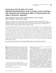
The time-course of MBP-AqpZ expression was by determining biomass and expression level after inducing the culture of C43 (DE3)/pMAL-AqpZ. As shown in Fig. 6a, under the optimized culture and induction conditions (30°C, 0.4 mM IPTG induction at pre-exponential phase), the growing profiles of C43 (DE3)/pMAL-AqpZ with and without IPTG induction were quite similar, and the OD600 value of the culture after the induction was only a little lower than those of the un-induced cultures, suggesting that the overexpression of AqpZ fusion protein did not significantly exert negative effect on cell growth. Corresponding with this, the expression level of AqpZ increased up to 10 h after induction, and the maximal concentration of MBP-AqpZ (about 200 mg/l) was obtained (Fig. 6b). Further extension of expression time more than 10 h did not increase the expression level presumably because of the degradation of the AqpZ fusion protein.
Discussion The lack of a simple, light, portable water recovery device is one of the limiting factors in extending space flight currently. The high selectivity and permeability make aquaporins ideal candidates for water recovery. However, the development of aquaporin-based water bio-filter is hampered by the availability of sufficient amount of water channel proteins since the overexpression of integral membrane proteins may exhibit toxicity towards the host due to the highly hydrophobic nature. Expression of hydrophobic membrane proteins as fusions with soluble partner followed by selective protease cleavage may provide a generally useful expression and purification strategy (Chen and Gouaux 1996; Douglas et al. 2005). It was expected that fusion of a hydrophilic
OD600
Effect of post-induction time on AqpZ expression
6
300
5
250
4
200
3
150
2
100
1
50
0 0
2
4
6
8
10
12
14
16
18
MBP-AqpZ (mg/l)
468
0 20
Time (h)
a Marker
1
2
3
4
5
6
7
8
66
45 35
b Fig. 6 Effect of post-induction duration on the expression level in C43 (DE3)/pMAL-AqpZ. a Growing profile of C43 (DE3)/pMALAqpZ with (open circle) and without (filled circle) induction and the corresponding time-course of MBP-AqpZ expression (filled diamond). Arrow indicates the induction timing. Data represent the average of triplicates. b SDS-PAGE analyses of target protein. Lane 1, 2, 3, 4, 5, 6, 7, 8 are inclusion bodies from samples taken in 1 h, 2 h, 4 h, 6 h, 8 h, 10 h, 13 h, and 16 h after induction, respectively
domain to the membrane protein of interest could improve both the expression level and the stability of the target protein. This was achieved by (1) expressing protein in cytoplasm rather than targeting the protein into the membrane to minimize toxicity via reducing perturbation of the cell membrane and the machinery for membrane protein insertion (Kiefer et al. 2000; Tate 2001); (2) taking advantage of the larger volume of the cytoplasm compared to the cell membrane; and (3) reducing degradation through the formation of insoluble, protease-resistant inclusion
Table 2 Expression level of AqpZ fusion proteins Marker
1
2
3
4
5
Partner
Membrane associated
Inclusion bodies
Reference
10-His 6-His TrxA DsbA GST MBP
2.5 mg/l 9.05 mg/l U U U U
NA 2.35 mg/l 5 mg/l 25 mg/l 30 mg/l 200 mg/l
(Borgnia et al. 1999a) (Lian et al. 2008) (Lian et al. 2008) This study This study This study
66
45 35
Fig. 5 SDS-PAGE analyses of MBP-AqpZ expression with different IPTG concentration. Lane 1, 2, 3, 4, 5 are inclusion bodies when induced by 0.2 mM, 0.4 mM, 0.6 mM, 0.8 mM, and 1.0 mM, respectively
NA represents data not available, and U represents undetectable due to low expression level
Appl Microbiol Biotechnol (2009) 82:463–470
bodies. However, relatively few transmembrane proteins have been tested for efficient expression as fusion proteins with hydrophilic partners (Buck et al. 2003; Chen and Gouaux 1996; Grisshammer et al. 1994; Grisshammer and Tate 1995; Laage and Langosch 2001; Pompejus et al. 1993; Stanasila et al. 1999). In our previous work, the toxicity of the overexpression of defensins in E. coli was greatly reduced by fusion to the C-terminus of thioredoxin (TrxA; Huang et al. 2007; Peng et al. 2004; Xu et al. 2006; Zhong et al. 2006), but the similar fusion of AqpZ with TrxA only brought about low-level expression of AqpZ fusion protein (ca. 5 mg/l; Table 2). The possible reason was that, as a low molecular weight fusion partner, TrxA could not neutralize the hydrophobicity of AqpZ because of six transmembrane domains in its 3-D structure. Therefore, three larger and more hydrophilic fusion partners, DsbA, GST and MBP, were selected as substitutes to improve the expression level of AqpZ. As expected, much higher expression level of fusion proteins was achieved with the aid of these partners than that of TrxA-AqpZ (Table 2). Especially, the productivity of AqpZ fusion protein was dramatically improved by using MBP as the fusion partner, and the highest expression level of MBP-AqpZ could reach up to 200 mg/L after the optimization of culture and induction conditions. It was also found in the present study that many culture and induction conditions, such as temperature, IPTG concentration, induction timing and post-induction duration, significantly affected the expression level of AqpZ fusion protein. The optimized conditions could reduce the negative effect of the membrane protein expression on cell growth greatly, which was confirmed by similar growth profiles of recombinant E. coli with and without IPTG induction (Fig. 6b). This suggested that the high-level expression of channel proteins could be achieved in E. coli with suitable fusion partners and subsequent systematical optimization of the expression conditions. In conclusion, the hydrophobic and toxic channel protein (AqpZ) could be expressed efficiently in E. coli by fusion with DsbA, GST and MBP respectively. The highest productivity of AqpZ fusion protein (200 mg/l) was achieved with fusion partner MBP after systematic optimization of the expression conditions. The present work demonstrated an efficient strategy to improve the expression level of membrane proteins in E. coli, which is especially important to study the function and 3D structure of membrane proteins. Further study of fusion protein purification would be warranted for functional study of the protein. Acknowledgments This work was financially supported by The National Natural Science Foundation of China (Grant No.20736008, 20676115), The Ministry of Science and Technology (Grant No 2007AA021702) and The Ministry of Education (Grant No. 20060335085), The People’s Republic of China.
469
References Borgnia MJ, Kozono D, Calamita G, Maloney PC, Agre P (1999a) Functional Reconstitution and Characterization of AqpZ, the E. coli Water Channel Protein. J Mol Biol 291:1169–1179 Borgnia MJ, Nielsen M, Engel A, Agre P (1999b) Cellular and molecular biology of the aquaporin water channels. Annu Rev Biochem 68:425–458 Bruno M, John EW (1996) Over-production of proteins in Escherichia coli: mutant hosts that allow synthesis of some membrane proteins and globular proteins at high levels. J Mol Biol 260:289–298 Buck B, Zamoon J, Kirby TL, DeSilva TM, Karim C, Thomas D, Veglia G (2003) Overexpression, purification, and characterization of recombinant Ca-ATPase regulators for high-resolution solution and solid-state NMR studies. Protein Expres Purif 30:253–261 Calamita G (2000) The Escherichia coli aquaporin-Z water channel. Mol Microbiol 37(2):254–262 Calamita G, Bishai WR, Preston GM, Guggion WB, Agre P (1995) Molecular Cloning and Characerization of AqpZ, a Water Channel from Escherichia coli. J Biol Chem 270:29063–29066 Chen GQ, Gouaux JE (1996) Overexpression of bacterio-opsin in Escherichia coli as a water-soluble fusion to maltose binding protein: Efficient regeneration of the fusion protein and selective cleavage with trypsin. Protein Sci 5:456–467 Douglas JL, Trieber CA, Afara M, Young HS (2005) Rapid, highyield expression and purification of Ca2+-ATPase regulatory proteins for high-resolution structural studies. Protein Expres Purif 40:118–125 Grisshammer R, Tate CG (1995) Overexpression of integral membrane proteins for structural studies. Q Rev Biophys 28:315–422 Grisshammer R, Little J, Aharony D (1994) Expression of rat NK-2 (neurokinin A) receptor in E. coli. Recept Channels 2:295–302 Huang L, Xu ZN, Zhong ZX, Peng L, Chen HQ, Cen PL (2007) Enhance expression and primary purification of soluble HBD3 fusion protein in Escherichia coli. Appl Biochem Biotechnol 142 (2):139–147 Kiefer H, Krieger J, Olszewski JD, Heijne G, Prestwich GD, Breer H (1996) Expression of an Olfactory Receptor in Escherichia coli: purification, reconstitution, and ligand binding. Biochemistry 35:16077–16084 Kiefer H, Vogel R, Mailer K (2000) Bacterial expression of G-proteincoupled receptors: prediction of expression levels from sequence. Recept Channels 7:109–119 Kozono D, Ding XD, Iwasaki I, Meng XY, Kamagata Y, Agre P, Kitagawa Y (2003) Functional expression and characterization of an Archaeal aquaporin: AqpM from Methanothermobacter marburgensis. J Biol Chem 278(12):10649–10656 Laage R, Langosch D (2001) Strategies for prokaryotic expression of eukaryotic membrane proteins. Traffic 2:99–104 LaVallie ER, DiBlasio EA, Kovacic S, Grant KL, Schendel PF, McCoy JM (1993) A thioredoxin gene fusion expression system that circumvents inclusion body formation in the E. coli cytoplasm. Biotechnology 11:187–193 Lian JZ, Fang XM, Cai J, Chen QX, Zheng Q, Kai L, Xu ZN (2008) Efficient expression of membrane-bound water channel protein (Aquaporin Z) in Escherichia coli. Protein Pept Lett 15:687–691 Maduke M, Pheasant DJ, Miller C (1999) High-level expression, functional reconstitution, and quaternary structure of a prokaryotic ClC-type chloride channel. J Gen Physiol 114:713–722 Makrides SC (1996) Strategies for achieving high-level expression of genes in Escherichia coli. Microbiol Rev 60(3):512–538 Nygren PA, Stahl S, Uhlen M (1994) Engineering proteins to facilitate bioprocessing. Trends Biotechnol 12:184–188
470 Peng L, Xu ZN, Fang XM, Wang F, Cen PL (2004) High-level expression of soluble human Beta-Defensin-2 in E. coli. Process Biochem 39:2199–2205 Pompejus M, Friedrich K, Teufel M, Fritz HJ (1993) High-yield production of bacteriorhodopsin via expression of a synthetic gene in Escherichia coli. Eur J Biochem 211:27–35 Power RF, Conneely OM, McDonnell DP, Clark JH, Butt TR, Schrader WT, O’Malley BW (1990) High level expression of a truncated chicken progesterone receptor in Escherichia coli. J Biol Chem 265:1419–1424 Pryor KD, Leiting B (1997) High-level expression of soluble protein in Escherichia coli using a His6-tag and maltose-bindingprotein double-affinity fusion system. Protein Expres Purif 10:309–319 Samuelsson E, Moks T, Nilsson B, Uhlen M (1994) Enhanced in vitro refolding of insulin-like growth factor I using a solubilizing fusion partner. Biochemistry 33:4207–4211 Stanasila L, Massotte D, KieVer BL, Pattus F (1999) Expression of delta, kappa and mu human opioid receptors in Escherichia coli and reconstitution of the high-affinity state for agonist with heterotrimeric G proteins. Eur J Biochem 260:430–438 Swartz JR (2006) Developing cell-free biology for industrial applications. J Ind Microbiol Biotechnol 33:476–485
Appl Microbiol Biotechnol (2009) 82:463–470 Tate CG (2001) Overexpression of mammalian integral membrane proteins for structural studies. FEBS Lett 504(3):94–98 Voges D, Jap BK (1998) Recombinant expression, purification and characterization of Kch, a putative Escherichia coli potassium channel protein. FEBS Lett 429:104–108 Wang DN, Safferling M, Lemieux MJ, Griffith H, Chen Y, Li XD (2003) Practical aspects of overexpressing bacterial secondary membrane transporters for structural studies. Biochim Biophys Acta 1610:23–36 Xu ZN, Zhong ZX, Huang L, Peng L, Wang F, Cen PL (2006) High-level production of bioactive human beta-defensin-4 in Escherichia coli by soluble fusion expression. Appl Microbiol Biotechnol 72:471–479 Yao Q, Bevan JL, Weaver RF, Bigelow DJ (1996) Purification of porcine phospholamban expressed in Escherichia coli. Protein Expres Purif 8:463–468 Zhang Y, Olsen DR, Nguyen KB, Olson PS, Rhodes ET, Mascarenhas D (1998) Expression of eukaryotic proteins in soluble form in Escherichia coli. Protein Expres Purif 12:159–165 Zhong ZX, Xu ZN, Peng L, Huang L, Fang XM, Cen PL (2006) Tandem repeat mhBD2 gene enhance the soluble fusion expression of hBD2 in Escherichia coli. Appl Microbiol Biotechnol 71(5):661–667