Feb 24, 1987 - WILLIAM C. BARRETTE, JR., J. MICHAEL ALBRICH, AND JAMES K. HURST*. Department of .... Chemical Co. Catalase was purchased from Sigma Chemical. Co. All other ...... J. Clin. Invest. 78:177-184. 3. Albrich, J. M., and J. K. Hurst. 1982. Oxidative .... Niven, D. F., P. A. Collins, and C. J. Knowles. 1977.
INFECTION AND IMMUNITY, OCt. 1987, p. 2518-2525 0019-9567/87/102518-08$02.00/0 Copyright C) 1987, American Society for Microbiology
Vol. 55, No. 10
Hypochlorous Acid-Promoted Loss of Metabolic Energy in Escherichia coli WILLIAM C. BARRETTE, JR., J. MICHAEL ALBRICH, AND JAMES K. HURST* Department of Chemical and Biological Sciences, Oregon Graduate Center, Beaverton, Oregon 97006-1999
Received 24 February 1987/Accepted 29 June 1987
Oxidation of Escherichia coli by hypochlorous acid (HOCI) or chloramine (NH2Cl) gives rise to massive hydrolysis of cytosolic nucleotide phosphoanhydride bonds, although no immediate change occurs in either the nucleotide pool size or the concentrations of extracellular end products of AMP catabolism. Titrimetric curves of the extent of hydrolysis coincide with curves for loss of cell viability, e.g., reduction in the adenylate energy charge from 0.8 to 0.1-0.2 accompanies loss of 99% of the bacterial CFU. The oxidative damage caused by HOCI is irreversible within 100 ms of exposure of the organism, although nucleotide phosphate bond hydrolysis requires several minutes to reach completion. Neither HOCI nor NH2Cl reacts directly with nucleotides to hydrolyze phosphoanhydride bonds. Loss of viability is also accompanied by inhibition of induction of B-galactosidase. The proton motive force, determined from the distribution of 14C-radiolabeled lipophilic ions, declines with incremental addition of HOCI or NH2Cl after loss of respiratory function; severalfold more oxidant is required for the dissipation of the proton motive force than for loss of viability. These observations establish a causal link between loss of metabolic energy and cellular death and indicate that the mechanisms of oxidant-induced nucleotide phosphate bond hydrolysis are indirect and that they probably involve damage to the energy-transducing and transport proteins located in the bacterial plasma membrane.
Stimulated neutrophils generate the potent microbicide hypochlorous acid (HOCI) by myeloperoxidase-catalyzed peroxidation of chloride ion (18, 20, 21, 25, 48, 49). The role of HOCI in the microbicidal action of neutrophils is not well defined, however, primarily because myeloperoxidasedeficient neutrophils generate other oxidants which are potentially toxic and because neutrophil granules contain other components that are capable of inactivating microbes by nonoxidative mechanisms (15, 27, 38). As an approach to assessing the contribution of HOCI to neutrophilic disinfection, we have sought to identify the molecular mechanisms by which it kills bacteria. At first glance this may seem unduly difficult, given the broad range of lethal reactions that might conceivably occur within a microbial cell. However, upon consideration of both the temporal response of bacteria exposed to HOCI and the principles governing HOCl reactivity, it is possible to conclude that the sites of lethal lesions involve electron-rich molecules or functional groups located within the cellular envelope. Escherichia coli need be exposed to bactericidal concentration levels of HOCI for less than 100 ms to lose all capacity to undergo colonial growth (3). Only molecules that possess highly nucleophilic sites can react this rapidly with HOCI (23, 24). Among cellular components, these include porphyrins and hemes, ferredoxin-like iron-sulfur centers, purine and pyrimidine bases, conjugated polyenes, amines, and amino acids in their neutral, deprotonated forms, and sulfhydryl groups (4). Furthermore, the bacterial cytosol is apparently not the locus of attack since highly HOC1sensitive cytosolic biomolecules (2, 4, 40) are not damaged until the bacteria are exposed to quantities of HOCI severalfold in excess of that required for killing (2, 8). In contrast, a variety of specific reactions of HOCI or the cell-free myeloperoxidase-H202-Cl system with cell wall (33, 39, 47), periplasmic (43), and membrane (4, 46) compo*
nents have been described, all involving nucleophilic reaction sites. Some of these reactions have been shown to coincide with loss of physiological function, e.g., cytochrome bleaching parallels loss of aerobic respiration in E. coli (4). Hypochlorous acid oxidation of E. coli also causes profound alteration of its transmembrane metabolite transport capabilities (8, 47). In a quantitative study of this phenomenon, we found that accumulation of several amino acids and sugars is inhibited in parallel with loss of cell viability (2). This observation is particularly significant because transport loss is the only metabolic dysfunction identified to date that correlates with cellular death in E. coli and therefore gives direction to the search for the underlying lethal molecular events. Loss of transport capabilities might be a consequence of lysis of the plasma membrane, which would render the cell incapable of maintaining any concentration gradients, selective inactivation of membrane-localized transport proteins, or dissipation of the metabolic energy required for active transport. We have shown that the membranes of HOCI-oxidized E. coli retain their characteristic impermeability to small molecules and ions, eliminating membrane lysis as a possible mechanism for transport loss (2). We report herein studies for which both proton motive force and nucleotide concentration levels in E. coli are determined in relation to cell viabilities. Since evidence has been advanced indicating that chloramine (NH2CI) and Nchloramines formed from reaction of endogenous amines with HOCI might act as intermediary oxidants in neutrophilic intraphagosomal reactions (21, 44), we have also examined the effects of NH2Cl upon the metabolic energy of E. coli. From the results obtained, mechanisms of transport loss and the underlying reactions are more closely defined. Furthermore, a remarkable dissipation of cellular adenylate energy reserves is uncovered, which might be the manifestation of a set of reactions comprising universal bactericidal and cytocidal mechanisms.
Corresponding author. 2518
VOL. 55, 1987
INACTIVATION OF E. COLI BY HOCI
MATERIALS AND METHODS
Reagents. HOCI and NH2CI solutions were prepared and analyzed according to previously described procedures (2, 3). Radiolabeled [3H]tetraphenylphosphonium bromide and [14C]salicylic acid were obtained from New England Nuclear Corp. High-performance liquid chromatography-grade ammonium phosphate (monobasic) was obtained from Baker Chemical Co. Catalase was purchased from Sigma Chemical Co. All other chemicals and biochemicals were reagent grade and used without further purification. Microorganisms. E. coli ATCC 25922 was obtained from the Microbiology Laboratory, Good Samaritan Hospital, Portland, Oreg. Bacterial cell concentrations were measured with a Cary 15 spectrophotometer. An A540 reading of 1.0 (1-cm-path-length cells) corresponded to 3 x 10⁸ to 5 x 10⁸ CFU/ml as measured by quantitative pour plate analysis and to 0.33 mg (dry weight) of E. coli per ml. The internal volume (Vi) of these organisms was taken to be 1.0 ,ul for 1.0 mg (dry weight) of E. coli (7). For all experiments, E. coli ATCC 25922 was grown overnight aerobically at 37°C in 50 ml of nutrient broth (8 g/liter; Difco Laboratories). This was used as the inoculum for 1 liter of the same medium, which was allowed to incubate aerobically until late log phase (4 h), at which point the cells were harvested by centrifugation at 7,000 x g for 10 min at 4°C with an Ivan Sorvall, Inc., RC2-B centrifuge. The cells were washed twice with cold 10 mM phosphate-154 mM NaCl buffer, pH 7.4, and suspended in cold 100 mM sodium phosphate-154 mM NaCl buffer, pH 7.4. The pH was adjusted by addition of fresh concentrated solutions of NaOH and measured with a Radiometer model PHM84 pH meter equipped with a Radiometer combination electrode. After adjustment of concentrations, cells were kept at 4°C until use. Immediately before use, 10- to 20-ml suspensions of cells were warmed for 6 min in an open vial with either aeration by bubbling or vigorous shaking (320 rpm) in a combination shaker-water bath maintained at 23°C. The cells were then flow mixed with HOCI or NH2Cl solutions of various concentrations as previously described (2, 3). This procedure is necessary because the reactions of HOCl with nucleophilic biochemicals are faster than mixing times when the oxidant is added as a bolus to mechanically stirred solutions (4). Under these conditions, transient, locally high concentrations of HOCl existing during the mixing process could give artifactual degradative reactions. The cells were returned to the shaker-water bath as described above for another 10 min. At this point, 25 mM sodium succinate and excess thiosulfate were added, unless otherwise indicated. For cells immediately quenched with S2032, the three-syringe configuration of the flow mixer was used (3). A 100-,ul portion of these cells was then removed for pour plate analysis of viability. Bacterial nucleotide concentrations. Nucleotide concentration levels were measured by chromatographically analyzing perchloric acid extracts of suspensions of E. coli. A 4-ml spring-loaded syringe (4, 32) containing 1 ml of 2 M HC04 was used to rapidly draw up 3 ml of cell suspension at 2.0 mg/ml (dry weight). These procedures are required because the intracellular turnover rate for ATP is high (9). The acid-quenched solution was allowed to stand on ice for 30 min, centrifuged at 12,000 x g for 10 min, and then neutralized by using concentrated KOH to precipitate the perchlorate ion. After neutralization, 500 ,ul was placed in 1.5-ml centrifuge tubes and spun at 4°C in a Micro Centaur microcentrifuge (Accurate Chemical and Scientific Corp.) at 13,500 x g for 1 min. A 300-,ul portion of the supernatant was
2519
then removed and stored frozen until chromatographic analysis. Samples stored in this fashion showed essentially no conversion of nucleoside triphosphates to nucleoside di- or monophosphates for periods greater than 1 month. Perchloric acid extraction of reference solutions of adenine nucleotides by using identical procedures gave 95 to 100% recovery of the nucleotides initially present. Chromatographic analyses were done on a Waters Associates, Inc., chromatography system consisting of a U6K injector, a model 6000A solvent injection system, a 441 fixed UV (254 nm) detector, and a ,uBondapak 4.6-by-250-mm C-18, 10-,um reverse-phase column. The mobile phase was 0.2 M ammonium phosphate (monobasic), pH 5.6, adjusted with concentrated ammonium hydroxide. Typically, injection volumes of 1 to 5 p.l of 1 mM nucleotide standards gave a full-scale (0.01 absorbance unit) response, whereas 100 p.l of cell extract was required. General procedures have been described previously (36, 42). Concentrations of ATP, ADP, and AMP were determined by comparing peak areas with reference standards run under identical conditions. The total adenylate nucleotide pool concentration was taken as the sum of concentrations of individual nucleotides. The adenylates have retention times that do not closely resemble those of any other nucleotides or components with the exception that ADP and hypoxanthine overlap slightly. This overlap requires that a base line be approximated in order to determine the ADP concentration. The separation is such that maximally a 10% error is introduced to the ADP concentration by this approximation. The concentration of ADP is usually much lower than the sum of adenine nucleotide concentrations, so that the error in estimating the adenylate pool size is small. The other nucleotides are not as well resolved as the adenine nucleotides, although resolution is sufficient to provide qualitative information about their presence and behavior. Proton motive force. The proton motive force, defined as Ap = Ati - 59. ApH (equation 1), consists of an electrical gradient (Aip, negative potential inside the cell) and a chemical proton gradient (ApH, alkaline inside the cell) and is expressed as an electrochemical potential (Ap) in millivolts (31). The proton motive force was determined by the method of uptake of radiolabeled permeant acids (ApH) and cations (A*) of Booth and co-workers (1, 35). Either 0.5 ,uCi of [3H]tetraphenylphosphonium ion (A*) or 3.0 ,uCi of [14C]salicylate (ApH) and 175 ,ug of catalase were added to a 700-,ul cell suspension at 2.0 mg/ml and allowed to incubate for 30 to 40 min with shaking at 320 rpm. Aeration during incubation was unnecessary, since measured transmembrane electrical and chemical potentials were unchanged when this procedure was omitted. A 500-1ul sample was then withdrawn and placed in a 1.5-ml centrifuge tube containing 10 p.l of 20% H202. Catalase-catalyzed disproportionation of H202 produces 02 to maintain aeration during the centrifugation process. A similar tube was prepared with nonradioactive reagents. Both tubes were centrifuged at 13,500 x g for 30 s; the supernatant from the unlabeled tube was removed by vacuum aspiration and replaced with 100 ,ul of supernatant from the labeled tube. The rest of the supernatant from the labeled tube was aspirated off, and the pellets were both diluted to 300 p.l total volume. This procedure provides a means of compensating equally for luminescence quenching of scintillation fluids by bacteria and the medium in radioassaying pellet and supernatant fractions. The pellets were suspended, diluted to 8 ml with New England Nuclear Corp. Aquasol 2 scintillation cocktail, and counted in a Beckman LS-3133P scintillation counter. The reproducibility in
2520
INFECT. IMMUN.
BARRETTE ET AL.
0
B.
12.0
100
8
140
min (ml)
160
180
200
22.0
24.0
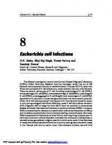
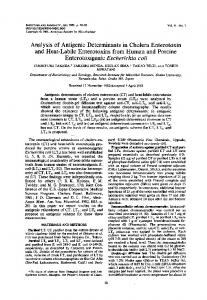
Chromatograms of E. coli ATCC 25922 nucleotides from perchloric acid extracts. (A) Unoxidized E. coli ATCC 25922. (B) E. coli ATCC 25922 oxidized by 100 ,umol of HOCI per g of E. coli. Abs., Absorbance. FIG. 1.
quenching from sample to sample
was sufficiently high that counts per minute were not converted to disintegrations per minute. The counts obtained from the unlabeled pellet solution represent the amount of label present in the external medium, and the counts obtained from the labeled pellet represent the amount of label taken up by the cells. Background counts caused by nonspecific adsorption of the labels
determined by treating cells with 7% butanol or 2% toluene, which render them unable to accumulate small molecules (26). These counts, typically 3% of the total counts present in solution, were subtracted from all measurements of labeled pellets. The parameters Aj and ApH were calculated as Aq4 (or ApH) = 59 log (P,J10 ViSJ) (equation 2), where Pi, SO, and Vi are the corrected counts per minute from the tube containing the labeled pellet, from the tube containing the unlabeled pellet, and the internal volume of the cell suspension, respectively. The proton motive force was calculated from equation 1, and the internal cellular pH was calculated by subtracting ApH from the measured value of eXternal pH. Respiration. Respiration was measured by monitoring the consumption of 02 by the bacteria with a Yellow Springs Instrument Co. 5750 Clark-type oxygen electrode. Three to four milliliters of a 2.0-mg/ml (dry weight) bacterial suspension, which had been incubated with aeration for 10 min with 25 mM sodium suceinate as an energy source, were placed in a water-jacketed cell similar to that previously described (23, 24). The oxygen electrode response was monitored with a Linear Instruments Corp. strip chart recorder. The slope of were
oxygen electrode response lated and
as a
function of time
was
calcu-
compared with slopes at various levels of HOCI
oxidation. The data are presented as percent respiration relative to untreated cells. Induction of ,I-galactosidase. Protein synthesis was estimated by measuring ,3-galactosidase activity after induction with isopropyl-p-D-thiogalactopyranoside (IPTG) (29). Bacteria at 2.0 mg/ml (dry weight) exposed to various concentrations of HOCI or NH2CI were incubated for 80 min at 37°C in a buffer containing 5 g of tryptone per liter, 10 mM IPTG, 100 mM phosphate, 154 mM NaCl, and 10 mM sodium succinate. The cells were diluted sixfold, and a 3.0-ml portion was placed in a 1.0-cm optical cuvette on ice and then ultrasonically disrupted by pulsed operation of a Heat Systems-Ultrasonics W-225 instrument at 25% output for 150 s by using the microtip probe. The P-galactosidase activity was measured by monitoring spectrophotometrically the hydrolysis of o-nitrophenylgalactosidase to o-nitrophenol at 420 nm (40). Initial rates were compared with those of unoxidized cells and presented as percent induction; the activity of cells grown in the absence of IPTG was less than 2% of the activity of induced cells. Data analysis. As is typical for this type of experiment (2), the amount of HOCI required to inactivate cells varied as much as 30% in successive growth cultures, precluding quantitative comparison of separate analyses of different bacterial suspensions. For this reason, all of the variables compared in a given set of experiments, e.g., viability, respiration, adenylate energy charge (EC), and proton motive force in Fig. 1, were made on a single growth culture. This procedure ensures that changes in the variables attending titrimetric addition of HOCI are accurately scaled to viability losses. However, because the titrimetric break point was not constant from culture to culture, a relatively large data scatter arose when results from separate experiments were averaged, the range being as large as +50% for individual points in the regions of greatest change. Nonetheless, the data reported as averages of several sets of determinations also accurately portray the relationships between viability loss and other variables found in the individual runs. RESULTS HOCI reactions with adenine nucleotides. High-performance liquid chromatography of solutions obtained by flow mixing HOCI with ATP, ADP, or AMP showed that AMP or ADP was not produced by reaction of ATP with the oxidant. However, bathochromic shifts in the nucleotide wavelength maxima occurred progressively from 260 to 270 nm upon the addition of increasing amounts of HOCI up to a concentration ratio of 1:1. Continued addition of HOCI beyond this ratio led to bleaching of the chromophore at 270 nm. These optical changes have been interpreted to indicate chloramine formation or ring chlorination, followed by oxidative disruption of the ring at higher HOCI concentrations (4). No chromatographic differences were observed at ratios under 1:1, but at higher ratios the relative peak areas declined in accord with the observed spectral bleaching. Thus, it is possible to distinguish between nucleotide and monochlorinated nucleotide spectroscopically but not on the basis of high-performance liquid chromatography retention times. E. coli nucleotide concentration levels. A typical chromatogram of the acid-soluble fraction of E. coli ATCC 25922 cells is shown in Fig. 1A, which is similar to published chromatograms (42). The adenine nucleotide pool consists primarily of ATP with smaller amounts of ADP and AMP. The total pool concentration and EC, defined (5) as EC = (ATP + 1/2
INACTIVATION OF E. COLI BY HOCI
VOL. 55, 1987
11150
i
120
L)
90
L
-V e - E;
z 60E 0
L 30
0
/.±mol
HOCI/g E col
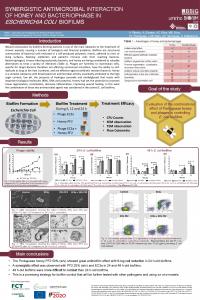
FIG. 2. Effect of HOCl on viability (0), proton motive force (0), respiration (A), and EC (A) of E. coli ATCC 25922. Data represented are the average of four sets of determinations in which 100%o control is (1.5 0.4) x 109 CFU/ml and respiratory rates for untreated cells are 0.8 to 1.0 nmol of 02 per min-mg (dry weight). Under the experimental conditions, pH equals 7.4, so that 59. ApH 0 and Ap A+l. =
ADP)/(ATP + ADP + AMP) (equation 3), were determined from the chromatograms to be approximately 5 mM and 0.7 to 0.8, respectively; these values are consistent with earlier measurements (28). EC values determined from replicate extractions of samples of cell suspensions were reproducible to +5 to 10%. The pool size varied by 50%o from day to day but did not appear to be a function of time of measurement after harvesting. The EC was dependent on the age of the cellular suspension and on the presence of an energy source. Resting cells that were sampled 15 min after warming showed a decreased EC compared with cells sampled within 5 min after warming (0.40 versus 0.65, respectively). However, the EC measured for cells 5 min after warming remained constant for at least 4 h after harvesting. Subsequent addition of an energy source with aeration (5 mM glucose or 25 mM succinate) to E. coli caused the EC to rise to 0.75 to 0.85, consistent with previously described behavior (28). All measurements were made within 3 h after harvesting by using cells that were suspended in buffer and shaken at 320 rpm to maintain aeration. These conditions yielded reproducible values for the EC and adenine nucleotide pool concentrations. The chromatogram in Fig. 1A also shows the presence of other nucleotides, nucleosides, and bases. Cytosine, uracil, and guanine nucleotides were detected at retention times of 3 to 5 min, respectively. Because the individual peaks overlapped extensively, it was not generally possible to make unique assignments. The nucleosides cytidine, uridine, inosine, and guanosine gave rise to peaks at 9.0, 12, 29, and 33 min, respectively, and the bases uracil, hypoxanthine, xanthine, and adenine were assigned to peaks at 6.2, 11, 13, and 26 min, respectively. The peak at 22 min has not been identified. The areas of this peak and the peak at 50 min (NAD+) were not perturbed by HOCl oxidation or dependent upon metabolic state but were affected only by dilution and hence served as internal markers that allowed comparison between different samples and provided a means to check for dilution errors. Areas of these peaks did not vary by more than 10% among samples of a typical experimental set.
Only uracil, hypoxanthine, xanthine, and adenine were detected in the supernatant fractions of centrifuged cell suspensions. This extracellular location suggests that they are the excreted metabolic end products of nucleotide catab-
2521
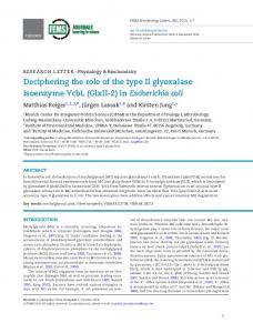
olism (30). With the exception of adenine, whose concentration was unchanged, their concentration levels increased slowly with the age of resting cell suspensions, being typically 1 to 10 ,uM on the basis of the solution volumes used. Nucleotide levels in E. coli after HOCI oxidation. The effects upon the nucleotide pool after exposure of E. coli to lethal levels of HOCI are shown in Fig. 1B. Comparison with Fig. 1A shows that concentrations of the nucleotide triphosphates, most evident for ATP, had decreased dramatically, whereas concentrations of the nucleotide monophosphates, most evident for AMP, had risen proportionately. Identification of this peak as AMP was confirmed spectrophotometrically. The UV absorption spectrum of pooled isolated fractions was identical to that of authentic samples (Xmax = 259 nm) but differed from that of chlorinated derivatives (Xmax = 265 nm). Heights of the GMP peak at 7.8 min, and unresolved peaks containing the nucleoside monophosphates UMP, at 5.0 min, and CMP, at 4.0 min, also increased markedly relative to the corresponding di- and triphosphate peaks, consistent with extensive phosphoanhydride bond cleavage in the other nucleotide pools. No new peaks were evident, nor was there any increase in adenine, adenosine, inosine, or hypoxanthine, species which would arise from AMP degradation (30). This latter result indicates that EC regulation by AMP catabolism (7, 28, 30) does not occur in HOCI-oxidized E. coli. The effects of various concentrations of HOCI upon viability and EC are presented in Fig. 2. Viability and EC decreased together as the cells were oxidized by increasing amounts of HOCI. When the degree of oxidation reached the point where 80% of the cells had been killed, the EC had dropped below 0.5. This relationship is most evident from Fig. 3, in which EC is plotted against the other variables. EC values were 0.1 to 0.2 after exposure to HOCl at concentration levels sufficient to kill 99% of the organisms. Changes in the total adenine nucleotide pool were dependent on the amount of added oxidant. At concentration levels up to six to seven times that required for inactivation, the pool size slowly increased with time until after about 20 min it had increased to two- to threefold over the level in unoxidized cells. Beyond this point, the immediately measured pool size dropped rapidly upon exposure to increasing concentrations of HOCl. The latter effect is undoubtedly caused by direct oxidative bleaching of the nucleotides (4). Cells treated identically except for the omission of HOCl did not show any changes in viability or EC, except for the small, timedependent effects discussed in the preceding section.
100 80
T
60 40
20-
0
0.16
0.32
0.48
0.64
0.80
EC
FIG. 3. Relationship between EC and other physiological variables. Data were taken from Fig. 2; symbols are defined in the legend to Fig. 2.
2522
INFECT. IMMUN.
BARRETTE ET AL. 0.80 0.64 In
-0.48 °
60
L)
'a
>>
9 u
.
.° 40
C0.32 'Dc
I
0
0.16 z
n2 20
40
tLmol
60
NH2CI/g.
0.0
80 E .coli
1smol oxidant/g. E.coli
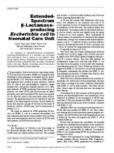
FIG. 4. Effect of NH2Cl on viability (0, 0) and energy charge (U, O) of E. coli ATCC 25922. Open symbols, Exposure to oxidant followed by immediate addition of S2032- ion; closed symbols, S2032- addition delayed 10 min. Data are for a single set of determinations; 100% control corresponds to 1.0 x 10⁹ CFU/ml.
Scavenging of HOCI by thiosulfate ion, added either immediately by quench-flow procedures (3) or 10 min after the mixing of E. coli and the oxidant, provided no protection for the bacteria and was unable to influence the extent of nucleotide phosphate bond hydrolysis. Hence, oxidative damage was complete within the first second of exposure of bacteria to HOCI. Nonetheless, hydrolysis of nucleoside diand triphosphates occurred more slowly, with rates that were dependent on the applied HOCI concentration. At 25 ,umol of HOCI per g of E. coli, the initial half-life (t012) for decay of EC to the final value shown in Fig. 2 was 90 s, and at 40 ixmol HOCI per g of E. coli, t1/2 60 s. =
Proton motive force of E. coli after HOCI oxidation. The effect of HOCI oxidation of E. coli upon the proton motive force at pH 7.4 is shown in Fig. 2. Under these conditions, ApH 0, indicating that the internal pH of the cells was also about 7.4; correspondingly, Ap was composed almost entirely of the electrical potential, A+i. Both potentials were pH dependent; Ai\ decreased by 38 mV/pH unit, and ApH increased by 50 mV/pH unit over the range pH 7.4 to 5.5, so that Ap increased only slightly as the pH decreased. The magnitudes of these parameters and their acid dependencies are typical for E. coli and other bacteria (see, e.g., reference 22). The results presented in Fig. 2 show that Ap decreased from 140 mV to 0 mV as the E. coli was oxidized by HOCI but that the level of oxidation required to collapse the gradient was two to three times the level required to reduce viability to 1% of that of the original suspension. At pH 5.5, at which a significant ApH exists, the same results were obtained, namely, ApH, Aij, and Ap drop to 0 mV at oxidation levels two to three times that required to kill the organisms. Under both sets of conditions, respiratory inhibition also lagged behind loss of viability (Fig. 2) (2). Oxidation of E. coli by chloramine. The effect of cellular oxidation by NH2CI upon adenylate energy reserves is shown in Fig. 4. In solutions to which thiosulfate ion was added 10 min after initiation of the reaction, EC decreased coincidentally with loss in viability in a manner analogous to HOCl-oxidized E. coli. However, in contrast to HOCI oxidation, if NH2Cl was scavenged with thiosulfate by the quench-flow method, no loss of adenylate energy charge or viability was observed. Induction of 1-galactosidase. The effect of HOCI and NH2Cl oxidation upon IPTG-promoted induction of pgalactosidase in E. coli is given in Fig. 5; the ,-galactosidase activity in uninduced bacteria is also given for comparative =
FIG. 5. Effect of HOCl (0, *, A) and NH2Cl (0, O, A) on IPTG-promoted induction of P-galactosidase in E. coli ATCC 25922. Circles, Viability; squares, intracellular ,B-galactosidase activity induced by IPTG; triangles, intracellular 3-galactosidase activity in the absence of added IPTG. Values are averages of 12 sets of determination for HOCI and 4 sets for NH2CI; 100% control is (2.0 + 0.5) x 109 CFU/ml for HOCl and (2.4 ± 0.9) x 109 CFU for NH2Cl. Typical initial specific rates for untreated IPTG-induced bacteria were 125 ± 25 nmol of o-nitrophenyl-13-D-galactopyranoside per min per mg (dry weight).
purposes. Induction of enzyme activity was lost upon adding small amounts of either oxidant, being completely inhibited at EC values below about 0.5 (Fig. 4). The effect was highly medium dependent, with severalfold greater amounts of HOCI required when amine buffers were present (Fig. 6a). Nonetheless, loss of 13-galactosidase induction paralleled loss of viability under all experimental conditions (Fig. 6a and b). DISCUSSION Transport inhibition. We have previously found that active transport of nutrients that derive energy either from Ap or coupled ATP hydrolysis (34) are inhibited equally well by 100
a.
b2
100
80
80
6
Z 60
>
2 40
20-
20-
0
0
40-
100
200
0
300
0
100
200
300
,4mol HOCI/g. E coli
FIG. 6. Effect of HOCI on IPTG-promoted induction of ,Bgalactosidase (a) and viabilities (b) of E. coli ATCC 25922 in various media. Buffers contained 100 mM sodium phosphate (H), 80 mM ammonium phosphate-100 mM sodium phosphate (0), 40 mM Tris hydrochloride (A), 100 mM triethanolamine (O), or 40 mM N-2hydroxyethylpiperazine-N'-2-ethanesulfonic acid (HEPES) (0). The solutions also contained 154 mM NaCl; acidities were adjusted to pH 7.4. Reaction was quenched at 10 min by adding Na2S203; 10 mM sodium succinate was also added at this time to provide an energy source.
VOL. 55, 1987
HOCI oxidation and that, in titrimetric comparison, loss of nutrient transport coincides with loss of ability of the cells to sustain colonial growth (2). Since the present studies demonstrate that the transmembrane electrochemical gradient is maintained until cellular respiration is inhibited, well beyond the point of loss of viability, the inability to accumulate metabolites by proton-coupled symport mechanisms cannot be attributed to loss of the driving force. It is also evident from these measurements and earlier studies on proton conductances and glycerol permeabilities (2) that the plasma membranes in HOCl-oxidized bacteria are capable of maintaining metabolite concentration gradients. The only simple alternative is that the membrane-localized transport proteins react directly with HOCI. Oxidative inactivation by HOCI is reasonable because transport proteins functioning by proton cotransport are thought to carry critical sulfhydryl groups (19) that can be expected to be highly susceptible to HOCl oxidation (4, 43). On the other hand, because ATP hydrolysis in HOCI-oxidized E. coli parallels loss of ATP-dependent metabolite transport (2) and ultimately goes essentially to completion (Fig. 1), an alternative mechanism for inactivation of these systems is loss of the driving force. No conclusions regarding possible direct oxidative inactivation of transport proteins in the ATP-dependent systems can therefore be reached. Metabolic regulation. The concept of adenylate energy charge was introduced (5) in recognition of the central role played by adenylate phosphorylation levels in regulating the activity of key enzymes controlling cellular metabolism (9, 28). Although the quantitative significance of the EC remains controversial (Trends Biochem. Sci. 2:N198-N200, 1977; 3:N39-41, 1978), it appears to be a useful index of the general well-being of cells. According to its proponents (10, 41), when the EC is greater than 0.7 to 0.8 the E. coli cells are fully active and will synthesize protein at a normal rate and undergo cellular division. At EC values between 0.5 and 0.7, the E. coli cells are described as resting (i.e., viable), but they no longer synthesize protein or grow. Finally, when the EC value decreases below 0.5, as in prolonged starvation, viability is irreversibly lost. This state is thought to represent such severe di-lption of biochemical homeostasis that the -r cells cannct r Considering Lhe ii portance of energy production and regulation to cellular function and growth, any effects of HOCI leading to loss of cellular energy reserves would be expected to be detrimental to the organism. The results illustrated in Fig. 2, 4, and 5 demonstrate that exposure of E. coli to lethal amounts of HOCI or NH2Cl causes the EC to decrease to extremely low levels (0.1 to 0.2). Furthermore, the oxidized cells did not respond to addition of glucose or succinate to the medium, unlike untreated E. coli, which gave an immediate step-up to EC values of 0.8 to 0.9, characteristic of growing cells (W. C. Barrette, Jr., unpublished observations). The HOCI-induced changes occur without decreasing the adenine nucleotide pool size or the proton motive force and are attributable to phosphoanhydride bond hydrolysis. Our observations are consistent with other reports noting that oxidative inactivation of E. coli by HOCl (2) and oxidation of erythrocytes by taurine chloramine (45) give rise to extensive intracellular ATP hydrolysis, although quantitative relationships were not examined in these earlier studies. When faced with conditions that lead de facto to lowered EC values, healthy bacteria can restore proper nucleotide concentration ratios by increasing formation or decreasing utilization of ATP (or both) or by decreasing the concentra-
INACTIVATION OF E. COLI BY HOCI
2523
tion of intracellular AMP (9, 28). Cessation of glycogen production (11) and protein synthesis (41) with stimulation of respiration and glycolysis increase ATP concentration levels. Excretion of AMP or its catabolism (30) and subsequent excretion of metabolic end products into the medium or net phosphorylation arising from adenylate kinase and nucleoside diphosphate kinase catalyzed reactions are means for lowering cytosolic AMP concentration levels. Direct elimination of AMP from the cytosol has been demonstrated not to occur in E. coli (28). Our chromatographic analyses of supernatant and pellet fractions show that all detectable nucleotides are localized in the cytosol and that bases are extracellularly localized. Since the concentration levels of catabolites and the adenine nucleotide pool size are unchanged after treatment with bactericidal amounts of HOCI or NH2Cl, inactivated E. coli cells are unable to control adenylate phosphorylation ratios by eliminating AMP from the cytosol. AMP inhibits many ATP-consuming reactions and stimulates many other ATP-producing reactions (9, 28), so one would expect glycolysis and the respiration rates to increase until the EC regains a normal value of 0.7 to 0.8. Because the respiration rate does not increase and the EC remains low, the oxidized cell also cannot maintain appropriate adenylate phosphorylation levels by decreasing consumption and increasing production of ATP. Thus, oxidized E. coli appears to have lost all capability of exerting metabolic control by regulating adenylate phosphorylation levels. Microbicidal reactions. Nucleotide phosphate bond hydrolysis is not a consequence of direct oxidative attack by HOCI, implying that the oxidative reactions disrupt metabolic pathways involved with ATP utilization or generation. This process most likely does not include inactivation of regulatory enzymes located in the cytosol since other cytosolic enzymes that are HOCl sensitive, such as aldolase (4) and ,-galactosidase (2, 49), are unaffected by bactericidal levels of chlorine oxidants (2, 8) and, as described in this study, the highly reactive adenine nucleotides are not oxidatively bleached until HOCl or NH2Cl in considerable excess of the amount required for killing is added to E. coli
suspensions. Two types of mechanisms by which HOCl oxidation of membrane components could lead to rapid ATP hydrolysis are direct attack on a membrane-bound ATP-dependent enzyme or transport protein or attack upon a membranebound protein not requiring ATP itself but resulting in ATP hydrolysis by an ATP-dependent system that attempts to compensate for metabolic imbalances caused by the damage. An example of the first type is modification of the protontranslocating ATP synthetase (17). In contrast to an earlier report (46) that ATP hydrolase activity is unimpaired in membrane fragments from HOCl-inactivated E. coli, we have now determined that the hydrolase activity of solubilized F1 subunits (17) decreases in parallel with viability (W. C. Barrette, Jr., unpublished observations). The synthetase activity of the intact enzyme is almost certainly also lost, although this remains to be established. ATP synthetase inhibition constitutes complete interruption of oxidative phosphorylation which, in the absence of other changes, could account for ATP depletion in respiring cells. A hypothetical example of the second type is loss of the gating mechanism in an ion transport system, e.g., potassium or phosphate, such that uncontrolled efflux of ions occurs. Other, ATP-dependent, potassium or phosphate ion transport systems (16) could become engaged in a "futile cycle" (37) to attempt to compensate for the leak. Preliminary studies (W. C. Barrette, Jr., unpublished observations)
2524
INFECT. IMMUN.
BARRETTE ET AL.
indicate that potassium ion efflux from E. coli also parallels viability losses. HOCI as a participant in neutrophilic bactericidal processes. The mechanism by which IPTG-inducible ,B-galactosidase activity is lost after the addition of bactericidal levels of chlorine oxidants (Fig. 4) has not been determined. Major contributing factors could include impaired transport of the inducer, loss of regulatory nucleotides, and loss of adenylate energy reserves, the last possibility arising because EC values fall far below 0.7 under these conditions (10, 41). Regardless of mechanism, the observation that HOCl inhibits P-galactosidase induction is significant because it contrasts sharply with results from studies on E. coli that had been phagocytosed by neutrophils or exposed to their granule components (6, 12-14). In these instances, the bacteria retained protein biosynthetic capabilities for periods extending far beyond cellular death. One is tempted to conclude that either exogenously added HOCl reacts differently from HOCl generated by myeloperoxidase catalysis or HOCI is not the primary neutrophil-generated toxin responsible for intraphagosomal killing. However, there is little doubt that stimulated neutrophils produce copious quantities of HOCI (18, 20, 21, 25, 48, 49), so that continued macromolecular biosynthesis by phagosome-entrapped E. coli is most remarkable. The incubation media used in the studies with neutrophils (6, 12-14) contained hydrophilic HOCl-reactive compounds (e.g., tris(hydroxymethyl)aminomethane, triethanolamine, Casamino Acids (Difco), maleate ion, and/or cycloheximide), however, which may have protected the bacteria by scavenging enzymatically generated HOCl (20, 44). This protective action is illustrated in Fig. 6, with respect both to viabilities and to P-galactosidase induction. The buffer concentration levels are comparable to those in the incubation media in the experiments with neutrophils and therefore provide a rough index of the magnitude of protection that might be afforded. These results raise the intriguing possibility that oxidative and nonoxidative microbicidal reactions occurring within the phagosome might be separable merely by judicious selection of medium buffers. Studies designed to investigate further the bioenergetic capabilities and metabolic state of neutrophil-phagocytosed E. coli are currently being pursued. ACKNOWLEDGMENTS We are indebted to E. R. Kashket for helpful advice concerning proton motive force measurements and to Kerry B. Callahan and Brian C. Patterson for excellent technical assistance. This work was supported by Public Health Service grant AI-15834 from the National Institute of Allergy and Infectious Diseases. LITERATURE CITED 1. Ahmed, S., and I. R. Booth. 1981. Quantitative measurements of proton-motive force and its relation to steady state lactose accumulation in Escherichia coli. Biochem. J. 200:573-581. 2. Albrich, J. M., J. H. Gilbaugh III, K. B. Callahan, and J. K. Hurst. 1986. Effects of the putative neutrophil-generated toxin, hypochlorous acid, on membrane permeability and transport systems of Escherichia coli. J. Clin. Invest. 78:177-184. 3. Albrich, J. M., and J. K. Hurst. 1982. Oxidative inactivation of Escherichia coli by hypochlorous acid: rates and differentiation of respiratory from other reaction sites. FEBS Lett. 144:157161. 4. Albrich, J. M., C. A. McCarthy, and J. K. Hurst. 1981. Biological reactivity of hypochlorous acid: implications for microbicidal mechanisms of leukocyte myeloperoxidase. Proc. Natl. Acad. Sci. USA 78:210-214. 5. Atkinson, D. E. 1968. The energy charge of the adenylate pool as
6.
7.
8. 9.
10.
11.
12. 13.
14.
15.
16. 17. 18. 19.
20.
21. 22. 23. 24.
25.
26.
a regulatory parameter: interaction with feedback modifiers. Biochemistry 7:4030-4034. Beckerdite, S., C. Mooney, J. Weiss, R. Franson, and P. Elsbach. 1974. Early and discrete changes in permeability of Escherichia coli and certain other gram-negative bacteria during killing by granulocytes. J. Exp. Med. 140:396-409. Booth, I. R., W. J. Mitchell, and W. A. Hamilton. 1979. Quantitative analysis of proton-linked transport systems: the lactose permease of Escherichia coli. Biochem. J. 182:687-696. Camper, A. K., and G. A. McFeters. 1979. Chlorine injury and the enumeration of waterborne coliform bacteria. Appl. Environ. Microbiol. 37:633-641. Chapman, A. G., and D. E. Atkinson. 1977. Adenine nucleotide concentrations and turnover rates: their correlation with biological activity in bacteria and yeast. Adv. Microb. Physiol. 15: 253-306. Chapman, A. G., L. Fall, and D. E. Atkinson. 1971. Adenylate energy charge in Escherichia coli during growth and starvation. J. Bacteriol. 108:1072-1086. Dietzler, D. N., M. P. Leckie, W. L. Sternheim, J. M. Ungar, D. L. Crimmins, and J. W. Lewis. 1979. Regulation of glycogen synthesis and glucose utilization in Escherichia coli during maintenance of the energy charge. J. Biol. Chem. 254: 8276-8287. Elsbach, P. 1973. On the interaction between phagocytes and micro-organisms. N. Engl. J. Med. 16:846-852. Elsbach, P., S. Beckerdite, P. Pettis, and R. Franson. 1974. Persistence of regulation of macromolecular synthesis by Escherichia coli during killing by disrupted rabbit granulocytes. Infect. Immun. 9:663-668. Elsbach, P., P. Pettis, S. Beckerdite, and R. Franson. 1973. Effects of phagocytosis by rabbit granulocytes on macromolecular synthesis and degradation in different species of bacteria. J. Bacteriol. 115:490-497. Elsbach, P., and J. Weiss. 1983. A reevaluation of the roles of the oxygen-dependent and oxygen-independent microbicidal systems of phagocytes. Rev. Infect. Dis. 5:843-853. Epstein, W., and L. Laimins. 1980. Potassium transport in Escherichia coli: diverse systems with common control by osmotic forces. Trends Biochem. Sci. 5:21-23. Fillingame, R. H. 1980. The proton-translocating pumps of oxidative phosphorylation. Annu. Rev. Biochem. 49:1079-1113. Foote, C. S., T. E. Goyne, and R. I. Lehrer. 1983. Assessment of chlorination by human neutrophils. Nature (London) 301:715716. Fox, C. F., and E. P. Kennedy. 1965. Specific laveling and partial purification of the M protein, a component of the J3-galactoside transport system of Escherichia coli. Proc. Natl. Acad. Sci. USA 54:891-899. Grisham, M. B., M. M. Jefferson, D. F. Melton, and E. L. Thomas. 1984. Chlorination of endogenous amines by isolated neutrophils: ammonia-dependent bactericidal, cytotoxic, and cytolytic activities of the chloramines. J. Biol. Chem. 259: 10404-10413. Grisham, M. B., M. M. Jefferson, and E. L. Thomas. 1984. Role of monochloroamine in the oxidation of erythrocyte hemoglobin by stimulated neutrophils. J. Biol. Chem. 259:6766-6772. Harold, F. M. 1986. The vital force: a study of bioenergetics, chapter 4. W. H. Freeman & Co., New York. Held, A. M., D. J. Halko, and J. K. Hurst. 1978. Mechanisms of chlorine oxidation of hydrogen peroxide. J. Am. Chem. Soc. 100:5732-5740. Hurst, J. K., P. A. G. Carr, F. E. Hovis, and R. J. Richardson. 1981. Hydrogen peroxide oxidation by chlorine compounds: reaction dynamics and singlet oxygen formation. Inorg. Chem. 20:2435-2438. Hurst, J. K., J. M. Albrich, T. R. Green, H. Rosen, and S. J. Klebanoff. 1984. Myeloperoxidase-dependent fluorescein chlorination by stimulated neutrophils. J. Biol. Chem. 259:48124821. Kashket, E. R. 1981. Effects of aerobiosis and nitrogen source on the proton motive force in growing Escherichia coli and Klebsiella pneumoniae cells. J. Bacteriol. 146:377-384.
INACTIVATION OF E. COLI BY HOCI
VOL. 55, 1987
27. Klebanoff, S. J., and R. A. Clark. 1978. The neutrophil: function and clinical disorders. North-Holland Publishing Co., Amsterdam. 28. Knowles, C. J. 1977. Microbial metabolic regulation by adenine nucleotide pools. Symp. Soc. Gen. Microbiol. 27:241-283. 29. Koch, A. L. 1963. Inactivation of the transport mechanism for a-galactosides of Escherichia coli under various physiological conditions. Ann. N.Y. Acad. Sci. 102:602-620. 30. Leung, H. B., and V. L. Schramm. 1980. Adenylate degradation in Escherichia coli. J. Biol. Chem. 255:10867-10874. 31. Mitchell, P. 1968. Chemiosmotic coupling and energy transduction. Glynn Research, Ltd., Bodmin, Cornwall, United Kingdom. 32. Niven, D. F., P. A. Collins, and C. J. Knowles. 1977. Adenylate energy charge during batch culture of Beneckea natriegens. J. Gen. Microbiol. 98:95-108. 33. Paul, B. B., A. A. Jacobs, R. R. Strauss, and A. J. Sbarra. 1970. Role of the phagocyte in host-parasite interactions. XXIV. Aldehyde generation by the myeloperoxidase-H202-chloride antimicrobial system: a possible in vivo mechanism of action. Infect. Immun. 2:414-418. 34. Rosen, B. P., and E. R. Kashket. 1978. Energetics of active transport, p. 559-620. In B. P. Rosen (ed.), Bacterial transport. Marcel Dekker, New York. 35. Rottenberg, H. 1979. The measurement of membrane potential and ApH in cells, organelles, and vesicles. Methods Enzymol. 55:547-569. 36. Schweinsberg, P. D., and T. L. Loo. 1981. Simultaneous analysis of ATP, ADP, AMP and other purines in human erythrocytes by high-performance liquid chromatography. J. Chromatogr. 181: 103-107. 37. Scrutton, M. C., and M. F. Utter. 1968. The regulation of glycolysis and gluconeogenesis in animal tissues. Annu. Rev. Biochem. 37:249-302. 38. Selsted, M. E., D. Szklarek, and R. I. Lehrer. 1984. Purification and antibacterial activity of antimicrobial peptides of rabbit granulocytes. Infect. Immun. 45:150-154. 39. Selvaraj, R. J., B. B. Paul, R. R. Strauss, A. A. Jacobs, and A. J.
40.
41.
42.
43.
44.
45.
46. 47.
48. 49.
2525
Sbarra. 1974. Oxidative peptide cleavage and decarboxylation by the MPO-H202-Cl antimicrobial system. Infect. Immun. 9:255-260. Sips, H. J., and M. N. Hamers. 1981. Mechanism of the bactericidal action of myeloperoxidase: increased permeability of the Escherichia coli cell envelope. Infect. Immun. 31:1116. Swedes, J. S., R. J. Sedo, and D. E. Atkinson. 1975. Relation of growth and protein synthesis to adenylate energy charge in an adenine-requiring mutant of Escherichia coli. J. Biol. Chem. 250:6930-6938. Taylor, M. W., H. V. Hershey, R. A. Levine, K. Coy, and S. Olivelle. 1981. Improved method of resolving nucleotides by reversed-phase high-performance liquid chromatography. J. Chromatogr. 219:133-139. Thomas, E. L. 1979. Myeloperoxidase, hydrogen peroxide, chloride antimicrobial system: nitrogen-chlorine derivatives of bacterial components in bactericidal action against Escherichia coli. Infect. Immun. 23:522-531. Thomas, E. L. 1979. Myeloperoxidase-hydrogen peroxidechloride antimicrobial system: effect of exogenous amines on antibacterial action against Escherichia coli. Infect. Immun. 25:110-116. Thomas, E. L., M. B. Grisham, D. F. Melton, and M. M. Jefferson. 1985. Evidence for a role of taurine in the in vitro oxidative toxicity of neutrophils towards erythrocytes. J. Biol. Chem. 260:3321-3329. Venkobachar, C., L. Iyengar, and A. V. S. P. Rao. 1975. Mechanism of disinfection. Water Res. 9:119-124. Venkobachar, C., L. Iyengar, and A. V. S. P. Rao. 1977. Mechanism of disinfection: effect of chlorine on cell membrane functions. Water Res. 11:727-729. Weiss, S. J., R. Klein, A. Slivka, and M. Wei. 1982. Chlorination of taurine by human neutrophils: evidence for hypochlorous acid generation. J. Clin. Invest. 70:598-607. Zglinczynski, J. M., and T. Stelmaszynska. 1975. Chlorinating ability of human phagocytizing leukocytes. Eur. J. Biochem. 56:157-162.