energy windows (8,9). This technique was applied to the quantification of myocardial tracer distribution in compari son with conventional acquired SPECT and ...
Estimation of Myocardial Perfusion and Viability Using Simultaneous 99mTc-Tetrofosmin-FDG Collimated SPECT Kazuki Fukuchi, Tetsuro Katafuchi, Kazuhito Fukushima, Yoriko Shimotsu, Masahiro Toba, Kohei Hayashida, Makoto Takamiya, and Yoshio Ishida Department of Radiology, National Cardiovascular Center, Suita, Osaka, Japan
This study was designed to elucidate the usefulness of crosstalk correction for dual-isotope simultaneous acquisition (DISA) with 99mTc-tetrofosminand FDG in estimating myocardial perfusion and viability. Methods: Eighteen patients with coronary artery disease were studied. First, SPECT was performed with a low-energy high-resolution collimator after a single injection of 99mTc-tetrofosmin (single 99mTc-tetrofosmin). Second, PET and DISA with an ultra-high-energy collimator were performed after glucose loading and an injection of FDG. DISA was designed to operate with simultaneous 3-channel acquisition, and weighted scatter correction of crosstalk from the 18Fphotopeak to the "mTc photopeak was performed by modification of an existing dualwindow technique. The FDG SPECT images were compared with the images obtained by PET. Both crosstalk-corrected and uncorrected 99mTc-tetrofosminimages were generated and com pared with the single 99mTc-tetrofosmin images. Results: Re
impaired because of chronic hypoperfusion; however, the metabolism is preserved, producing a perfusion-metabolism mismatch. Until now, perfusion-metabolism mismatching has been assessed mainly by PET, but a recent modification to SPECT enables it, with FDG, to provide clinical informa tion equivalent to that from PET (3-5). The advantage of FDG SPECT over PET is the use of dual-isotope simultaneous acquisition (DISA) by combining a 99mTc-labeled myocardial perfusion agent and FDG. The
benefit lies in the shorter duration of procedures, with an identical geometric registration of the different isotope images. The reduction in patient study time decreases the risk of artifacts caused by patient motion and improves patient throughput and comfort. Quantification of DISA imaging, however, is limited by the contribution of the movement of scattered and primary photons of 1 radionugional percentage uptake of FDG agreed well between DISA and PET. However, regional percentage uptake of 99mTc-tetrofosmin clide into the primary photopeak energy window of the other was generally higher on the uncorrected 99mTc-tetrofosminim radionuclide (crosstalk). Experimental data from previous ages than on the single 99mTc-tetrofosminimages, especially in studies using simulated myocardial distributions of 99mTc areas of low flow (percentage count of 99mTc-tetrofosmin> 50%). and 18Frevealed that the downscatter contribution of I8F on The crosstalk correction contributed to improving the agreement the "mTc images becomes theoretically significant in se between regional percentage uptakes and significantly improved verely hypoperfused areas (6,7). However, the efficacy of the detectability of myocardial perfusion-metabolism mismatch ing. Conclusion: With 3-channel acquisition and weightedcrosstalk correction for quantification of DISA has not, to scatter correction of crosstalk from the 18Fphotopeak to the 99mTc our knowledge, been evaluated in clinical viability studies. photopeak, DISA with 99mTc-tetrofosminand FDG is feasible for
assessing regional myocardial perfusion and viability. Key Words: FDG; SPECT; 99mTc-tetrofosmin;dual-isotope acquisition J NucÃ-Med 2000; 41:1318-1323
.he identification of injured but viable myocardium in patients with previous myocardial infarction and left ventricu lar dysfunction has become increasingly important as a means of predicting improvement in the ventricular perfor mance of hibernating myocardium after surgical revascularization (1,2). The contractility of hibernating myocardium is Received Apr. 26,1999; revision accepted Jan. 19, 2000. For correspondence or reprints contact: Yoshio Ishida, MD, PhD, Depart ment of Radiology, National Cardiovascular Center, Fujishiro-dai 5-7-1, Suita, Osaka 565-8565 Japan.
1318
In this study, we evaluated the effects of crosstalk correction on DISA with 99raTc-tetrofosmin and FDG. We used a modified version of a previously described method of crosstalk correction based on the simultaneous use of 3 energy windows (8,9). This technique was applied to the quantification of myocardial tracer distribution in compari son with conventional acquired SPECT and PET. MATERIALS AND METHODS Eighteen patients with coronary artery disease (CAD) and a history of myocardial infarction were included in this study (14 men, 4 women; age range, 52-76 y; mean age, 64 y). We included only patients with stable CAD; patients with recent myocardial infarction (^1 mo) were excluded. One patient had previously undergone revascularization. All patients had undergone coronary angiography and left ventriculography within 2 wk of a radionu clide study. CAD was defined as a reduction of at least 75% in the luminal diameter of at least 1 major epicardial coronary artery.
THEJOURNAL OFNUCLEAR MEDICINE • Vol. 41 • No. 8 • August 2000
18F-FDG (!
••nTc-Tetrofosmin
)12
(740MBq)75g loading1 5 min
" *glucose iI
min 1 30 min
IPET-tran
.)70MBq 40 min
|
30 min I
smission PET-emissionSingle Tetrofosmin
DISA-SPECT
SPECT
Significant stenosis was present in 3 vessels in 5 patients, 2 vessels in 5 patients, and 1 vessel in 8 patients (mean, 1.8 vessels per patient). The average left ventricular ejection fraction was 35.5% ± 8.6%. We carefully excluded patients with a fasting blood glucose level greater than 100 mg/dL. All patients gave written informed consent in accord with the ethical guidelines established by the institution.
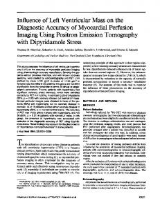
Imaging Protocol A schematic outline of the imaging protocol is shown in Figure 1. After an overnight fast, the resting patients were injected with 740 MBq "Tc-tetrofosmin. SPECT was performed using a dual-head system (Vertex Plus; ADAC Laboratories, Milpitas, CA) equipped with a low-energy high-resolution collimator. Thirty-two steps were acquired from each head in the photopeak energy window (140 keV ±10%) over 360°of a 64 X 64 matrix with a pixel size of 5.1 mm. Projection data were reconstructed using 12 maximum-likelihood expectation maximization iterations and Butterworth filtering (cutoff, 0.44 cycle/cm; order, 10). After "Tc-tetrofosmin acquisition, the patients were given a 75-g oral dose of glucose. PET transmission was performed using a line source of 68Ge and 68Ga followed by intravenous administra tion of 370 MBq FDG; 40 min later, PET emission was conducted for 10 min. Emission data were reconstructed consecutively, with measured attenuation correction based on the transmission data. Images were reconstructed by filtered backprojection using a Manning filter (0.5 cycle/cm), yielding 47 transverse slices having a thickness of 2.0 mm in a 128 X 128 matrix. An EC AT EXACT (Siemens Medical Systems, Inc., Hoffman Estates, IL) was used for the PET study. Immediately after PET emission, DISA acquisition was per formed in 30 steps in a 64 x 64 matrix over 180°(90 s/step) with an ultra-high-energy collimator using the same camera system for single "Tc-tetrofosmin SPECT. The system was designed to operate with 3 independent energy windows, allowing acquisition of separate scatter images (Fig. 2). The highest photopeak was
"Te
Photopeak Window
FIGURE 1. protocol.
Schematic outline of imaging
located at 511 keV ± 10% for acquisition of FDG images. For correction for the photopeak of 99mTc,a modification of an existing dual-window weighted-scatter correction technique was applied (10). It was based mainly on the assumption that a scintigraphic image measured in an energy window located beside the photopeak of"Tc(140keV ±10%) and shifted toward higher energy values (170 keV ±7.5%) represents the scatter component that blurs the photopeak image. The definition of this additional energy window and the scatter part under the photopeak were determined on the basis of-y energy spectra registered in the phantom experiment, and the scatter part in the photopeak window was found to be equivalent to 70% of the peak area of this scatter window for "Tc. Accordingly, crosstalk correction was established using this percent age as the correction factor for scatter image subtraction. The weighted projection data of the higher energy window (the scatter component) was taken from the projection data of the photopeak of 99mjc images were reconstructed using 12 maximum-likelihood expectation maximization iterative reconstructions (Butterworth filter; 0.42 cycle/cm, order 10, for the "Tc image; 0.38 cycle/cm, order 10, for the I8F image). We generated both crosstalk-corrected and crosstalk-uncorrected perfusion images with "Tc-tetrofosmin by DISA.
Quantitative Analysis To compare the regional myocardial tracer distribution obtained by each of the 3 imaging procedures (single "Tc-tetrofosmin and crosstalk-corrected and uncorrected "Tc-tetrofosmin by DISA), a previously described method based on regions of interest was used for quantitative evaluation (11,12). The region of interest with maximal tracer uptake among 20 regions of interest in short-axis cuts, corresponding to the region with the best individual perfusion, was used as the normal reference region, and "Tc-tetrofosmin uptake was expressed as percentage uptake in this reference region. FDG uptake normalized to this reference region was also compared between PET and FDG SPECT. A pattern of perfusion-metabolism mismatch was considered present when the relative "Te
11F Photopeak Window
(140 keV ±10%)
(511 keV ±10%)
V O O •
Scaner Window (170 keV ±7.5%)
FIGURE 2. -/-Ray energy spectrumregis tered in patient's data during "Tc-tetrofosmin-FDG dual-isotope acquisition. Vertical lines mark borders of the 3 energy windows.
QUANTIFICATION OF DUAL-ISOTOPE ACQUISITION SPECT • Fukuchi et al.
1319
tetrofosmin uptake was less than 70% of the maximal percentage activity and when the ratio of FDG to 99mTc-tetrofosmin exceeded 1.2 (13). A perfusion-metabolism match was considered present when concordance reduction (