Goldmann perimetry also was done to determine if defects found with the ring test were present with another method. Testing with Humphrey perimetry revealed ...
Investigative Ophthalmology & Visual Science, Vol. 32, No. 13, December 1991 Copyright © Association for Research in Vision and Ophthalmology
Evaluation of Sensitivity and Specificity of Spatial Resolution and Humphrey Automated Perimetry in Pseudotumor Cerebri Patients and Normal Subjects Michoel Wall,*t Mandi D. Conwoy,* Philip H. House, £ and Rosalie Allely* To determine the sensitivity and specificity of high-pass resolution perimetry ("ring test")) 18 patients with pseudotumor cerebri (PTC) and 18 age-matched controls were examined with the Humphrey program 24-2 and the ring test. Goldmann perimetry also was done to determine if defects found with the ring test were present with another method. Testing with Humphrey perimetry revealed defects in 15 PTC patients and four control subjects; with the ring test, 13 PTC patients and two control subjects had abnormalities. The disturbed areas in the control subjects with both automated tests were not reproducible. Humphrey perimetry had a sensitivity of 83% and the ring test, 72%. The specificities were Humphrey perimetry, 78% and the ring test, 89%. These differences were not statistically significant. Qualitative assessment of the presence and extent of damage using the pointwise probability plots and graphically displayed raw data showed good correlation of the tests in 11 of the 18 patients. The lack of correlation in four of the patients was caused by the presence of a generalized depression or a peripheral contraction on the Humphrey test; this defect, not present on retesting, may have been related to fatigue or poor motivation. The ring test is a sensitive and specific perimetric technique in patients with PTC. Invest Ophthalmol Vis Sci 32:3306-3312,1991
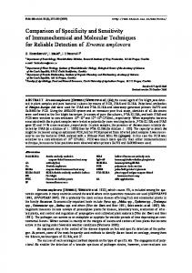
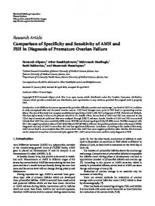
test").7"9 In this test, the stimuli areringsgenerated by processing doughnut-shaped targets with a high-pass filter (Fig. 1). This produces a spatial resolution target of low contrast and high spatial frequency with the average target luminance equal to the background. This target has the property of a "vanishing optotype," ie, detection and spatial resolution thresholds are theoretically equivalent. The targets are available in 14 sizes with a step factor of 1.26. The acuity at each of 50 test locations is scored as the smallest ring size the subject can discern; therefore, the better the acuity, the lower the score (Fig. 2). After ring presentation, each legal patient response is acknowledged by a black square image displayed on the screen at the tested location. The test takes approximately 5.5 min per eye. To determine its ability to distinguish accurately those with disease from those who are disease-free (sensitivity and specificity), we tested 18 patients with pseudotumor cerebri (PTC) and 18 normal subjects. In addition, we attempted to correlate qualitatively the extent and similarity of visual field defects detected by the ring test with those of other perimetric tests.
Differential light sensitivity threshold automated perimetry is more sensitive than Goldmann perimetry. ' Because the test is time consuming and monotonous, some patients become fatigued and change head position. This may lead to visual field defects from various artifacts or unreliable performance.2'3 It has been reported up to 45% of glaucoma patients and 30% of normal subjects tested did not meet the reliability criteria of a conventional automated perimeter.24 This source of measurement error can cause falsepositive results and led to a loss of specificity.5 Also, the simple discrimination of a white target on a white background may limit the sensitivity of each method and the correlation with the underlying anatomy.6 One attempt to overcome these problems was reported with high-pass resolution perimetry (the "ring From the Departments of ""Ophthalmology and fPsychiatry & Neurology, Tulane University School of Medicine, New Orleans, Louisiana, and the ^Department of Ophthalmology, Royal Perth Hospital, Perth, Western Australia. Supported in part by an unrestricted grant from Research to Prevent Blindness, Inc., New York, NY. Presented at the Annual Meeting of the Association for Research in Vision and Ophthalmology, Sarasota, Florida, April 30 to May 5, 1989. Submitted for publication: November 20, 1990; accepted July 1, 1991. Reprint requests: Michael Wall, MD, Tulane University School of Medicine, Department of Psychiatry & Neurology, 1430 Tulane Avenue, New Orleans, LA 70112.
Patients and Methods Eighteen patients with known PTC and 19 agematched normal subjects consented to participate in
3306
No. 13
SENSITIVITY AND SPECIFICITY IN PTC / Wall er al
Fig. 1. Examples of high-pass resolution targets. The intratarget contrast is equal to the background.
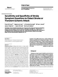
the study. The mean age of the patients was 37.9 ± 12.1 yr and of the control subjects, 34.2 ± 10.4 yr. The controls and patients all underwent a complete neuro-ophthalmologic examination. Normal subjects were recruited from Tulane Medical Center employees and residents. Potential normal subjects all underwent Goldmann perimetry. One had a visual field defect and was excluded. All patients first underwent Goldmann perimetry followed by the automated methods done in a random order. Both eyes were tested. Program 24-2 of the Humphrey field analyzer (Allergan-Humphrey, San Leandro, CA) was chosen because it has approximately the same number of test loci (52) as the ring test (50). However, the 24-2 test loci are separated equally in a 6° grid, and the ring test uses a nerve fiber bundle-related pattern and eliminates the pericecal points and test loci in the central 5° (Fig. 2). We did Goldmann perimetry using a modified ArmalyDrance strategy.10 The perimetrist did not know the results of automated perimetry. All tests were done on
0307
the same day with the examinations separated by at least 30 min to decrease the effect of fatigue. All patients fulfilled the modified Dandy criteria for PTC (Table 1)." The patients with PTC and five normal subjects were experienced in manual and automated perimetry. No patients or normal subjects were experienced in the ring test. However, ten patients and ten subjects were retested four times as part of another study; their second examination was used for our analysis. We administered the ring test according to the recommendations of the manufacturer's operating manual (Ophthimus system; HighTech Vision). The patients' appropriate near correction was given with care taken to prevent lens rim artifact. Patient fixation was monitored by the visual field technician and with intermittent blind-spot testing (Heijl-Krakau method). We examined the results of Goldmann perimetry in a masked fashion. Localized defects were tabulated by noting step defects of more than 15° in one isopter or more than 10° in two adjacent isopters; any scotomas (other than the physiologic blind spot) not eliminated by refraction were tabulated. We considered constriction present when isopters fell at least 10° from where they were expected (based on the data of Egge).12 We used the pointwise probabilities of the Humphrey Statpac to determine the presence of localized defects for the 24-2 fields. For the ring test, the determination was made using an age-corrected probability plot analysis based on values in 114 normal subjects.13 RINGPROB (a BASIC program written by MW) was used for the analysis (available free on request). Test loci scores were interpreted as abnormal if they fell outside the 95% confidence limits of the control population. The important differences between the design of these two probability plots are the Humphrey Statpac data base has approximately fourfold as many subjects and uses a formula that weights the values for the central points. This central weighting is not necessary for the ring test because variability does not rise with increasing eccentricity.13"15 We defined a localized defect for both automated testing methods as the presence of three disturbed
Table 1. Modified Dandy criteria for PTC
• Fig. 2. Ring test results of a right eye of a normal subject. The area surrounding the physiologic blind spot and the central five degrees are not tested. Note the gradually increasing ring size with increasing eccentricity. *
1. Signs and symptoms of increased intracranial pressure 2. Absence of localizing findings on neurologic examination 3. Absence of deformity, displacement, or obstruction of the ventricular system and otherwise normal neurodiagnostic studies, except for increased cerebrospinal fluid pressure 4. Awake and alert patient 5. No other cause of increased intracranial pressure present
3308
INVESTIGATIVE OPHTHALMOLOGY & VISUAL SCIENCE / December 1991
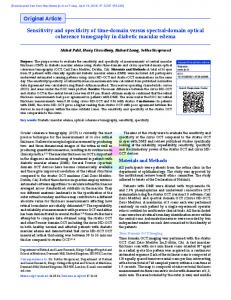
points in contiguity (P < 0.05) or one markedly disturbed point (P < 0.01) next to a suspicious point (0.01 < P < 0.05). We determined global loss by using the Statpac P < 0.05 confidence limits for the mean defect. A similar global deviation and its 95% confidence limits were was used for the ring test. We calculated the sensitivity and specificity for each test for each subject by determining if a visual field defect was present in either eye and compared this result with the subject's known diagnostic group (PTC or control). The visualfieldsthen were assessed qualitatively using graphic displays of the raw data and pointwise probability plots for the extent and possible similarity of the defects. Results Goldmann perimetry was abnormal in nine of 18 patients. Humphrey perimetry detected visual field loss in 15 of 18 patients; the ring test detected this in 13 patients. However, by the Humphrey 24-2 test, four of the patients had a peripheral contraction or a generalized depression that was not present with repeat testing within the next 6 weeks (Fig. 3). Humphrey perimetry disclosed a visual field distur-
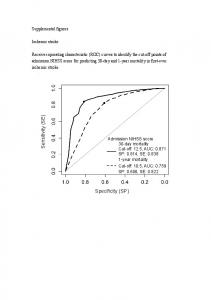
bance in four control subjects; two others had a disturbed field with the ring test. None of these defects were reproducible with repeat testing. The defects present according to test, subject, and group are found in Table 2. The extent and similarity of defects present in the patients is found in Table 3. In general, the defects were similar in shape and extent within subjects. Eleven patients had good correlation of the defects of the perimetric tests. Three patients had defects found with automated perimetry in clinically suspicious areas (visual field areas where defects are common in PTC) on only one of the two automated tests. The defects occurred in two visual fields in one patient with the ring test and two visualfieldsin two patients with the Humphrey perimeter. Four patients had a generalized depression or a peripheral contraction only on the Humphrey test; these defects were not present on retesting. Specificities and sensitivities are found in Table 4. The differences between the three groups in specificity and sensitivity were not significant (P > 0.3, by Fisher's exact test). Figure 4 shows a patient with PTC and an inferior nasal defect found with all three tests. The extent of
..:: r.v.'iViVivr.v.": ::::::::"
iiiisii::.. ::::::::::::::;.!-
::::::::::!
i::!::::: ::::::::::
:|:viiiii
1,
Vol. 32
:::::;?!:::::
I:::::::::-
Fig. 3. A peripheral contraction present on testing with Humphrey perimetry (left). The Goldmann visualfieldand theringtest were normal. The disturbance was not present on retest 1 week later (right). TOTfL OEVIPTIOH
TOTPL DEVIRTION . XfXf
• ":
• £
• '•: • • •:
S
B ' : 1 11 •
:: • •
k
PROGflBIinYSmS :=P< R CP< R 8 P < IX • P