CLINICAL AND VACCINE IMMUNOLOGY, Apr. 2010, p. 626–630 1556-6811/10/$12.00 doi:10.1128/CVI.00010-10 Copyright © 2010, American Society for Microbiology. All Rights Reserved.
Vol. 17, No. 4
Sensitivity, Specificity, and Confounding Factors of Novel Serological Tests Used for the Rapid Diagnosis of Bovine Tuberculosis in Farmed Red Deer (Cervus elaphus)䌤 Bryce M. Buddle,1* Tania Wilson,1 Michel Denis,1 Rena Greenwald,2 Javan Esfandiari,2 Konstantin P. Lyashchenko,2 Simon Liggett,3 and Colin G. Mackintosh4 AgResearch, Hopkirk Research Institute, Palmerston North, New Zealand1; Chembio Diagnostic Systems Inc., Medford, New York2; Disease Research Laboratory, University of Otago, Dunedin, New Zealand3; and AgResearch Invermay, P.O. Box 50034, Mosgiel, New Zealand4 Received 12 January 2010/Returned for modification 2 February 2010/Accepted 10 February 2010
In this study, novel serological tests were used to detect tuberculosis (TB) in groups of farmed red deer (Cervus elaphus) varying in disease status or possible confounding factors. Groups of deer naturally or experimentally infected with Mycobacterium bovis and animals vaccinated against paratuberculosis were studied, as were uninfected animals and animals naturally or experimentally infected with Mycobacterium avium subsp. paratuberculosis. Sera were assayed using two rapid lateral-flow tests, Chembio’s CervidTB STAT-PAK and DPP VetTB tests, and results were compared to those from tuberculin skin tests. Both serological tests had a high sensitivity, but specificity was adversely affected after animals had received a vaccine against paratuberculosis and were subsequently skin tested. The specificity of the DPP VetTB test was higher than that of the CervidTB STAT-PAK test, with natural infection with M. avium subsp. paratuberculosis adversely affecting the specificity of only the CervidTB STAT-PAK test. The sera from M. avium subsp. paratuberculosis-infected deer that produced false-positive reactions in the CervidTB STAT-PAK test were retested with a multiantigen print immunoassay (MAPIA), and some of these sera were shown to react with the MPB83 antigen. Combining the results from the serological tests and the skin tests showed only a slight increase in the sensitivity of detection of M. bovis-infected animals. It is concluded that both the CervidTB STAT-PAK and DPP VetTB tests offer rapid, convenient, and easy detection of bovine tuberculosis in deer, albeit with significant interference from paratuberculosis vaccination status and subsequent skin testing. The latter finding illustrates one of the limitations of currently available vaccines against paratuberculosis. proportion of false positives is likely to increase with the decreasing prevalence of M. bovis-infected deer herds in various parts of the world, as improved strategies to control TB in these animals are implemented. Hence, the development of novel tests with enhanced specificity is imperative for more accurate detection of bovine TB in deer. The CervidTB STAT-PAK lateral-flow test (Chembio Diagnostic Systems, Inc., Medford, NY) has been evaluated using a number of different species of free-ranging wild deer (3, 9, 14). Although the test showed a high sensitivity for detecting M. bovis infection, it is not clear whether the test specificity is affected when testing sera from M. avium subsp. paratuberculosis-infected deer. More recently, a new point-of-care test for bovine TB that uses the innovative dual-path platform (DPP) technology has been designed by Chembio. The DPP VetTB test was demonstrated to have the potential for enhanced specificity (4). These rapid tests have practical advantages over other diagnostic tools, as they are easy to perform and can be undertaken on the farm and results are obtained typically within 20 min (9). The aim of the present work was to assess the performance of the two new Chembio tests in a blind, coded study by using serum samples from various groups of red deer (Cervus elaphus), including animals naturally or experimentally infected with M. bovis, as well as animals infected with M. avium subsp. paratuberculosis or vaccinated against paratuberculosis. Our data suggest that the serological assays may be useful in the
Infections with Mycobacterium bovis in farmed or wild deer continue to be a problem in many areas of the world, and novel, more efficient tests aimed at diagnosis of this disease are needed (1, 5). One of the main tools used as a diagnostic approach is the skin test procedure using purified protein derivative (PPD) from M. bovis (1). Unfortunately, the tuberculin skin test in deer has a relatively low specificity, and it is considered prudent to retest skin test-positive animals by using a confirmatory test to avoid the slaughter of false-positive responders (2, 11). It has been suggested that the high prevalence of paratuberculosis in farmed deer causes the cross-reactivity in the skin test, leading to a high rate of false positives (2, 11). Alternative detection strategies have been explored, but these alternatives are costly or time-consuming (15, 16, 17). An IgG1 enzyme-linked immunosorbent assay (ELISA) for detection of tuberculosis (TB) in deer has been developed, but results were confounded by infection with Mycobacterium avium subsp. paratuberculosis, the causative agent of paratuberculosis (12). Vaccination against paratuberculosis also confounds tests for TB in deer (13), and it is important to assess whether the use of any new serological tests may circumvent this problem. The
* Corresponding author. Mailing address: AgResearch, Hopkirk Research Institute, Grasslands Research Centre, Tennent Drive, Private 1008, Palmerston North 4442, New Zealand. Phone: 64-6-3518679. Fax: 646-353-7853. E-mail:
[email protected]. 䌤 Published ahead of print on 17 February 2010. 626
VOL. 17, 2010
RAPID SEROLOGICAL TESTS FOR BOVINE TB IN DEER
TABLE 1. Groups of deer tested in the serological survey Infection or vaccination status Group
No. of deer in group
1 2 3 4A
24 15 32 14
4B
14
4C 4D
15 15
M. avium subsp. paratuberculosis
M. bovis
Naturally infected Naturally infected Experimentally infected Vaccinated against paratuberculosis Vaccinated against paratuberculosis
Experimentally infected Experimentally infected
rapid and accurate diagnosis of TB in deer but that vaccination against paratuberculosis may be a confounding factor. MATERIALS AND METHODS Animals. Sera were obtained from four different red deer studies. The groups of animals included for testing are described in Table 1. Group 1 consisted of 24 deer of various ages which were naturally infected with M. bovis (M. bovis culture confirmed). The animals were sourced from three farms on South Island, New Zealand, which TB was present. Twenty-two animals had gross tuberculous lesions detected following slaughter, and M. bovis was isolated from lesions or pooled lymph nodes (head, thoracic, or intestinal lymph nodes) from all 24 of these animals. Blood samples were collected 2 weeks after a midcervical tuberculin skin test (MCT). Group 2 consisted of 15 deer of various ages, naturally infected with M. avium subsp. paratuberculosis (culture confirmed) and sourced from a single herd which had no history of M. bovis infection. These animals had not been skin tested prior to collection of blood samples. Group 3 consisted of 32 rising-one-year-old female deer experimentally infected with M. avium subsp. paratuberculosis using a method previously described (12). The weaner deer were sourced from a farm that had no confirmed cases of TB or clinical cases of paratuberculosis and that had supplied weaner deer for experimental purposes for the past 20 years. M. avium subsp. paratuberculosis was isolated from the mesenteric lymph nodes of all 32 of these animals. Blood samples were collected 2 weeks after a comparative cervical test (CCT) and 49 weeks after experimental infection. The deer from group 4 consisted of 60 3-month-old, newly weaned deer sourced from a property with no history of TB or paratuberculosis. These animals were randomly divided into two groups, with one group receiving a 1-ml subcutaneous injection of a commercially available whole-cell paratuberculosis vaccine containing killed M. avium subsp. paratuberculosis with a mineral oil adjuvant (Silirum; Pfizer Animal Health Limited) and the other group remaining unvaccinated. The animals were transferred to a farm with an animal containment facility and were grazed together for the next 5 months. At week 20, each group was subdivided into two subgroups of 15 animals. One vaccinated subgroup and one nonvaccinated subgroup were challenged with M. bovis as described previously (10). Briefly, a 0.2-ml volume containing 500 CFU of M. bovis was deposited into the left tonsillar crypt of an anesthetized deer. The subgroups 4A and 4C, which had not been infected with M. bovis, were grazed together on one area of the farm, while the subgroups 4B and 4D, experimentally infected with M. bovis, were grazed together on another area of the farm. Two animals were excluded from the study; one from group 4A died as a result of an accident, and one from group 4B remained uninfected with M. bovis. For the remaining 29 animals from groups 4B and 4D, M. bovis was isolated from gross lesions or pooled lymph nodes (head, thoracic, or intestinal lymph node samples) collected at necropsy at 27 weeks after the experimental challenge with M. bovis. No M. bovis was isolated from pooled lymph nodes of the nonchallenged deer (groups 4A and 4C). Blood samples were collected at three time points in the trial: (i) immediately prior to challenge with M. bovis; (ii) 14 weeks after M. bovis challenge, 2 weeks after an MCT; and (iii) 26 weeks after M. bovis challenge, 2 weeks after a CCT. Blood samples from the various studies were collected and serum separated, and sera were stored at ⫺20°C until the serological tests were undertaken. Necropsy and bacterial culture. Animals were examined for the presence of tuberculous lesions following slaughter. For bacterial culture, samples were col-
627
lected from gross tuberculous lesions, or if no lesions were observed, pools of lymph nodes were collected from the head, thorax, and intestinal tract for culture of M. bovis and from the mesenteric lymph nodes for culture of M. avium subsp. paratuberculosis. Detailed descriptions of bacteriological culture for M. bovis and M. avium subsp. paratuberculosis are presented elsewhere (10, 11, 12). CervidTB STAT-PAK test. The CervidTB STAT-PAK test is a single-directional lateral-flow serological test which can provide a quick determination of the presence of M. bovis antibody (7, 9). The test uses colored latex-based lateralflow technology and a cocktail of selected M. bovis antigens, including ESAT-6, CFP10, and MPB83. The test required one drop of serum sample (30 l) and 3 drops of sample buffer, which were added sequentially to the sample pad. Results were read after 20 min. Any visible band in the test area in addition to the control line was considered an antibody-positive result, whereas no band in the test area in addition to the visible control line was considered a negative result. All sera were coded and tested blindly by staff from AgResearch. DPP VetTB test. The DPP technology involves two nitrocellulose strips which are connected in a “T” shape inside the cassette device (4). This allows independent delivery of the test sample and the antibody-detecting reagent, in contrast to the single-strip format used in the CervidTB STAT-PAK test. The DPP VetTB test required 5 l of serum sample and provided a result in 20 min. The presence and intensity of either of the two separate test lines (MPB83 antigen and CFP10/ESAT-6 fusion protein) were evaluated visually and by a DPP optical reader, as described previously (4). All sera were coded and tested blindly at Chembio. MAPIA. A multiantigen print immunoassay (MAPIA) was performed as previously described (8, 9). The panel of M. bovis antigens included ESAT-6, CFP10, MPB59, MPB64, MPB70, MPB83, the 16-kDa protein, the 38-kDa protein, two fusion proteins comprising CFP10/ESAT-6 and the 16-kDa protein/MPB83, and two native antigens, bovine PPD and M. bovis culture filtrate. Skin testing for TB. An MCT was undertaken by injecting 0.1 volume containing 0.1 mg (5,000 IU) bovine PPD tuberculin (AsureQuality, Upper Hutt, New Zealand) intradermally into the midsection of the neck. The skin test was read by comparing the double skin thicknesses with a digital caliper at 0 and 72 h after injection. A positive result was an increase in skin thickness of ⱖ2 mm. The CCT used 0.1-ml intradermal injections of 5,000 IU bovine PPD and 2,500 IU avian PPD (AsureQuality) at two closely clipped sites on the neck, with double skin thickness measurements taken prior to injection and 72 h later. A positive result was defined as an increase in the skin thickness at the bovine PPD injection site minus an increase in the skin thickness at the avian PPD injection site of ⱖ2 mm. The schedule of the skin testing is indicated in “Animals” above. When the CCT was undertaken, the result for the MCT was calculated using the bovine PPD component of the CCT. Statistical analyses. The test sensitivity was determined by dividing the number of M. bovis culture-positive, test-positive animals by the total number of M. bovis culture-positive animals, with the result expressed as a percentage. The test specificity was determined by dividing the number of TB-free, test-negative animals by the total number of TB-free animals, with the result expressed as a percentage. The proportions used for calculating the sensitivities and specificities of the two serological tests were compared using Fisher’s exact test. The 95% confidence limits for the findings were calculated using Minitab 15.1.0.0 (2006).
RESULTS Serological tests. The diagnostic performances obtained with the different treatment groups are shown in Table 2. The overall sensitivities of the two lateral-flow serological tests were similar, although the specificity of the DPP VetTB test (91.4%) was slightly higher than that of the CervidTB STATPAK test (83.8%). The specificity of the CervidTB STAT-PAK test was compromised for animals experimentally and naturally infected with M. avium subsp. paratuberculosis, whereas the specificity of the DPP VetTB test was adversely affected only for animals experimentally infected with M. avium subsp. paratuberculosis. The only significant difference between the results for the two serological tests was for the specificity of deer naturally infected with M. avium subsp. paratuberculosis, where the DPP VetTB test had a significantly lower proportion of false-positive reactions than the CervidTB STAT-PAK test (P ⬍ 0.05).
628
BUDDLE ET AL.
CLIN. VACCINE IMMUNOL.
TABLE 2. Sensitivities and specificities of the CervidTB STAT-PAK and DPP VetTB tests for different groups of deer Sensitivity or specificitya of: Group(s) CervidTB STAT-PAK test
Combination of groups naturally and experimentally infected with M. bovis (groups 1, 4B, and 4D at 26 weeks postinfection) Group naturally infected with M. bovis (group 1) Groups experimentally infected with M. bovis (groups 4B and 4D) 26 weeks postinfection 14 weeks postinfection Combination of all groups not infected with M. bovis (groups 2 and 3 and, prior to M. bovis challenge, 4A to D) 8-month-old deer calvesb (groups 4A to D prior to M. bovis challenge) Group naturally infected with M. avium subsp. paratuberculosis (group 2) Group experimentally infected with M. avium subsp. paratuberculosis (group 3)
DPP VetTB test
86.5 (45/52); 74.2, 94.4
84.6 (44/52); 71.9, 93.1
75.0 (18/24); 53.3, 90.2
75.0 (18/24); 53.3, 90.2
96.4 (27/28); 81.7, 99.9 82.8 (24/29); 64.2, 94.2
92.9 (26/28); 76.5, 99.1 69.0 (20/29); 49.2, 84.7
83.8 (88/105); 75.3, 90.3
91.4 (96/105); 84.4, 96.0
98.3 (57/58); 90.8, 99.9 60.0 (9/15); 32.3, 83.7 68.7 (22/32); 50.0, 83.9
98.3 (57/58); 90.8, 99.9 100 (15/15); 81.9, 100 75.0 (24/32); 56.6, 88.5
a Values shown are percentages (number of animals producing a positive response/total number tested 关for sensitivity兴 or number of animals producing a negative response/total number tested 关for specificity兴)and 95% confidence intervals. Values given for the first four comparisons are sensitivities, and values given for the last four comparisons are specificities. b Includes 29 deer vaccinated against paratuberculosis.
Test performance in deer vaccinated against Johne’s disease. To dissect the impact of vaccination against paratuberculosis on the specificity of the serological tests, vaccinated and nonvaccinated subgroups were compared (Table 3). For both serological tests, the number of false positives among the TBfree deer calves vaccinated against paratuberculosis (group 4A) increased from zero or one of the 14 animals before any skin tests (20 weeks after vaccination and immediately prior to the time of M. bovis challenge) to nine of 14 at 2 weeks following the CCT (26 weeks later). In contrast, for group 4C deer, which had been neither vaccinated nor challenged, the number of false positives increased only slightly, from zero or one of the 15 animals prior to any skin tests to zero or three by 2 weeks following the CCT. These two subgroups of animals were kept separate from those challenged with M. bovis, and M. bovis was not cultured from any pooled lymph nodes of these animals collected at necropsy. We cannot exclude the possibility that some of these animals were naturally exposed to environmental mycobacteria during the latter part of the study, although no other Mycobacteria sp. was isolated from their tissues on medium suitable for M. bovis and M. avium, which is also suitable for the isolation of most common environmental mycobacteria.
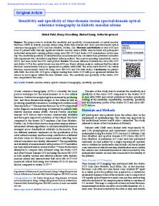
MAPIA. The sera from 16 animals from groups 2 and 3, which had been infected with M. avium subsp. paratuberculosis and had false-positive reactions in the CervidTB STAT-PAK test, were further evaluated by MAPIA to identify M. bovis antigens that may have been associated with these reactions. The MAPIA did not show any responses to recombinant antigens in sera from deer naturally infected with M. avium subsp. paratuberculosis, although there were some bands with M. bovis culture filtrate (data not shown). However, there was some MPB83 and MPB70 (and 16-kDa protein/MPB83) reactivity in the experimentally infected group, as shown in Fig. 1. The cross-reactivity appeared to be associated mostly with the MPB83 protein. All of the sera from the experimentally infected group showed strong responses to M. bovis culture filtrate and some to bovine PPD. Skin tests. For the 24 animals naturally infected with M. bovis (group 1), 18 were positive in the MCT. Combining the MCT and the serological tests to detect M. bovis infection status resulted in 21 of 24 positive (for the MCT/CervidTB STAT-PAK test combination) or 20 of 24 positive (for the MCT/DPP VetTB test combination). A total of 16 of the 32 deer experimentally infected with M. avium subsp. paratuberculosis (group 3) reacted positively in the MCT (bovine PPD
TABLE 3. Proportions of deer reacting positively via the serological tests and tuberculin skin tests following vaccination against paratuberculosis and/or experimental challenge with M. bovis
Vaccination status and group
Challenged and culture positive for M. bovis
No. of deer with positive reaction/total no. tested CervidTB STAT-PAK test
DPP VetTB test
MCT
0 wk
14 wk
26 wk
0 wk
14 wk
26 wk
12 wk
24 wk
CCT at 24 wk
a
Vaccinated against paratuberculosis Group 4A Group 4B
No Yes
0/14 0/14
3/14 12/14
9/14 13/13b
1/14 0/14
1/14 11/14
9/14 13/13b
14/14 14/14
12/14 14/14
1/14 5/14
Nonvaccinated Group 4C Group 4D
No Yes
1/15 0/15
4/15 12/15
3/15 14/15
0/15 0/15
0/15 9/15
0/15 13/15
0/15 15/15
0/15 15/15
0/15 15/15
a b
Weeks after M. bovis challenge. No serum sample was collected from one animal at 26 weeks after M. bovis challenge.
VOL. 17, 2010
RAPID SEROLOGICAL TESTS FOR BOVINE TB IN DEER
FIG. 1. MAPIA of 10 Mycobacterium bovis proteins or fusion proteins plus bovine PPD (B-PPD) and M. bovis culture filtrate (MBCF), performed using sera from 10 TB-free deer which had been experimentally infected with Mycobacterium avium subsp. paratuberculosis. All of these sera had previously reacted false positively in the CervidTB STAT-PAK test.
response alone), while all were negative in the CCT, with the avian PPD responses greater than the bovine PPD responses for all animals. The results of the skin tests for group 4 animals are shown in Table 3. For deer vaccinated against paratuberculosis but not challenged with M. bovis (group 4A), all or the majority of the animals reacted false positively in the skin test at 12 and 24 weeks after M. bovis challenge, while only one false-positive response was observed in the CCT at 24 weeks after M. bovis challenge. The MCT correctly identified all of the M. bovis-infected animals which had also been vaccinated against paratuberculosis (group 4B), but the CCT correctly identified only 5 of 14 at 24 weeks after M. bovis challenge. Both the MCT and the CCT correctly identified all of the M. bovis-infected and noninfected deer which had not been vaccinated (groups 4C and 4D). DISCUSSION The MCT is the primary screening test for diagnosis of TB in deer in New Zealand, but there are problems when animals are vaccinated against or infected with M. avium subsp. paratuberculosis (12, 13). As seen from the results of the current study, the majority of the TB-free animals which were vaccinated against paratuberculosis or experimentally infected with M. avium subsp. paratuberculosis reacted in the MCT, producing false-positive reactions. When the CCT was used as a confirmatory test, only one of the 14 vaccinated animals which was not infected with M. bovis produced a false-positive result, but when the CCT was used on animals vaccinated against paratuberculosis and subsequently infected with M. bovis, only five of 14 animals were correctly identified as M. bovis infected. The false-negative reactions resulted from boosting of immune
629
responses to avian PPD. There were no false-positive reactions in the CCT for the 32 deer experimentally infected with M. avium subsp. paratuberculosis. The current study was aimed to test the suitability of two rapid serological tests to serve as confirmatory assays for the diagnosis of TB in deer in the face of confounding problems such as vaccination against or infection with paratuberculosis. The two serological tests had the same sensitivity as that found for the MCT (75%) in deer naturally infected with M. bovis. Combining the skin and serological test results produced a small improvement in sensitivity, but the specificity of this approach was compromised due to the high rates of skin test false-positive reactions. The sensitivities of the serological tests were highest in the group of animals experimentally infected with M. bovis. The DPP VetTB test demonstrated a higher specificity than did the CervidTB STAT-PAK test when evaluated in deer naturally infected with M. avium subsp. paratuberculosis. It should be acknowledged, however, that blood samples from these animals were not collected in association with skin testing. Skin testing may boost cross-reactive antibody responses in deer naturally infected with M. avium subsp. paratuberculosis, possibly resulting in some false-positive responses in the DPP VetTB test. Sera need to be collected following skin testing from these types of animals and assayed using the DPP VetTB test to determine whether this is the case. The specificities of the two serological tests for deer vaccinated against Johne’s disease were relatively poor, and the number of false-positive responses increased markedly following the CCT. The high number of false-positive responses in the group only vaccinated against Johne’s disease (group 4B) at the final bleed is likely to have resulted from the boosting of antibody levels following skin testing with bovine PPD. It is recognized that tuberculin testing can boost antibody responses to certain mycobacterial antigens in M. bovis-infected animals (6, 7, 19). These results reemphasize the limitations of the currently available vaccines against paratuberculosis when confirmatory serological tests for bovine TB are used following skin testing. The MAPIA revealed that some of the sera from deer experimentally infected with M. avium subsp. paratuberculosis which produced false-positive responses in the CervidTB STAT-PAK test reacted with MPB83, a key antigen in the specific antibody detection during M. bovis infection (7, 9). This unexpected finding is difficult to explain, because the MPB83 protein is not produced by M. avium subsp. paratuberculosis (19) and it remains unclear how this antigen could elicit antibody responses in deer with paratuberculosis. A possible explanation of why only some of the sera that produced false-positive responses in the lateral-flow tests reacted with MPB83 in the MAPIA was that the MAPIA detects only IgG, while the two lateral-flow tests detect IgG and IgM. We cannot exclude the possibility that some of the false-positive responses in the TB-free deer were due to exposure to environmental mycobacteria, such as M. kansasii, which has been shown to produce MPB83 protein and confound serological tests for M. bovis infection (18). The CervidTB STAT-PAK test has previously been evaluated using deer, and the test sensitivities were comparable to values determined in the current study, i.e., sensitivities of 85.7% (3) for wild deer in the United Kingdom and 54.5% (14) and 75% (9) for white-tailed deer in the United States. In the
630
BUDDLE ET AL.
absence of apparent confounding factors, the test specificities were higher than those in the current studies, ranging from 94.8 to 98.9% (3, 9, 14). There may be differences in sensitivity between species of deer due to different affinities of test reagents with antibodies from these deer, especially between Old World Cervus sp. and New World Odocoileus sp. Numerous factors need to be taken into account when determining the optimal test to diagnose TB in deer. These factors include cost, availability, ease of use, reproducibility, and emphasis of the test procedure on high sensitivity or high specificity, ideally both. The animal-side serological tests have opened new avenues for the rapid, efficient, and accurate determination of TB status in a variety of veterinary species (4, 9) and could have particular application as an on-farm TB confirmatory test or an animal-side test for captured wild deer. Their use on the farm could reduce the number of farm visits and thus substantially reduce costs and accelerate the decisionmaking process. Given the current situation with farmed deer, novel serological tests should be aimed at providing results with a high degree of specificity, inasmuch as the MCT will continue to be used as the primary screening tool and serology as a secondary test in countries such as New Zealand. The present study has shown that paratuberculosis vaccination or M. avium subsp. paratuberculosis infection status can affect the diagnostic accuracy of the two lateral-flow serological assays, although improved specificity could be achieved with the DPP VetTB test. Confounding factors, such as infection with Johne’s disease or exposure to environmental mycobacteria, may be less of a problem in countries with lower M. avium subsp. paratuberculosis prevalences and/or in the use of serological tests for recently captured wild deer, where potentially cross-reactive immune responses would not be boosted by skin testing.
CLIN. VACCINE IMMUNOL.
4.
5. 6. 7.
8.
9.
10.
11.
12.
13.
14.
15.
ACKNOWLEDGMENTS We thank Allison McCarthy for expert technical assistance and Geoff de Lisle and Gary Yates for the bacteriological culture of tissues. This work was contracted by the New Zealand Animal Health Board.
16.
17.
REFERENCES 1. Cousins, D. V., and N. Florisson. 2005. A review of tests available for use in the diagnosis of tuberculosis in non-bovine species. Rev. Sci. Tech. 24:1039– 1059. 2. de Lisle, G. W., C. G. Mackintosh, and R. G. Bengis. 2001. Mycobacterium bovis in free-living and captive wildlife, including farmed deer. Rev. Sci. Tech. 20:86–111. 3. Gowtage-Sequeira, S., A. Paterson, K. P. Lyashchenko, S. Lesellier, and M. A. Chambers. 2009. Evaluation of the CervidTB STAT-PAK for the
18.
19.
detection of Mycobacterium bovis infection in wild deer in Great Britain. Clin. Vaccine Immunol. 16:1449–1452. Greenwald, R., O. Lyashchenko, J. Esfandiari, M. Miller, S. Mikota, J. H. Olsen, R. Ball, G. Dumonceaux, D. Schmitt, T. Moller, J. B. Payeur, B. Harris, D. Sofranko, W. R. Waters, and K. P. Lyashchenko. 2009. Highly accurate antibody assays for early and rapid detection of tuberculosis in African and Asian elephants. Clin. Vaccine Immunol. 16:605–612. Griffin, J. F., and G. S. Buchan. 1994. Aetiology, pathogenesis and diagnosis of Mycobacterium bovis in deer. Vet. Microbiol. 40:193–205. Griffin, J. F. T., and C. G. Mackintosh. 2000. Tuberculosis in deer: perceptions, problems and progress. Vet. J. 160:202–219. Harrington, N. P., O. P. Surujballi, J. F. Prescott, J. R. Duncan, W. R. Waters, K. Lyashchenko, and R. Greenwald. 2008. Antibody responses of cervids (Cervus elaphus) following experimental Mycobacterium bovis infection and the implications for immunodiagnosis. Clin. Vaccine Immunol. 15:1650–1658. Lyashchenko, K. P., M. Singh, R. Colangeli, and M. L. Gennaro. 2000. A multi-print immunoassay for the diagnosis of infectious diseases. J. Immunol. Methods 242:91–100. Lyashchenko, K. P., R. Greenwald, J. Esfandiari, M. A. Chambers, J. Vicente, C. Gortazar, N. Santos, M. Correia-Neves, B. M. Buddle, R. Jackson, D. J. O’Brien, S. Schmitt, M. V. Palmer, R. J. Delahay, and W. R. Waters. 2008. Animal-side serologic assay for rapid detection of Mycobacterium bovis infection in multiple species of free-ranging wildlife. Vet. Microbiol. 132: 283–292. Mackintosh, C. G., T. Qureshi, K. Waldrup, R. E. Labes, K. G. Dodds, and J. F. Griffin. 2000. Genetic resistance to experimental infection with Mycobacterium bovis in red deer (Cervus elaphus). Infect. Immun. 68:1620–1625. Mackintosh, C. G., R. P. Littlejohn, and B. R. Thompson. 2007. Improving the tuberculin test in red deer (Cervus elaphus). Proc. Deer Branch N. Z. Vet. Assoc. 24:83–86. Mackintosh, C. G., R. E. Labes, B. R. Thompson, R. G. Clark, G. W. de Lisle, P. D. Johnstone, and J. F. Griffin. 2008. Efficacy, immune responses and side-effects of vaccines against Johne’s disease in young red deer (Cervus elaphus) experimentally challenged with Mycobacterium avium subsp. paratuberculosis. N. Z. Vet. J. 56:1–9. Mackintosh, C. G., R. E. Labes, and J. F. Griffin. 2005. The effect of Johne’s vaccination on tuberculin testing in farmed red deer (Cervus elaphus). N. Z. Vet. J. 53:216–222. O’Brien, D. J., S. M. Schmitt, K. P. Lyashchenko, W. R. Waters, D. E. Berry, M. V. Palmer, J. McNair, R. Greenwald, J. Esfandiari, and M. K. Cosgrove. 2009. Evaluation of blood assays for detection of Mycobacterium bovis in white-tailed deer (Odocoileus virginianus) in Michigan. J. Wildl. Dis. 45:153– 164. Surujballi, O., C. Lutze-Wallace, C. Turcotte, M. Savic, D. Stevenson, A. Romanowska, W. Monagle, G. Berlie-Surujballi, and E. Tangorra. 2009. Sensitive diagnosis of bovine tuberculosis in a farmed cervid herd with use of an MPB70 protein fluorescence polarization assay. Can. J. Vet. Res. 73:161– 166. Waters, W. R., M. V. Palmer, B. A. Pesch, S. C. Olsen, M. J. Wannemuehler, and D. L. Whipple. 2000. MHC class II-restricted, CD4(⫹) T-cell proliferative responses of peripheral blood mononuclear cells from Mycobacterium bovis-infected white-tailed deer. Vet. Immunol. Immunopathol. 76:215–229. Waters, W. R., M. V. Palmer, and D. L. Whipple. 2002. Mycobacterium bovis-infected white-tailed deer (Odocoileus virginianus): detection of immunoglobulin specific to crude mycobacterial antigens by ELISA. J. Vet. Diagn. Invest. 14:470–475. Waters, W. R., M. V. Palmer, T. C. Thacker, J. B. Payeur, N. B. Harris, F. C. Minion, R. Greenwald, J. Esfandiari, P. Andersen, J. McNair, J. M. Pollock, and K. P. Lyashchenko. 2006. Immune responses to defined antigens of Mycobacterium bovis in cattle experimentally infected with Mycobacterium kansasii. Clin. Vaccine Immunol. 13:611–619. Wiker, H. G. 2009. MPB70 and MPB83—major antigens of Mycobacterium bovis. Scand. J. Immunol. 69:492–499.