ity by release of plantar fascia, spring ligament, long and short ... perimental model of the flatfoot by releasing plantar liga- .... Plantar ligaments rise from facies.
Experimental modeling and biomechanical measurement of flatfoot deformity Wenxin Niu1,3, Yunfeng Yang2, Yubo Fan3, Zuquan Ding1, Guangrong Yu2 1
2
School of Life Science & Technology, Tongji University, Shanghai, China Orthopaedic Department, Tongji Hospital , Tongji University, Shanghai, China 3 Bioengineering Department, Beihang University, Beijing, China
Abstract — Treatment of the flatfoot requires a quantitative understanding of the biomechanical factor on this common deformity. Seven unembalmed foot-ankle complex specimens were used in this study to model experimental flatfoot deformity by release of plantar fascia, spring ligament, long and short plantar ligaments. Digital speckle correlation method (DSCM) and strain gauges were applied to measure displacements and strains of specimens before and after ligaments resection. Acquired data were compared with each other and statistically analyzed. Results of this experiment showed different appearance of foot-arch according to release of different ligament or complex. Obvious flatfoot deformity appeared only after at least three of the four main ligaments were cut off. Under the condition that four ligaments were completely released, the arch height decreased, longitudinal arch prolonged, forefoot abduced, force in various tissues redistributed, and center of bearing shifted forwards. This method is suitable for constructing experimental model of flatfoot deformity. This study accumulated large amounts of data for further research. Keywords — Flatfoot; Experimental model; Ligament resection; Foot-ankle biomechanics.
I. INTRODUCTION Flatfoot is a common disease, whose main clinical manifestations are plantar medial rotation of talus, decrease in the medial arch height, supination and abduction of the forefoot, even joint and soft tissue lesions, resulting in lower extremity pain, weakness and walking inconvenience [1,2]. Many factors may lead to flatfoot, including insufficiency of posterior tibial tendon, inflammatory joint disease, trauma (Lisfranc joint injuries, fractures of the calcaneus), Charcot foot, Achilles tendon contracture, mid-foot instability, imbalance of foot neuromuscular and so on [2]. Although biomechanical research on flatfoot is in the ascendant, it is hard to construct a good experimental model for this disease, and the knowledge of biomechanical environment under plat foot condition is also insufficient [3]. In light of these problems, this study was to construct an experimental model of the flatfoot by releasing plantar ligaments on cadaver specimens, and compare mechanical parameters of displacement, strain of various foot bones between normal and flat foot in normal standing state.
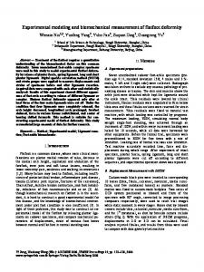
II. METHODS A. Experiment preparations Seven unembalmed cadaver foot-ankle specimens (median age 41.4, standard deviation 15.8; 4 males and 3 females; 4 left and 3 right side) were collected. Radiograph was taken on them to exclude any osseous pathology of preexisting disease or trauma. The skin and muscles above the ankle joint were detached while kept the ligaments around the ankle intact. Tibia residuals were levered by clinical instrument, fibulas residuals were amputated to 6cm below tibia ones and these fibulas sections were reserved for strain measurement. Tibia residuals were fixed to material test machine, with which loading was controlled by programs. The maximum loading, 700N, simulating normal body weight single-foot standing, was achieved through an increment of 100N, and within every increment loading was halted for 10 seconds, while all data were harvested by other equipments. Before the formal trial, specimens were preconditioned to 300N for five times with a rate of 2mm/min. Loading rate of formal trial was also 2mm/min. Test machine accurately recorded time, force and displacement during whole range. After experiment of intact specimen, plantar fascia, spring ligament, long and short plantar ligaments were cut off according to different sequences, and experimental operation above was repeated. B. Displacement Measurement with DSCM Custom-made black pins were inserted to corresponding 10 bones (tibia, fibula, calcaneal, navicular, media cuneiform, and five metatarsal bones) as markers. Standard marker was fixed to custom-made bedplate. Two series of CCD system positioned at inboard and front of the specimen with 90° angle, connected to image processing computers respectively, were used to catch trial images while static moment in every stages. Bones shot by inboard camera include tibia, calcaneal, navicular, media cuneiform, and the 1st metatarsal, as shot by foreside camera include others (Fig.1). With the application of DSCM programs, displacements in 2-D or 3-D can be calculated. Finial analysis rejected fibulas, and results from tibias were used to compare with data given by test machine.
Yi Peng, Xiaohong Weng (Eds.): APCMBE 2008, IFMBE Proceedings 19, pp. 133–138, 2008 www.springerlink.com © Springer-Verlag Berlin Heidelberg 2008
134
Wenxin Niu,, Yunfeng Yang, Yubo Fan, Zuquan Ding, Guangrong Yu
Fig. 1 Experimental images under 700N axial loading (Left-top: normal specimen in medial view; Right-top: normal specimen in foreside view; Left-below: ligaments released specimen in medial view; Right-below: ligaments released specimen in foreside view.)
C. Strain Measurement with Strain Gauge Strain gauges used to measure bone surface strain were connected to statical strain indicator (DH-3818) with 1/4 bridge converter and common compensating gauge. For every specimen, 10 gauges were pasted with 502-glue to the bones surface nearby DSCM markers. Before this operation, bones surface was trimmed, lipid was removed with acetone, and drying operation was carried out with anhydrous ethanol. Temperature compensating gauge was pasted to fibula section mentioned before. D. Statistical analysis All data were demonstrated quantitatively as mean ± standard deviation ( x ± s ), and statistically analyzed in
_______________________________________________________________
SPSS13.0 software. Analysis of variance was employed in significance test of different groups, followed by S-N-K test with equal variances and Dunnett's test with unequal variances. III. RESULTS A. Displacement Measurement Results For intact specimens, tibias displacement in vertical dimension is 4.66±1.42mm under 400N loading, and 5.55±0.74mm under 700N loading. For specimens off 4 main ligaments, this value is 9.00±2.48mm under 400N loading and 11.27±1.77mm under 700N lading. After ligaments resection, it could be visible that specimens took a typical of flatfoot deformity, arrangement of different bones
IFMBE Proceedings Vol. 19
_________________________________________________________________
Vertical Displacement (mm)
Experimental modeling and biomechanical measurement of flatfoot deformity
135
30 25 20 15 10 5 0 Ca
Na
MC
M1
M2
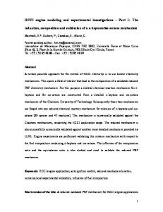
16 M-L Displacement (mm)
P-A Displacement (mm)
14 12 10 8 6 4 2 0 -2
Ca
Na
MC
M1
-4
14 12 10 8 6 4 2 0 -2 -4 -6 -8
M3
M1
M4
M2
M5
M3
M4
M5
Fig. 2 Three-dimension Displacements of various bones (Int=Intact; LR=Ligaments release; Ca=Calcaneal; Na=Naviculare; MC=Medial Cuneiform; M1~M5: 1st~5th Metatarsal.) in the structure changed, especially in the longitudinal arch, and abduction of the forefoot was obvious. Except that calcaneal bones in ligaments resection specimens retracted, all bones in media longitudinal arch had antedisplacements in different degrees under loading. Difference of these antedisplacements lead to an increasing prolongation of the medial longitudinal arch, which was about 3.7mm under 400N loading and 5.0mm under 700N loading for intact specimens, about 11.4mm under 400N loading and 12.6mm under 700N loading for ligaments resection specimens. Under intact state, the 1st metatarsal had an inward displacement with load increasing, while the other four metatarsals abducted, and the degree of abduction increased from the inside outwards. After ligaments resection, all metatarsals abducted, the most obvious change was in the 1st metatarsal. Measurement data of the displacements in all planes are shown as Figure 2. There is significant difference between the data before and after ligament resection with exception
_______________________________________________________________
of vertical displacements of M4 under both loading, M5 under 700N. B. Strain measurement results Measurement results of strain gauges increased with loading force increasing, regardless of tensile or compressive strain. Because of low strain rates under lower load, there was greater measurement error, and it is difficult to discover regularity, so only values of various bones under 700N load were shown in Fig.3. Because of strictly controlled experimental conditions, the distribution of these data is very ideal. All bones, except naviculare bone, displayed compressive strain at the measurement location. After ligaments resection, strain of naviculare, the 1st, 2nd, and 4th metatarsal increased significantly, and calcaneus bone had a decreased trend.
IFMBE Proceedings Vol. 19
_________________________________________________________________
Strain of bone surface (×0.001)
136
Wenxin Niu,, Yunfeng Yang, Yubo Fan, Zuquan Ding, Guangrong Yu
200
INTACT LR
100 0 -100
Ca
Na
MC
M1
M2
M3
M4
M5
-200 -300 -400 -500
Fig. 3 Strains of different gauges
IV. DISCUSSION A. The effect of ligaments injury on foot-arch structure Connection and encasement of ligaments is important for maintaining the foot structure. Many researches have been developed on the change of foot-arch height after relaxation and complete release of ligaments. This study concerned plantar fascia, spring ligament, long and short plantar ligaments. Many researches on the importance of plantar fascia to the foot-arch are based on mathematical models [4-7]. These models were mainly based on the theory raised by Hicks [8], that stabilizing role of plantar fascia on foot-arch structure was considered a windlass mechanism, and plantar fascia is more like a bow string, which braced both ends of the longitudinal arch, preventing it from separation and collapse. More accurate finite element model also confirmed the effect of plantar fascia relaxation on structure [912]. Through passively dorsal flexing toes, tension of plantar fascia can be seen by naked eyes [13]. Through cadaver experiment Huang et al [14] considered that resection of plantar fascia decreased the height of foot-arch by 25%. In some experiments [15], resection of plantar fascia lead to decreasing of arch height, increasing of length, abduction of the forefoot, and medial rotation of talus. Murphy et al [16] used metal markers to label foot structure, monitored them with X-ray while axial loading, and found that complete release of plantar fascia, compared to partly release, would lead to collapse of media and lateral longitudinal arch by 62~100%. There is no supporting structure between the calcaneus and navicular, therefore, caput tali is supported by spring ligaments fibrocartilago complex and posterior tibial tendon at the below. Rupture of the spring ligaments fibrocartilago complex may lead to insufficiency of posterior tibial tendon, support losing and collapse of caput tali, occurrence of flatfoot disease finally [17-19]. Clinical reports have identified it [20,21]. Plantar ligaments rise from facies
_______________________________________________________________
plantaris and anterior tubercle of calcaneus, its deep fibers, the short plantar ligament, extend to cuboid bone crest at plantar surface, and tuberosity of cuboid bone, and superficial layers, the plantar long ligament, extend to the 4th and 5th metatarsals. Plantar ligaments are thick and tough to maintain lateral longitudinal arch. It is usually difficult to distinguish between them, and they are often described as an entirety [14]. From the view of position or functions, plantar ligaments can be seen important complement of plantar fascia [22]. Huang et al [14] studied the relationship between effects of skeletal structure and ligaments on maintaining foot arch structure, and considered that 63% contribution was offered by skeletal structure, and 37% was offered by spring ligament, long and short plantar ligaments, plantar fascia. In this study, for sequences inconsistency of ligaments resection, there was little statistical significance on the effects of single ligament or combinations of two or three ligaments, but it was still observed that the effect of plantar fascia is the most obvious. With the help of finite element analysis [10,11], this study considered that foot structure is a dynamic and stable system maintained by coordination of bones, muscles, ligaments, and so on. Contribution of multifactors on maintaining the structure has coupling effect, is not a simple addition of many single factors. So it is incorrect to quantify contribution of single factor and multiple factors to the form of percentage. Injury of ligaments leads to a phenomenon of disorganization, decreasing of arch height, prolongation of arch length, and abduction of the forefoot. Disorganization will deteriorate the mechanical environment, make the center of bearing moving forward, and redistribute the force in the transverse arch. It is worth noting that, strains of metatarsals in complete ligaments release state were higher than that in intact state, but not higher than that in state that three of the four ligaments were released. It proved that complete ligaments resection severely damaged the structure, and metatarsals gradually lost their supporting role. For location and direction dependence of the strain gauge measurement, and lack of reference of related research, this study should be enhanced by improving the method and optimizing experimental design. B. Experimental modeling of the flatfoot deformity Flatfoot is series of diseases featured as lower or disappear of foot arch, including developmental and acquired flatfoot. Developmental flatfoot may be caused by maldevelopment of bones or joints, and systemic relaxation of the ligaments, such as Manfan's disease. Acquired flatfoot often associated with posterior tibial tendon insufficiency, overstress of musculus triceps surae, relaxation of mid-foot, supination and abduction of the forefoot, eversion of hind-
IFMBE Proceedings Vol. 19
_________________________________________________________________
Experimental modeling and biomechanical measurement of flatfoot deformity
foot, dislocation of talus, traumatic deformity, plantar fascia rupture, Charcot's foot, and neuromuscular imbalance diseases (such as polio, cerebral palsy, closed injury of brain, etc.) [23]. Causes of flatfoot are divergent. There is no uniform view on the development of this disease, and no quantitative indicator to define it in clinic. Clinical research focused on it with visible symptomatic deformity, when changes of mechanical environment have emerged, and simple surgery on soft tissue can not correct deformities or prevent disease recurrence. Most researches on flatfoot were confined to analysis of clinical observation, and mathematical model is hard to construct for lack of knowledge. Cadaver experimental was used some studies [24-26]. But there was no recognized modeling method on experimental model of flatfoot deformity. A simple and practical method is through incision of joint capsules and release of ligaments on normal foot specimens [27]. Others constructed model by eliminating the force in the tibialis posterior muscle to simulate a complete tear of the tibialis posterior tendon and reducing by 50% the strength of the spring ligament, the short plantar ligament and portions of the long plantar ligament, and the plantar fascia [28]. However, no detailed measurement data based on the flatfoot model was described before, and it is hard to formulate different modeling method for deformity in different degrees and types. This study showed different biomechanical performance of foot-arch after different ligaments were released. Obvious deformity appeared only after three of four main ligaments were released. Though there is no statistical analysis on the state that less than four ligaments were released, more data and experience have been accumulated to further study in expanding samples.
REFERENCES 1. 2. 3. 4. 5. 6. 7. 8. 9. 10. 11.
12. 13. 14. 15. 16.
V. CONCLUSION
17.
Ligaments resection is an effective method to model flatfoot deformity in different types and degrees. The cause of flatfoot deformity is multifactorial, and various factors have coupling effect with each other. Quantitative research with advanced methods can offer advice for clinical operations.
18. 19. 20. 21. 22. 23.
_______________________________________________________________
137
Friedman MA, Draganich LF, Toolan B, et al. (2000) The effects of adult acquired flatfoot deformity on tibiotalar joint contact characteristics. Foot Ankle Int. 22:241-246 McCormack AP, Ching RP, Sangeorzan BJ. (2001) Biomechanics of procedures used in adult flatfoot deformity. Foot Ankle Clin. 6:15-23. Richie DH. (2007) Biomechanics and clinical analysis of the adult acquired flatfoot. Clin Podiatr Med Surg. 24: 617-644 Wangdo K, Arkady SV. (1995) Role of plantar fascia in the load bearing capacity of the human foot. J Biomech. 28:1025-1033. Salathe EP, Aranglo GA. (2002) A biomechanical model of the foot: the role of muscles, tendons, and ligaments. J Biomech Eng. 124:281-287 Gefen A, Megido-Ravid M, Itzchak Y, et al. (2000) Biomechanical analysis of the three-dimensional foot structure during gait: a basic tool for clinical applications. J Biomech Eng. 122:630-639 Arangio GA, Chen C, Salathe EP. (1998) Effect of varying arch height with and without the plantar fascia on the mechanical properties of the foot. Foot Ankle Int. 19:705-709 Hicks JH. (1955) The foot as a support. Acta Anat. 25:34-45. Gefen A. (2002) Stress analysis of the standing foot following surgical plantar fascia release. Yang YF, Yu GR, Huang SP, et al. (2007) Effect of the plantar fasciotomy on the movement of the foot arch. The 1st International Conference on Bioinformatics and Biomedical Engineering. 486-489. Yang YF, Yu GR, Niu WX, et al. (2007) Effect of the plantar ligaments injury on the longitudinal arch height of the human foot. Life System Modeling and Simulation, International Conference, LSMS 2007, Lecture Notes in Computer Science. 4689:111-119 Cheung JT, Zhang M, An KN. (2004) Effects of plantar fascia stiffness on the biomechanical responses of the ankle-foot complex. Clin Biomech. 19: 839-846 Viel E, Esnault M. (1989) The effect of increased tension in the plantar fascia: a biomechanical analysis. Physiotheraphy Practice. 5:69-73 Huang CK, Kitaoka HB, An KN, et al. (1993) Biomechanical evaluation of longitudinal arch stability. Foot Ankle. 14:353-357. Thordarson DB, Kumar PJ, Hedman TP, et al. (1997) Effect of partial versus complete plantar fasciotomy on the windlass mechanism. Foot Ankle Int. 18: 16-20. Murphy GA, Pneumaticos SG, Kamaric E, et al. (1998) Biomechanical consequences of sequential plantar fascia release. Foot Ankle Int. 19:149-152. Mann RA, Thompson FM. (1985) Rupture of the posterior tibial tendon causing flat foot. Surgical treatment. J Bone Joint Surg Am. 67:556-561 Gazdag AR, Cracchiolo A 3rd. (1997) Evaluation of injury of the spring ligament and clinical assessment of tendon transfer and ligament repair. J Bone Joint Surg Am. 79;675-681. Deland JT. (2001) The adult acquired flatfoot and spring ligament complex: Pathology and implications for treatment. Foot Ankle Clin. 6: 129-135. Davis WH, Sobel M, Dicarlo EF, et al. (1996) Gross, histological, and microvascular anatomy and biomechanical testing of the spring ligament complex. Foot Ankle Int. 17:95-102. Choi K, Lee S, Otis JC, et al. (2003) Anatomical reconstruction of the spring ligament using peroneus longus tendon graft. Foot Ankle Int. 24: 430-436. Crary JL, Hollis JM, Manoli A. (2003) The effect of plantar fascia release on strain in the sping and long plantar ligaments. Foot Ankle Int. 24:245-250. McCormack AP, Ching RP, Sangeorzan BJ. (2001) Biomechanics of procedures used in adult flatfoot deformity. Foot Ankle Clin. 6:15-23.
IFMBE Proceedings Vol. 19
_________________________________________________________________
138
Wenxin Niu,, Yunfeng Yang, Yubo Fan, Zuquan Ding, Guangrong Yu
24. Logel KJ, Parks BG, Schon LC. (2007) Calcaneoculboid distraction arthrodesis and first metatarsocuneiform arthrodesis for correction of acquired flatfoot deformity in a cadaver model. Foot Ankle Int. 28:435-440. 25. Vora AM, Tien TR, Parks BG, et al. (2006) Correction of moderate and severe acquired flexible flatfoot with medializing calcaneal osteotomy and flexor digitorum longus transfer. J Bone Joint Surg Am. 88A:1726-1734.
_______________________________________________________________
26. Havenhill TG, Toolan BC, Draganich LF. (2005) Effects of a UCBL orthosis and a calcaneal osteotomy on tibiotalar contact characteristics in a cadaver flatfoot model. Foot Ankle Int. 26:607-613. 27. Thordarson DB, Schmotzer H, Chon J, et al. (1995) Dynamic support of the human longitudinal arch. A biomechanical evaluation. Clin Orthop Relat Res. 316:165-172. 28. Arangio GA, Reinert KL, Salathe EP. (2004) A biomechanical model of the effect of subtalar artheoerisis on the adult flexible flat foot. Clin Biomech. 19:847-852
IFMBE Proceedings Vol. 19
_________________________________________________________________