www.ietdl.org Published in IET Microwaves, Antennas & Propagation Received on 9th November 2011 doi: 10.1049/iet-map.2012.0436
ISSN 1751-8725
Exposure of the human brain to an electromagnetic plane wave in the 100–1000 MHz frequency range for potential treatment of neurodegenerative diseases A. Khaleghi1,2 M.S. Eslampanah Sendi3 R. Cha´vez-Santiago1,4,5 F. Mesiti4 I. Balasingham1,4,5 1
The Intervention Center, Oslo University Hospital, Oslo, Norway Department of Electrical and Computer Engineering, K. N. Toosi University of Technology, Tehran, Iran 3 Department of Electrical Engineering, Sharif University of Technology, Tehran, Iran 4 Department of Electronics and Telecommunications, Norwegian University of Science and Technology, Trondheim, Norway 5 Institute of Clinical Medicine, University of Oslo, Oslo, Norway E-mail:
[email protected] 2
Abstract: Radio signals can induce an electric field inside the brain, which might be potentially beneficial in the treatment of neurodegenerative diseases. For instance, a new method for the treatment of Alzheimer’s disease in mice through the exposure to the radiation of mobile phones has been successfully demonstrated. In the light of these results, studying the induction of an electric field in the human brain through the controlled exposure to radio signals is of paramount importance for the eventual development of similar treatment techniques in humans. In this study, the authors study the radio signals in 100– 1000 MHz as a means for inducing an electric field into the human brain in a non-invasive fashion. A voxel representation of the human body was used for numerical simulations, and the induced electric fields in the brain tissues (white and grey matters) were calculated for different incoming wave polarisations. A quantitative measure of the electric field inside the brain tissues is presented together with the average specific absorption rate. It is shown that the average electric field intensity in the brain has two local maxima at 300 and 600 MHz. These results are important for defining the optimal frequency band and wave polarisation.
1
Introduction
The exposure of the human body to a radiofrequency (RF) electromagnetic field (EMF) at different frequencies, different power levels and different modulation types including pulsed modulation or wireless communicationrelated modulations can produce different biological effects [1]. The most well-known and parameterised study of these effects belong to the specific absorption rate (SAR), which is related to thermal effects [2]. Several computational and experimental tools have been developed to measure these effects. Two of the most influential guidelines, International Commission on Non-ionising Radiation Protection (INCNIRP) [1] and Institute of Electrical and Electronics Engineers (IEEE) [3], are designed to avoid the identified health hazards related to excessive heating of human body tissues. For the related thermal effects, neither the frequency nor the modulation type is an important issue. In recent years, much attention has been devoted to the study of the interaction of modulated radio signals with living tissues [4, 5]. Different biological effects have been distinguished, which can be explained by electrical,
IET Microw. Antennas Propag., 2012, Vol. 6, Iss. 14, pp. 1565– 1572 doi: 10.1049/iet-map.2012.0436
physical and chemical phenomena [5 – 10]. Recent results reported by Arendash et al. and Dragicevic et al. [11, 12] indicated that the exposure of transgenic mice to mobile phone signals shows positive effects in the treatment of Alzheimer’s disease in these animals. This has been explained by the enhanced brain mitochondrial function caused by the induced electric field in the brain. These observations open great possibilities for the development of similar therapeutic treatments of Alzheimer’s disease in humans. However, the reported beneficial effects of EMF exposure in mice may be possible because of the small thickness of a mouse’s skin and skull, which allows the 900 MHz mobile phone signals to penetrate deeply inside the animal’s brain. Contrarily, in the case of human beings it is likely that the signal penetration is limited to the superficial parts of the brain in this frequency band. A number of research works available in the literature have studied the exposure of human beings to the frequency bands associated with mobile phones and mobile base stations [13 – 16]. The main motivation for these studies was the growing use of handheld communication devices and the society’s concern about the safety of RF exposure. Other
1565
& The Institution of Engineering and Technology 2012
www.ietdl.org works have studied the exposure to the radiation from wireless implantable devices inside the human body [17] and the whole body exposure to a wide frequency range [18]. In these works, detailed analyses of the brain exposure were not included. To the best of our knowledge, the exposure to RF for a wide frequency range, different polarisations, and different brain matters by using a heterogeneous anatomical model has not yet been thoroughly investigated. Furthermore, the specific frequency and polarisation requirements for controlled exposure of the brain to radio signals aimed at developing new therapeutic treatments of neurodegenerative diseases have not been previously identified; the results presented in this paper address this issue. Hereinafter, we consider an RF wave as the means to induce an appropriate level of electric field in the brain for possible interaction with human brain cells, that is, neurons. We assume that the RF is modulated in a way that it influences the brain activity. Defining the modulation type and the power level of the RF signal for possible interaction with the brain tissues needs further investigation and is beyond the scope of this work. Under these assumptions, the main contribution of this paper is the analysis of radio wave exposure within a wide range of carrier frequencies (100 – 1000 MHz) to identify the exact frequency band that allows the maximum induction of electric field intensity, and consequently maximum current density, inside the human brain with the least possible SAR value. For this sake, a voxel model of the human body that includes the frequency-dependent material properties of the biological tissues was used for computation. The rest of the paper is organised as follows: in Section 2, the interaction of an EMF with biological tissues and the effects on the connectivity of brain cells are briefly described. In Section 3, the simulation scenario in a heterogeneous voxel anatomical model is presented. In Section 4, the results of the numerical simulations are analysed and the intensity of the electric field and SAR values at different frequencies and wave polarisations are presented for the white and grey matters in the brain. Section 5 concludes the paper. For further information of this work’s implications, an Appendix is included with a discussion on the possible neuronal activity caused by the exposure of the brain to an electromagnetic wave.
2
Interaction of EMFs with neurons
The anatomical and functional structure of the cerebral cortex is of great importance for the correct accomplishment of the main brain functions such as learning, cognition, memory and motor abilities [18, 19]. Recent studies [20] demonstrated that the connectivity map of brains affected by Alzheimer’s disease is characterised by the appearance of broken links between the functional clusters of neurons, thereby preventing the proper communication of neuronal signals. The signalling activity of the neurons has a strong impact on the development of peculiar connectivity structures and a precise stimulation, applied on neurons, can allow the emergence of alternative pathways and the enhancement of the neuronal activity where they have been reduced or interrupted by brain diseases. To this end, the use of RF to promote the electromagnetic induction of stimulation signals on the cerebral cortex can support the design of advanced non-invasive treatment techniques. The electromagnetic exposure of a dispersive dielectric material can trigger a proportional electromagnetic induction of 1566 & The Institution of Engineering and Technology 2012
currents on the surface of the exposed material, that is, the grey and white brain matters in this case, thus providing a possible explanation to the observed benefits on the mice brains [11, 12]. Neurons in the cerebral cortex can be seen as biological communication devices connected to each other in a network. Each neuron receives and processes thousands of electrochemical input signals from other cells, producing output signals that propagate in the neuronal network [21]. The information exchanged between connected neurons is represented by sequences of instantaneous peaks in the membrane electric potential of the cell, called action potentials (AP), or spikes, generated according to the received inputs and the current state of the neuron (more details are provided in the Appendix section). The dynamic behaviour of the potential has been investigated in the past and several mathematical models, based on differential equations, have been proposed. Among others, the Integrate and Fire and the Hodgkin – Huxley [22] models are the most popular and describe the potential with different levels of approximation. At the molecular level, when an input signal is received, the synaptic junction on the receiving neuron enables the opening of ion channels on the membrane, allowing the flow of ion currents that depolarise (increase) or hyperpolarise (decrease) the potential. If the additive effect of all the incoming input raises the potential above a physiological threshold, an AP is generated (fired). In this respect, it is clear that an external action on the neuronal membrane can produce variations on the potential, with strong impact on the firing activity of neurons and, consequently, on the information processing of the cortex. The external stimulation of the neuron cell can take action at different levels: † At the molecular and ionic level by changing the electrical characteristics of the cell (conductivity and permittivity) and the behaviour of the ion flows through the membrane. This can be conducted possibly by a modulated signal that changes the cell characteristics through time-varying modulated heating. † At the macro-scale level by direct injection or induction of external currents on the neuronal membrane, altering the membrane potential; this can be possibly introduced by the interaction of a radio wave with the ions and the plasma boundary of the cells. A combination of both actions can also occur in a realistic scenario. The main aim of the cortex stimulation is to allow an accurate and controlled enhancement or reduction of the overall neuronal activity when the spontaneous activity is weakened by brain diseases. Furthermore, a proper stimulation strategy can enable the appearance of oscillatory behaviours and synchronisation phenomena in global neuronal response.
3
Simulation scenario
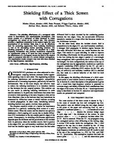
In order to investigate the influence of RF on the possible stimulation of the brain (which might be caused by an induced electric field that generates an electrical current or by the heating of the tissues), a voxel model of the human body embedded in an electromagnetic simulation tool was used. This heterogeneous anatomical model of an adult male is illustrated in Fig. 1. The model, with 1 mm of spatial resolution and 24 different tissues, was taken from the National Library of Medicine and it differentiates IET Microw. Antennas Propag., 2012, Vol. 6, Iss. 14, pp. 1565–1572 doi: 10.1049/iet-map.2012.0436
www.ietdl.org material properties were incorporated in the simulation tool, a single time-domain simulation run provided the electric field and the current density inside the human head tissues at different frequencies.
4
EMF simulations
The electric field inside the human head model was calculated through numerical simulations. Owing to the dispersive material properties of the human tissues (see Fig. 2), an electrical current density can be generated inside the brain by the induced electric field, which can be expressed as Fig. 1 Simulation scenario for the exposure of a human head to an electromagnetic plane wave
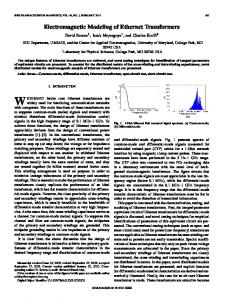
between white and grey matters in the brain. CST Microwave TM Studio , which is an electromagnetic simulation tool that solves Maxwell’s equations numerically through the timedomain finite integration technique, was used for our computation. The heterogeneous anatomical model is essential for these simulations because the different layers of tissues and their shape can significantly alter the calculated electric field intensity inside the brain because of the standing wave generated inside the tissues. The exposure source in our simulations was a plane wave radiated downward from above the head top (z-axis) as indicated in Fig. 1. The plane wave simulates the radiation from an antenna source located in the far field. Two different directions of the electric field, along the x-axis and y-axis, were considered. The radiated electric field intensity was set to 1 V/m. Different frequencies within 100 – 1000 MHz with 50 MHz steps were used for the excitation of the plane wave. The perfectly matched layer absorbing boundary condition was used for the simulations, thus the body environment reflections were ignored. The part of the human model considered for our simulations was above the shoulders, thus the wave penetration from the bottom side towards the brain can be neglected because of the high loss for this path. This assumption reduced the computation time significantly. For the sake of ensuring the reliability of the data generated using the aforementioned numerical simulations, a similar scenario as previously described was implemented with a model of a human male obtained through magnetic resonance imaging. This model was embedded in SEMCAD X, a simulation tool that uses the finitedifference time-domain (FDTD) numerical analysis technique. This alternative model, however, cannot separate the white and grey matters of the brain. The results obtained with both scenarios were fairly similar. The dielectric properties, conductivity s (S/m) and relative permittivity (1r) of the tissue materials are frequency-dependent and are provided by Gabriel [23]. Fig. 2 shows the variation of the conductivity and relative permittivity against frequency for some common tissues including the white and grey matters of the brain. The frequency dependency of the materials was expressed by the Cole – Cole model [23]. We used a simplified model of the frequency dependency, which can be expressed by a second-order polynomial for the 100– 1000 MHz frequency range. The details of this approximation can be found in [24]. The permeability of human tissues, mr , has a unity value. Since all the IET Microw. Antennas Propag., 2012, Vol. 6, Iss. 14, pp. 1565– 1572 doi: 10.1049/iet-map.2012.0436
J = sE(A/m2 )
(1)
where J is the current density (A/m2) vector and E (V/m) is the electric field vector in the computation area. The SAR, a standard parameter expressed in W/kg that measures the amount of absorbed energy by the biological tissues, is calculated from the electric field by using SAR =
s|E|2 (W/kg) 2r
(2)
where E is the peak electric field strength and r is the mass density (kg/m3) of the tissues. In order to assess the absorption in each tissue, the IEEE C95.3 standard was used for calculating the SAR 1 g contiguous. This is estimated by averaging the local maximum SAR,
Fig. 2 Variation of the conductivity and relative permittivity against frequency for some head tissues a Conductivity (S/m) b Relative permittivity against frequency for some head tissues 1567
& The Institution of Engineering and Technology 2012
www.ietdl.org
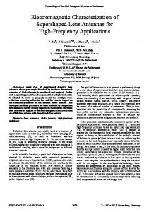
Fig. 3 Average SAR 1 g (dBuW/kg) distribution inside the brain for the exposure to an x-axis polarised plane wave excited with a 1 V/m electric field at different frequencies (see figure in colour)
integrating the highest SAR volume in a given tissue until a mass of 1 g is reached. As the first quantitative measure of RF exposure, the SAR value was calculated in our scenario. Fig. 3 shows the sagittal( yz) plane distribution of 1 g averaged SAR in the brain tissues for 100 MHz up to 900 MHz. These calculations were done for electric field polarisation along the x-axis direction. As shown, different SAR values and distributions for different frequencies can be observed in the brain tissues. Similarly, Fig. 4 shows the sagittal-plane distribution of 1 g averaged SAR for electric field polarisation in the y-axis direction. The same SAR scale was used for both Figs. 3 and 4. The SAR distribution and values are different for each polarisation. Larger SAR values can be observed for the y-axis polarised wave. This difference can be explained by the curvature of the head and the brain tissues and by the different boundary conditions seen by different polarisations as the wave propagates through the voxels of the anatomical model. This observation is in agreement with our previous investigations, in which different penetration properties of 1568 & The Institution of Engineering and Technology 2012
an electromagnetic wave in the human chest were observed when the wave polarisation changed [24]. In addition, the SAR values for both polarisations increase and then decrease with frequency. We note that the increment of the SAR values with frequency is caused not only by the increase of the electric field intensity inside the brain but also because of the increase of the conductivity of the tissues with frequency (see Fig. 2a). We can also note brain regions where the SAR reaches peak values in both Figs. 3 and 4. This can be explained by the different paths the radio wave follows to reach the brain tissues. Owing to this multipath propagation, in certain parts of the brain the wave replicas arrive in co-phase mode and sum-up coherently, which elevates the electric field intensity and the SAR value. In addition, the tissue layering causes a standing wave that creates the focus points for SAR values. In order to provide a view of the overall SAR in the brain tissues, the average SAR over the whole brain was calculated for the white and grey matters. Fig. 5 shows this average SAR for 1 V/m radiation of the electric field for both polarisations IET Microw. Antennas Propag., 2012, Vol. 6, Iss. 14, pp. 1565–1572 doi: 10.1049/iet-map.2012.0436
www.ietdl.org
Fig. 4 Average SAR 1 g (dBuW/kg) distribution inside the brain for the exposure to an y-axis polarised plane wave excited with a 1 V/m electric field at different frequencies (see figure in colour)
against frequency. As can be seen, the average SAR increases with frequency and has two main local peak values at 300 MHz and about 600 MHz. There is a local minimum SAR at 400 MHz, and above 600 MHz the SAR values decrease smoothly. In spite of the lower conductivity of the white matter compared with the grey matter and the similar mass density of both tissues, the average SAR for the white matter is larger than for the grey. This means a larger induced electric field in the white matter. The average SAR for the y-axis polarised electric field for all the frequencies is larger than for the x-axis electric field polarisation. By normalising the whole average SAR at all the different frequencies to the average SAR at 100 MHz, differences of 8.5 and 9.5 dB SAR at 300 and 600 MHz, IET Microw. Antennas Propag., 2012, Vol. 6, Iss. 14, pp. 1565– 1572 doi: 10.1049/iet-map.2012.0436
respectively, are observed for the y-axis polarised electric field. For the x-axis polarisation, the whole average SAR increases by 9.2 and 11 dB for the aforementioned frequencies when compared with the SAR at 100 MHz. The whole average electric field intensities in the grey and white matters of the brain are shown in Fig. 6. Note the existence of two peaks of the average electric fields at 300 MHz and about 600 MHz. Both peaks have approximately the same amplitude in each polarisation and brain matter cases. However, we notice higher electric field intensity in the white matter than in the grey one. Moreover, the y-axis polarised wave produces higher electric field intensity inside the brain tissues; the higher electric field results in higher current density too. 1569
& The Institution of Engineering and Technology 2012
www.ietdl.org
Fig. 5 SAR averaged over the whole brain (grey and white matters) against frequency for the x-axis and y-axis electric field polarisations
For the potential application of RF radiation to the treatment of neurodegenerative diseases, inducing high electric field inside the brain while keeping the SAR value as low as possible is the best compromise. From the results in Figs. 5 and 6 we propose using the 250 – 300 MHz frequency band for this emerging medical application. The proposed frequency band exhibits maximum electric field intensity inside the human brain with lesser SAR than the frequencies around 600 MHz and above. In addition, we
recommend using the y-axis wave polarisation, which induces more electric field inside the brain with the same 1 V/m exposure source. The results described in this section are in fair agreement with those that were obtained by using the FDTD technique. The maximum electric field and SAR of the whole brain were obtained with this other tool (SEMCAD X). The effects of the different polarisations were the same in this alternative scenario, which confirmed the validity of the
Fig. 6 Electric field intensity (V/m) averaged over the whole brain (grey and white matters) against frequency for the x-axis and y-axis electric field polarisations 1570 & The Institution of Engineering and Technology 2012
IET Microw. Antennas Propag., 2012, Vol. 6, Iss. 14, pp. 1565–1572 doi: 10.1049/iet-map.2012.0436
www.ietdl.org above results. The relative values of the electric field and SAR were comparable, but the absolute values differed. This discrepancy appears because of the different human models used in the two scenarios. Moreover, as mentioned before, the SEMCAD X human model cannot separate the white and grey matters in the brain.
5
Conclusions
We simulated the exposure of a human head to a plane-wave radiation within 100 – 1000 MHz using a heterogeneous anatomical model. The plane wave was radiated from above the head top with two different orthogonal polarisations. SAR calculations at different frequencies and polarisations were presented. The wave polarisation aligned to the sagittal-plane produced a larger average SAR and average electric field in the brain than the coronal-plane polarised wave. A local maximum SAR in the brain was observed at 300 MHz whereas the average maximum SAR was found at 600 MHz. We noticed that the brain white matter exhibited larger average SAR than the grey matter despite the lower conductivity of the former. On the other hand, the average electric field intensity is maximal at 300 and 600 MHz for all the polarisations and brain matters. Based on these results, we conclude that the optimal frequency range to induce the largest electric field intensity with the lowest SAR inside the human brain is 250– 300 MHz. Larger electric field intensity can carry larger current density and consequently better signalling inside the brain tissues. The propagation of enhanced neuronal activities in a population of tens of thousands of neurons can give rise to the appearance of interesting spiking patterns, correlation and synchronisation between clusters of neurons. Such a scenario may be desirable for an eventual treatment of Alzheimer’s disease in humans based on the exposure of the brain to an EMF radiation.
6
References
1 International Commission on Non-Ionizing Radiation Protection: ‘Exposure to High Frequency Electromagnetic Fields, Biological Effects and Health Consequences (100 KHz– 300 GHz) – Review of the Scientific Evidence and Health Consequences’, 2009 2 Foster, K.R., Repacholi, M.H.: ‘Biological effects of radiofrequency fields: does modulation matter?’, Radiat. Res., 2004, 162, (2), pp. 219–225 3 ‘IEEE standards coordinating committee 28 on non-ionizing radiation hazards’. Standard for Safe Levels with Respect to Human Exposure to Radio Frequency Electromagnetic Fields, 3 kHz to 300 GHz, ANSI/IEEE, Washington, DC, 1999 4 Juutilainen, J., Yto, A.H., Kumlin, T., Naarala, J.: ‘Review of possible modulation-dependent biological effects of radiofrequency fields’, Bioelectromagnetics, 2011, 32, (7), pp. 511–534 5 Balzano, Q., Sheppard, A.: ‘RF nonlinear interactions in living cells – I: nonequilibrium thermodynamic theory’, Bioelectromagnetics, 2003, 24, (7), pp. 473– 482 6 Sharma, R.P., Batra, K., Excell, P.S.: ‘Plasma effects in electromagnetic field interaction with biological tissue’, J. Plasma Phys., 2011, 77, (01), pp. 117–132 7 Blackman, C.F., Benane, S.G., House, D.E., Joines, W.T.: ‘Effects of ELF (1–120 Hz) and modulated (50 Hz) RF fields on the efflux of calcium ions from brain tissue in vitro’, Bioelectromagnetics, 1985, 6, (01), pp. 1 –11 8 Heida, T., Wagenaar, J.B.M., Rutten, W.L.C., Marani, E.: ‘Investigating membrane breakdown of neuronal cells exposed to nonuniform electric fields by finite-element modeling and experiments’, IEEE Trans. Biomed. Eng., 2002, 49, (10), pp. 1195–1203 9 Apollonio, F., Liberti, M., D’Inzeo, G., Tarricone, L.: ‘Integrated models for the analysis of biological effects of EM fields used for mobile communications’, IEEE Trans. Microw. Theory Tech., 2000, 48, (11), pp. 2082– 2093 IET Microw. Antennas Propag., 2012, Vol. 6, Iss. 14, pp. 1565– 1572 doi: 10.1049/iet-map.2012.0436
10 Volkow, N.D., Tomasi, D., Wang, G.-J., et al.: ‘Effects of cell phone radiofrequency signal exposure on brain glucose metabolism’, J. Am. Med. Assoc., 2011, 305, (8), pp. 808– 813 11 Arendash, G.W., Sanchez-Ramos, J., Mori, T., et al.: ‘Electromagnetic field treatment protects against and reverses cognitive impairment in Alzheimer’s disease mice’, J. Alzheimers Dis., 2010, 19, (1), pp. 191–210 12 Dragicevic, N., Bradshaw, P.C., Mamcarz, M., et al.: ‘Long-term electromagnetic field treatment enhances brain mitochondrial function of both Alzheimer’s transgenic mice and normal mice: a mechanism for electromagnetic field-induced cognitive benefit?’, Neuroscience, 2011, 185, pp. 135– 149 13 Kantartzis, N.V., Tsiboukis, T.D.: ‘Modern EMC analysis techniques volume II: models and applications’ (Morgan and Claypool Publishers, 2008) 14 Iskra, S., McKenzie, R., Cosic, I.: ‘Absorption in human body at 900 MHz for oblique incidence of plane wave’, Electron. Lett., 2009, 45, (12), pp. 602–604 15 Conil, E., Hadjem, A., El Habachi, A., Wiart, J.: ‘Whole body exposure at 2100 MHz induced by plane wave of random incidences in a population’, C. R. Phys., 2010, 11, (9–10), pp. 531– 540 16 Hadjem, A., Conil, E., Gati, A., Wong, M.-F., Wiart, J.: ‘Analysis of power absorbed by children’s head as a result of new usages of mobile phone’, IEEE Trans. Electromagn. Compat., 2010, 52, (4), pp. 812–819 17 Xu, L.S., Meng, M.Q.-H., Hu, C.: ‘Effects of dielectric values of human body on specific absorption rate following 430, 800, and 1200 MHz RF exposure to ingestible wireless device’, IEEE Trans. Inf. Technol. Biomed., 2010, 14, (1), pp. 52– 59 18 Uusitupa, T., Laakso, I., Ilvonen, S., Nikoskinen, K.: ‘SAR variation study from 300 to 5000 MHz for 15 voxel models including different postures’, Phys. Med. Biol., 2010, 55, (4), pp. 1157–1176 19 Sporns, O.: ‘Networks of the brain’ (MIT Press, 2011) 20 Stam, C., Jones, B., Nolte, G., Breakspear, M., Scheltens, P.: ‘Smallworld networks and functional connectivity in Alzheimer’s disease’, Cereb. Cortex, 2007, 17, (1), pp. 92– 99 21 Mesiti, F., Balasingham, I.: ‘Novel treatment strategies for neurodegenerative diseases based on RF exposure’. Proc. Fourth Int. Symp. Applied Sciences in Biomedical and Communication Technologies (ISABEL), Barcelona, Spain, 26– 29 October 2011, DOI: 10:1145/2093698.2093798 22 Dayan, P., Abbot, L.F.: ‘Theoretical neuroscience: computational and mathematical modeling of neural systems’ (MIT Press, 2001) 23 Gabriel, C.: ‘Compilation of the dielectric properties of body tissues at RF and microwave frequencies’. Brooks Air Force, N.AL/OE-TR1996-0037, San Antonio, TX, 1996 24 Khaleghi, A., Balasingham, I., Cha´vez-Santiago, R.: ‘Computational study of ultra-wideband wave propagation into the human chest’, IET Microw. Antennas Propag., 2011, 5, (5), pp. 559–567 25 Mesiti, F., Floor, P.A., Kim, A.N., Balasingham, I.: ‘On the modeling and analysis of the RF exposure on biological systems: a potential treatment strategy for neurodegenerative diseases’, Nano Commun. Netw., 2012, 3, pp. 103– 115
7 7.1
Appendix Impact on the neuronal activities
Upon the considerations of Section 3 we can assess the impact of the current induced by the electric field, (1), on the firing activity of a sample neuron. As stated in Section 2, the neuronal activity can be described by a mathematical model. For the sake of simplifying the analysis, let us consider a simple spiking model, the Leaky-Integrate and Fire (LIF) [22]. In the LIF model, the neuron membrane is represented with a passive electrical RC-circuit with output potential um(t) driven by an input current equal to the sum of the currents because of the capacitance and resistance of the membrane, identified as Cm and Rm , respectively I(t) = IC (t) + IR (t) = Cm
dum (t) um (t) + dt Rm
(3)
From (3), we derive the LIF expression of the membrane 1571
& The Institution of Engineering and Technology 2012
www.ietdl.org potential as a function of the physiological characteristics of the membrane
tm
dum (t) = −(um (t) − EL ) + I(t) dt
(4)
where tm ¼ CmRm is the time constant of the membrane and EL is the equilibrium potential of the membrane at rest conditions [22]. In the proposed scenario, we can assume a stationary field inducing a constant current density JRF (A/ m2) on the cellular membrane of an isolated neuron (not connected with the network) in order to evaluate the single contribution of the induced component of the stimulation. For this scenario, the analytic expression of the potential is the following um (t) = EL + Rm IRF [1 − exp(−t/tm )] + (u0 − EL )exp(−t/tm )
(5)
where u0 ¼ um(t ¼ 0) ¼ EL is the initial condition of the potential and the total induced current IRF is derived from the current density J times the total membrane surface, AC , that is, IRF ¼ J × AC . When the potential reaches a physiological threshold, uth , an action potential is generated and the potential is reset to the initial value EL , following the threshold condition if um (tk )uth ⇒ um (tk+ ) = u0 = EL
(6)
with tk representing the firing time of the last action potential.
To evaluate the spiking activity of the neurons as a function of an arbitrary input current IRF induced by the electric field, we use a simple model. Let us recall the considerations of Section 3 where a field within 250 – 300 MHz with y-axis polarisation and variable field intensity is suggested for safe stimulation of the brain. In Fig. 7, the membrane potential of a neuron is depicted as a function of time and for different realistic values of the input current IRF generated in the considered exposure scenario. As we can note, the firing rate highly depends on the value of the current, the higher the current, the higher the frequency, with physiological constraints because of the refractory period of the specific cell and the membrane time constant tm , which refers to the charge and discharge rate of the current, similar to an RC circuit. In the same figure, the theoretical firing rate (neglecting the refractory period) as a function of the input current, is depicted. As we can note, the number of action potentials fired in a unit of time increase with the input current, highlighting once more the fact that a direct functional connection between current and neuronal activity exists [25]. In a wider perspective, when a selected area of the neurons or a large part of the cerebral cortex is exposed to RF, the propagation of enhanced neuronal activities in a population of tens of thousands of neurons can give rise to the appearance of interesting spiking patterns, correlation and synchronisation between clusters of neurons. In this respect, the investigation of the existing relationships between a proper electromagnetic exposure, the currents induced on the cortex surface and the consequent change of the brain connectivity map, are of leading importance for the understanding of the observed benefits under RF exposure of mice brains affected by neurodegenerative diseases.
Fig. 7 Impact on the neuronal activities a Membrane potential of a neuron receiving a variable input current IRF induced by an RF exposure b Theoretical firing rate as a function of the input current, with the minimum value to trigger the firing
1572
& The Institution of Engineering and Technology 2012
IET Microw. Antennas Propag., 2012, Vol. 6, Iss. 14, pp. 1565–1572 doi: 10.1049/iet-map.2012.0436