ful discussions on α-hemolysin, and Marcia Glatt and other members of .... Tobkes, N., Wallace, B. A. & Bayley, H. (1985) Biochemistry 24, 1915â1920. 37.
THE JOURNAL OF BIOLOGICAL CHEMISTRY © 1996 by The American Society for Biochemistry and Molecular Biology, Inc.
Vol. 271, No. 1, Issue of January 5, pp. 568 –573, 1996 Printed in U.S.A.
Expression and Characterization of Recombinant Caveolin PURIFICATION BY POLYHISTIDINE TAGGING AND CHOLESTEROL-DEPENDENT INCORPORATION INTO DEFINED LIPID MEMBRANES* (Received for publication, October 10, 1995, and in revised form, November 6, 1995)
Shengwen Li, Kenneth S. Song, and Michael P. Lisanti‡ From the Whitehead Institute for Biomedical Research, Cambridge, Massachusetts 02142-1479
Caveolin, a 22–24-kDa integral membrane protein, is a principal component of caveolar membranes in vivo. Caveolin has been proposed to function as a scaffolding protein to organize and concentrate signaling molecules within caveolae. Because of its unusual membrane topology, both the N- and C-terminal domains of caveolin remain entirely cytoplasmic and are not subject to luminal modifications that are accessible to other integral membrane proteins. Under certain conditions, caveolin also exists in a soluble form as a cytosolic protein in vivo. These properties make caveolin an attractive candidate for recombinant expression in Escherichia coli. Here, we successfully expressed recombinant full-length caveolin in E. coli. A polyhistidine tag was placed at its extreme C terminus for purification by Ni21-nitrilotriacetic acid affinity chromatography. Specific antibody probes demonstrated that recombinant caveolin contained a complete N and C terminus. Recombinant caveolin remained soluble in solutions containing the detergent octyl glucoside and formed high molecular mass oligomers like endogenous caveolin. By electron microscopy, recombinant caveolin homo-oligomers appeared as individual spherical particles that were indistinguishable from endogenous caveolin homo-oligomers visualized by the same technique. As recombinant caveolin behaved as expected for endogenous caveolin, this provides an indication that recombinant caveolin can be used to dissect the structural and functional interaction of caveolin with other protein and lipid molecules in vitro. Recombinant caveolin was efficiently incorporated into lipid membranes as assessed by floatation in sucrose density gradients. This allowed us to use defined lipid components to assess the possible requirements for insertion of caveolin into membranes. Using a purified synthetic form of phosphatidylcholine (1,2-dioleoylphosphorylcholine), we observed that incorporation of caveolin into membranes was cholesterol-dependent; the addition of cholesterol dramatically increased the incorporation of caveolin into these phosphatidylcholine-based membranes by ;25–30-fold. This fits well with in vivo studies demonstrating that cholesterol plays an essential role in maintaining the structure and function of caveolae. Further functional analysis of these reconstituted caveolin-containing membranes showed that they were capable of recruiting a soluble * This work was supported in part by National Institutes of Health FIRST Award GM-50443 (to M. P. L.) and a grant from the W. M. Keck Foundation (to the Whitehead Fellows Program). The costs of publication of this article were defrayed in part by the payment of page charges. This article must therefore be hereby marked “advertisement” in accordance with 18 U.S.C. Section 1734 solely to indicate this fact. ‡ To whom correspondence should be addressed: Whitehead Inst. for Biomedical Research, Nine Cambridge Center, Cambridge, MA 02142-1479. Tel.: 617-258-5225; Fax: 617-258-9872; E-mail: lisanti@ wi.mit.edu.
recombinant form of Gi2a. This is in accordance with previous studies demonstrating that caveolin specifically interacts directly with multiple G protein a-subunits. Thus, recombinant caveolin incorporated into defined lipid membranes provides an experimental system in which the structure, function, and biogenesis of caveolin-rich membrane domains can be dissected in vitro.
Caveolae are specialized domains of the plasma membrane that are found in most cell types (1, 2). However, they are most abundant in adipocytes, endothelial cells, fibroblasts, and muscle cells (3). In adipocytes, caveolae represent up to 20% of the total plasma membrane surface area (4). Functionally, caveolae have been implicated in endothelial transcytosis (5), potocytosis (1), and signal transduction (6 –12). The “caveolae signaling hypothesis” states that caveolar localization of certain lipidmodified signaling molecules could provide a means for integrating certain transmembrane signaling events (13–15). Caveolin, a 22–24-kDa protein, is an integral membrane component of caveolar membranes (16). It has been proposed that caveolin may function as a scaffolding protein for organizing and concentrating caveolin-interacting molecules within caveolae (17). In this regard, caveolin appears to be very important for the formation of caveolar membranes. Caveolin expression levels correlate very well with the biochemical and morphological appearance of caveolae. For example, (i) caveolin is most abundant in cell types that contain numerous caveolae, i.e. adipocytes, endothelial cells, smooth muscle cells, and fibroblasts (18); (ii) caveolin and caveolae are both induced ;10 – 25-fold during the differentiation of 3T3-L1 fibroblasts to the adipocyte form (4, 19); (iii) caveolin levels are dramatically reduced, and caveolae are morphologically absent in cells transformed by various activated oncogenes (v-abl, activated ras, and others) (20); and (iv) recombinant expression of caveolin in caveolin-negative cell lines results in the correct targeting of caveolin to caveolae-enriched membrane fractions and allows the de novo formation of caveolae (21, 22). These results suggest that caveolin represents an important structural protein for directing the formation of caveolar membranes. Several structural properties of caveolin are also consistent with a role for caveolin in organizing caveolar domains. Both the N- and C-terminal domains of caveolin face the cytoplasm and are thus accessible for interactions with cytoplasmically oriented molecules (23); caveolin copurifies with cytoplasmic signaling molecules including heterotrimeric G proteins, Src family tyrosine kinases, and Ras-related GTPases (6, 8, 9, 11, 12, 21); caveolin interacts directly with heterotrimeric G proteins and can functionally regulate their GTPase activity, holding the G protein in the inactive conformation (24); and caveolin exists within caveolar membranes as a high molecular mass homo-oligomer (17, 25). Thus, caveolin could serve as an oligo-
568
Reconstitution of Caveolin-rich Membranes meric docking site for organizing and concentrating inactive signaling molecules within caveolar membranes (17). To study the molecular interaction of caveolin with other molecules, we have recently expressed full-length caveolin and portions of caveolin as GST1 fusion proteins in Escherichia coli (17, 21, 24). However, preparations of GST-full-length caveolin also contained substantial amounts of GST cleaved to its active core as the complete fusion was somewhat unstable. The partial success of this approach prompted us to express full-length caveolin in E. coli without the GST moiety. Full-length caveolin without GST would more closely approximate the natural state of the caveolin molecule in vivo. Here, we report the purification and characterization of soluble recombinant full-length caveolin without GST and its interaction with lipid membranes of defined molecular composition. Despite that caveolin is an integral membrane protein, it was expressed well using this approach. This may be related to the observations that under certain conditions, caveolin also exists in a soluble form as a cytosolic protein in vivo (26), and both caveolin N- and Cterminal domains remain entirely cytoplasmic and are not subject to luminal modifications (21, 23, 27). EXPERIMENTAL PROCEDURES
Materials—mAb 2234 directed against caveolin was provided by John R. Glenney (Transduction Laboratories). The cDNA for canine caveolin was obtained as we described previously (6). Monoclonal antibody 9E10 was provided by the Harvard Monoclonal Antibody Facility (Cambridge, MA). Ni21-NTA-agarose for purification of polyhistidinetagged proteins was from QIAGEN Inc. The purified lipid extract from brain tissue was from Sigma (type VI, catalog No. B1877). Cholesterol and synthetic phosphatidylcholine (1,2-dioleoylphosphorylcholine) were from Matreya, Inc. and Avanti Polar Lipids, respectively. The GST-Gi2a construction, encoding the cDNA for bovine Gi2a, was the kind gift of Dr. Ikuo Nishimoto (Massachusetts General Hospital). Anti-Gi2a antibodies were from Dupont NEN. Construction of Recombinant Caveolin—To purify caveolin after expression in E. coli, we incorporated the polyhistidine tag into the C terminus of the Myc-tagged canine caveolin cDNA using PCR primers (caveolin-Myc-His7). The C-terminally Myc-tagged form of caveolin used as the template was as we described previously (21). The final construction was subcloned into the MCS (HindIII-BamHI) of the vector pET-17b (Novagen, Inc.) for expression in E. coli (strain BL21(DE3); Novagen, Inc.). Expression and Purification of Recombinant Caveolin—E. coli (strain BL21(DE3)) was transformed with the recombinant plasmid harboring the caveolin cDNA. A 1:100 dilution of an overnight starter culture was used to inoculate 600 ml of LB medium containing ampicillin (175 mg/ml), and the culture was incubated with shaking at 37 °C. After the absorbance at 600 nm reached 0.6, freshly dissolved isopropyl-1-thio-bD-galactopyranoside was added to a final concentration of 0.5 mM and cultured for an additional 3 h. Bacteria were collected by centrifugation in an SS34 rotor (6000 rpm for 10 min at 4 °C) and washed once with STE buffer (150 mM NaCl, 10 mM Tris, pH 8.0, 2 mM EDTA), and the bacterial pellet was snap-frozen in liquid nitrogen. After freezing, bacteria were resuspended in 15 ml of STE buffer and treated with lysozyme (100 mg/ml for 15 min on ice). The sample was then adjusted to 1.5% N-lauroylsarcosine (Sigma) and homogenized with a Polytron tissue grinder (Brinkmann Instruments; two 30-s bursts). The homogenate was clarified by centrifugation in an SS34 rotor (16,000 rpm for 15 min at 4 °C). After clarification, the supernatant was adjusted to 2% Triton X-100 and allowed to bind to Ni21-NTA-agarose for 4 h rotating at 4 °C. Note that 100 ml of Ni21-NTA-agarose was used per 100 ml of bacterial culture. After binding, Ni21-NTA-agarose was washed six times with STE buffer and eluted in STE buffer containing 200 mM imidazole and 60 mM octyl glucoside for 2 h at 4 °C. The soluble eluate was designated as recombinant caveolin. For long-term storage, the eluate was adjusted to 50% glycerol and stored at 220 °C. A yield of ;5 mg of purified recombinant caveolin was obtained from a 600-ml bacterial culture. It is important to note that purified recombinant caveolin 1 The abbreviations used are: GST, glutathione S-transferase; mAb, monoclonal antibody; NTA, nitrilotriacetic acid; PAGE, polyacrylamide gel electrophoresis; Mes, 4-morpholineethanesulfonic acid; MBS, Mesbuffered saline.
569
also remains soluble even after dialysis to remove octyl glucoside. Immunoblotting and Streptavidin Blotting—Samples were separated by SDS-PAGE (15% acrylamide) and transferred to nitrocellulose. After transfer, nitrocellulose sheets were subjected to immunoblotting with anti-caveolin IgG (mAb 2234; 1:400) or anti-Myc IgG (mAb 9E10; 1:1000) or to streptavidin blotting to visualize biotin-labeled proteins. For immunoblotting, incubation conditions were as described by the manufacturers (Promega and Amersham Corp.), except that we supplemented our blocking solution with both 1% bovine serum albumin and 1% non-fat dry milk (Carnation). Biotinylated proteins were detected by blotting with iodinated streptavidin and autoradiography with an intensifying screen as described (28). Velocity Gradient Centrifugation—Estimation of the molecular mass of recombinant caveolin was performed as we described previously for endogenous native caveolin (17). Briefly, samples were loaded atop a 5– 40% linear sucrose gradient (4.3 ml) and centrifuged at 50,000 rpm (;340,000 3 g) for 10 h in an SW 60 rotor (Beckman Instruments). Note that the entire gradient was prepared with MBS (25 mM Mes, pH 6.5, 0.15 M NaCl) plus 60 mM octyl glucoside. After centrifugation, gradient fractions were collected from above. Molecular mass standards for velocity gradient centrifugation were as follows: carbonic anhydrase (29 kDa), bovine serum albumin (66 kDa), alcohol dehydrogenase (150 kDa), b-amylase (200 kDa), and apoferritin (443 kDa) (Sigma). Low-angle Rotary Shadowing—Samples were sandwiched between two pieces of freshly cleaved mica. Mica sheets were separated and placed on the stage of a Denton 502A vacuum evaporator and dried for 20 min. Samples were shadowed with a platinum-palladium wire at an angle of 4° while rotating at 60 rpm and then coated with carbon at an angle of 90°. Replicas were removed by floating onto water and placed on uncoated 400-mesh copper grids. Micrographs of random fields were made with a 1200 CX JEOL electron microscope. Incorporation of Recombinant Caveolin into Lipid Membranes—The incorporation of recombinant caveolin into membranes was monitored by floatation in sucrose density gradients. Briefly, recombinant caveolin was mixed with purified lipid components in 2 ml of MBS containing 60 mM octyl glucoside and dialyzed overnight against MBS lacking detergent to allow the association of caveolin with lipids. More specifically, ;85 mg of recombinant caveolin was added either to 1.8 mg of the purified lipid extract dissolved in octyl glucoside or to 2.5 mg of synthetic phosphatidylcholine dissolved in octyl glucoside; experiments involving cholesterol were performed in a similar manner, except that phosphatidylcholine and cholesterol were mixed in a 1:1 molar ratio before the addition of caveolin. The dialysate was then adjusted to 40% sucrose by the addition of 2 ml of 80% sucrose prepared in MBS and placed at the bottom of an ultracentrifuge tube. A 5–30% discontinuous sucrose gradient was formed above (4 ml of 5% sucrose, 4 ml of 30% sucrose; both in MBS lacking detergent) and centrifuged at 39,000 rpm for 16 –20 h in an SW 41 rotor (Beckman Instruments). A light-scattering band confined to the 5–30% sucrose interface contained the associated lipids and formed regardless of whether or not caveolin was added. Fractions (1 ml each) were collected from the top and subjected to 15% SDS-PAGE/immunoblot analysis with anti-caveolin mAb 2234. Alternatively, the light-scattering band at the 5–30% sucrose interface was collected by centrifugation and subjected to immunoblot analysis. Recruitment of GST-Gi2a onto Reconstituted Caveolin-rich Membranes—Recombinant GST-Gi2a was purified as we previously described for other GST fusion proteins (24). Purified soluble GST-Gi2a (;50 –100 mg) was incubated with one-tenth of a single preparation of either reconstituted caveolin-rich membranes or caveolin-deficient membranes. After 2– 4 h at 4 °C rotating end-over-end, soluble unbound GST-Gi2a was removed by re-floatation in sucrose density gradients. Reconstituted membranes used as the substrate for binding were prepared with the purified lipid extract and recombinant caveolin as detailed above. Bound GST-Gi2a was visualized by immunoblotting with anti-Gi2a antibodies. RESULTS
Purification and Characterization of Soluble Recombinant Caveolin—A strategy was devised for recombinant expression and purification of soluble caveolin in E. coli. The cDNA for the entire coding sequence of canine caveolin was inserted into the multiple cloning site of a bacterial expression vector under control of the T7 promoter. A Myc epitope tag was placed at its C terminus with a polyhistidine tag following the Myc tag (Fig. 1A). The polyhistidine tag allows purification by affinity chromatography using Ni21-NTA-agarose. A yield of ;5 mg of
570
Reconstitution of Caveolin-rich Membranes
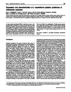
FIG. 1. Purification of recombinant caveolin. A, schematic diagram summarizing the construction of recombinant caveolin. A Myc epitope tag was placed at its C terminus with a polyhistidine tag following the Myc tag for affinity purification by Ni21-NTA-agarose chromatography. Note that mAb 2234 recognizes an epitope within caveolin residues 1–21; mAb 9E10 recognizes the Myc epitope (EQKLISEEDLN). B, characterization of recombinant caveolin by immunoblot analysis. Recombinant caveolin was biotinylated in vitro to assess its purity; it appeared as a single band by streptavidin blotting. In addition, the same band contains a complete N and C terminus as evidenced by immunoblot analysis with specific antibody probes (mAb 2234 and mAb 9E10).
purified recombinant caveolin was typically obtained from a 600-ml bacterial culture. Purified recombinant caveolin remained soluble in solutions containing the detergent octyl glucoside; it did not behave as an insoluble aggregate. Recombinant caveolin appeared relatively pure. As caveolin fails to stain with standard protein stains (21), purified preparations of recombinant caveolin were biotinylated in solution. This technique is more sensitive than silver staining; in vitro biotinylation allows the detection of picogram quantities of protein (29). Biotinylated proteins were detected after SDSPAGE and transfer to nitrocellulose by blotting with iodinated streptavidin. Caveolin appeared as a single band without any major contaminants or degradation products (Fig. 1B). However, it should be noted that the molecular mass of recombinant caveolin was slightly larger than that of endogenous caveolin. This reflects the addition of the Myc epitope and polyhistidine tags. To monitor the completeness of the N- and C-terminal ends of caveolin, two specific antibody probes were utilized: mAb 2234, which recognizes caveolin residues 1–21, and mAb 9E10, which detects the Myc epitope placed at the C-terminal end of caveolin. The single major band observed by in vitro biotinylation was immunoreactive with both these probes (Fig. 1B), indicating that recombinant caveolin contains a complete N and C terminus and therefore represents the full-length caveolin protein. Endogenous caveolin exists as a ;350-kDa homo-oligomer (containing ;14 –16 monomers/oligomer) as shown using several different approaches (17, 25). The oligomeric state of purified recombinant caveolin was next assessed by employing an established velocity gradient system developed previously to study homo-oligomers of endogenous caveolin (17). Several integral membrane proteins have been previously shown to migrate at their expected monomeric molecular mass in these
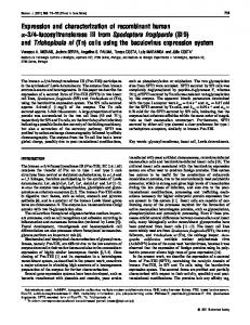
FIG. 2. Velocity gradient centrifugation of recombinant caveolin. A, purified recombinant caveolin was loaded atop a 5– 40% sucrose gradient containing octyl glucoside and subjected to centrifugation for 10 h. Fractions were analyzed on blots by incubation with anti-caveolin IgG (mAb 2234). Note that recombinant caveolin migrates as a distinct high molecular mass complex and peaks in fraction 8. No caveolin monomers were detectable. The migration of molecular mass standards is also shown for comparison. B, shown is a graphic representation of A quantitated by scanning densitometry as described previously (8, 44).
same velocity gradients (17). Fig. 2 shows that purified recombinant caveolin migrated as expected as a high molecular mass complex between 200- and 443-kDa molecular mass standards. This provides an indication that recombinant caveolin is correctly folded as it remains soluble in detergent solutions and forms high molecular mass oligomers of the same relative size as endogenous caveolin. Endogenous caveolin homo-oligomers appear as individual globular particles (;4 – 6 nm in diameter) by electron microscopy using a low-angle platinum shadowing technique (17). The same approach was used to examine the ultrastructure of recombinant caveolin. Fig. 3 shows that recombinant caveolin appeared as individual globular particles of the same diameter previously reported for endogenous caveolin homo-oligomers. Thus, recombinant caveolin behaves as expected for endogenous caveolin in its solubility properties, oligomeric state, and morphologic appearance. It is important to note that when the same construction used here was recombinantly expressed in mammalian cells, it cofractionated with endogenous caveolin (Fig. 4), an indication that the Myc epitope tag and the His tag do not interfere with the caveolar targeting of this engineered form of caveolin. Previous reports have also indicated that tagging of caveolin at either its N or C terminus with the Myc epitope does not affect its caveolar localization in vivo (21, 27, 30). Incorporation of Recombinant Caveolin into Lipid Mem-
Reconstitution of Caveolin-rich Membranes
FIG. 3. Low-angle platinum shadowing of recombinant caveolin homo-oligomers. Homo-oligomers appear as individual spherical particles with a diameter of ;4 – 6 nm. Bar 5 0.05 mm.
FIG. 4. Polyhistidine-tagged caveolin cofractionates with endogenous caveolin. Caveolin containing a C-terminal polyhistidine tag (see Fig. 1; caveolin-Myc-His7) was stably expressed in MadinDarby canine kidney cells as described previously for other caveolin constructs (21). Madin-Darby canine kidney cells were then fractionated using an established protocol that separates caveolin from the bulk of cellular membranes and cytosolic proteins. This fractionation scheme is based on the specific buoyant density of caveolin-rich domains and their resistance to solubilization by the non-ionic detergent Triton X-100 at low temperatures (6, 8, 12, 15, 19, 26, 45). The distributions of polyhistidine-tagged caveolin (caveolin-Myc-His7) and endogenous caveolin are shown. Fractions were collected from the top, separated by SDS-PAGE (15% acrylamide), and analyzed by immunoblotting. Immunoblot analysis was carried out with mAb 9E10 to selectively detect caveolin-Myc-His7 (not shown) and with anti-caveolin IgG (mAb 2297) to detect caveolin-Myc-His7 and endogenous caveolin. Note that both aand b-isoforms of endogenous caveolin are detected (21 and 24 kDa) and that they cofractionate with caveolin-Myc-His7 (29 kDa). The generation of b-caveolin results from an alternate translation initiation site at amino acid 32 within the full-length caveolin cDNA (21). We have previously shown, using this fractionation scheme, that endogenous caveolin (fractions 5 and 6) is purified ;2000-fold relative to total Madin-Darby canine kidney cell lysates (15, 24). In addition, caveolin is separated from .99.95% of total Madin-Darby canine kidney cellular proteins and compartment-specific markers for the endoplasmic reticulum, Golgi apparatus, lysosomes, mitochondria, and non-caveolar plasma membrane that remain in fractions 9 –13 of these bottom-loaded sucrose density gradients (6, 8, 15).
branes Is Cholesterol-dependent—As caveolin is an integral membrane protein, the ability of recombinant caveolin to be incorporated into lipid membranes was evaluated. Recombinant caveolin was added to an octyl glucoside-solubilized extract of purified lipids derived from brain. Incorporation of recombinant caveolin was monitored by floatation in sucrose density gradients. In this assay, octyl glucoside-dissociated lipids reassociated and migrated to the 5–30% sucrose interface of these gradients. Assembly of lipids was independent of the addition of caveolin. Under these conditions, caveolin was quantitatively incorporated into these membranes (Fig. 5). As expected, floatation of caveolin was strictly dependent upon the addition of lipids as caveolin remained at the bottom of these gradients if lipids were omitted; caveolin pellets in these gradients in the absence of lipids as it forms a high molecular mass oligomer (see above). As endogenous caveolin can be lipidmodified by palmitoylation of three cysteine residues within its C-terminal domain (27), these results also indicate that lipid modification of caveolin is not necessary for its incorporation into membranes as this modification does not occur in bacteria.
571
FIG. 5. Incorporation of recombinant caveolin into lipid membranes. The incorporation of recombinant caveolin into membranes was monitored by floatation in sucrose density gradients. Recombinant caveolin (;85 mg) was mixed with 1.8 mg of the purified lipid extract dissolved in octyl glucoside (see “Experimental Procedures”). The mixture was adjusted to 40% sucrose and placed at the bottom of an ultracentrifuge tube. A 5–30% discontinuous sucrose gradient (lacking detergent) was formed above and subjected to centrifugation for 20 h. A light-scattering band confined to the 5–30% sucrose interface (fractions 4 and 5) was observed and contained the associated lipids. This band formed regardless of whether or not caveolin was added. Fractions (1 ml) were collected from the top and subjected to immunoblot analysis with anti-caveolin mAb 2234. Note that caveolin pellets in the absence of lipids as it forms a high molecular mass oligomer (caveolin alone). In the presence of lipids, caveolin associates with membranes and attains buoyancy (caveolin plus lipids). Note that the lipid extract does not contain endogenous caveolin (lipids alone).
Additional control experiments were performed to rule out the possibility that caveolin is randomly trapped within these lipid membranes as they form. For this purpose, we also expressed and purified a recombinant cytosolic protein (GST) in E. coli. In contrast to recombinant caveolin, recombinant GST showed no incorporation into lipid membranes (data not shown). As a purified cytosolic protein is excluded during the process of membrane reconstitution, this indicates that caveolin is not simply trapped within the lumen of these lipid membranes. The lipid extract used in our initial experiments contained a complex mixture of naturally occurring lipids. To simplify the system, we next used a chemically synthetic form of pure phosphatidylcholine that is fluid at 4 °C. This lipid assembled into membranes as expected and migrated to the 5–30% sucrose interface. However, caveolin was very inefficiently incorporated under these conditions; little or no caveolin attained buoyancy. Several independent lines of evidence suggest that cholesterol plays an essential role in maintaining both the structure and function of caveolar membranes in vivo (16, 26, 31, 32). In addition, it has been postulated that caveolin may interact specifically with cholesterol, although no direct evidence has been presented to support this assertion (26). Thus, we supplemented chemically pure phosphatidylcholine with cholesterol (in a 1:1 molar ratio) and monitored the incorporation of caveolin into this two-component lipid system. Under these conditions, the addition of cholesterol dramatically increased the incorporation of caveolin into these phosphatidylcholine-based membranes by ;25–30-fold (Fig. 6). Thus, this model system (recombinant caveolin inserted into purified or synthetic lipid membranes) provides an experimental system in which the structure, function, and biogenesis of caveolin-rich membranes can be dissected in vitro. Recruitment of G Protein a-Subunits onto Reconstituted Caveolin-rich Membranes—G protein a-subunits are highly concentrated in preparations of caveolar membranes purified from diverse sources (6, 8, 9, 24, 33). Caveolin interacts directly with multiple G protein a-subunits, including Gs, Go, and Gi2 (24). This interaction does not require coexpression of bg-sub-
572
Reconstitution of Caveolin-rich Membranes
FIG. 7. Recruitment of a purified soluble form of Gi2a onto reconstituted caveolin-rich membranes. Caveolin was incorporated into membranes as described under “Experimental Procedures” and in the legend of Fig. 5. Equivalent amounts of reconstituted caveolin-rich membranes and caveolin-deficient membranes were then incubated in solution with soluble recombinant GST-Gi2a. To remove unbound GST-Gi2a, membranes were then isolated by refloatation in sucrose density gradients. After refloatation, fractions 4 and 5 of each gradient (corresponding to the light-scattering band at the 5–30% sucrose interface) were collected, pooled, and subjected to immunoblot analysis with anti-Gi2a IgG. Note that the binding of soluble GST-Gi2a to membranes is caveolin-dependent. GST-Gi2a migrates at ;68 kDa, as expected, as it should have the cumulative molecular mass of GST (26 –27 kDa) plus Gi2a (40 kDa). FIG. 6. Cholesterol-dependent incorporation of recombinant caveolin into phosphatidylcholine-based membranes. Recombinant caveolin (;85 mg) was mixed with 2.5 mg of synthetic phosphatidylcholine dissolved in octyl glucoside and treated as described in the legend of Fig. 5. In a second condition, phosphatidylcholine and cholesterol were mixed in a 1:1 molar ratio before the addition of caveolin. After floatation in sucrose density gradients, fractions 4 and 5 of each gradient (corresponding to the light-scattering band at the 5–30% sucrose interface) were collected, pooled, and subjected to immunoblot analysis with anti-caveolin IgG. Note that cholesterol addition increased the incorporation of caveolin into these phosphatidylcholinebased membranes by ;25–30-fold.
units (24). Based on the observation that caveolin is a cytoplasmically oriented integral membrane protein and that G protein a-subunits cycle on and off the membrane, we have proposed that caveolin could function to recruit G protein a-subunits onto caveolin-rich areas of the plasma membrane (17, 24). This hypothesis is further supported by the observation that mutational and pharmacologic activation of an epitope-tagged form of Gsa prevents its cofractionation with caveolin when recombinantly expressed in mammalian cells (24). To test this idea more directly, we evaluated whether recombinant caveolin could recruit soluble G protein a-subunits onto membranes in vitro. For this purpose, we expressed and purified a soluble recombinant form of Gi2a in bacteria (GST-Gi2a). Reconstituted lipid membranes containing recombinant caveolin were then briefly incubated with soluble recombinant GST-Gi2a. To stringently remove unbound GST-Gi2a, caveolincontaining lipid membranes were then reisolated by floatation in sucrose density gradients. Thus, bound GST-Gi2a must attain the buoyancy of lipid membranes in this system. As a control for nonspecific association of GST-Gi2a with membranes, an equivalent amount of reconstituted lipid membranes lacking recombinant caveolin was processed in parallel and subjected to the same binding and refloatation after incubation with soluble recombinant GST-Gi2a. Fig. 7 shows that GST-Gi2a bound to reconstituted caveolin-rich membranes, but not to caveolin-deficient membranes. This indicates that caveolin is sufficient to recruit GST-Gi2a onto lipid membranes. As Gi2a normally undergoes dual acylation (myristoylation and palmitoylation) when expressed in mammalian cells, our results indicate that these lipid modifications that do not occur in bacteria are not absolutely necessary for the caveolin-dependent recruitment of Gi2a onto membranes. However, such lipid modifications may still serve to modulate these interactions in vivo. To rule out the unlikely possibility that the interaction of GST-Gi2a with reconstituted caveolin-rich membranes is simply due to nonspecific protein-protein adsorption, we performed additional control experiments. For this purpose, we evaluated whether another soluble recombinant protein, GST, could interact with these membranes in a caveolin-dependent manner.
No binding of GST to either caveolin-containing membranes or caveolin-deficient membranes was observed (data not shown). This is consistent with previous studies demonstrating that the interaction of GST-caveolin with baculovirus-expressed G protein a-subunits is highly specific (24). DISCUSSION
Caveolin was first identified as a major v-Src substrate in Rous sarcoma virus-transformed cells and later as a caveolar marker protein (16, 18, 30, 34, 35). Cholesterol is thought to provide the “lipid glue” that holds caveolin within caveolar membranes and is essential for the proper biological functioning of caveolae (16, 26, 31). Increasing evidence indicates that caveolin may function within caveolar membranes as a scaffolding protein to organize and concentrate caveolin-interacting molecules (17). One class of caveolin-interacting proteins thus far identified is G protein a-subunits (24). Here, we have begun to systematically reconstitute caveolinrich membrane domains in vitro using purified recombinant proteins and synthetic lipid components. We expressed and purified a recombinant full-length form of caveolin from E. coli. Soluble recombinant caveolin contained a complete N and C terminus, formed high molecular mass oligomers of the correct size and shape as seen by velocity gradient centrifugation and electron microscopy, and could be inserted into lipid membranes after removal of the detergent octyl glucoside. Insertion of caveolin into lipid membranes appeared to depend on the presence of cholesterol, as shown using purified and synthetic lipid components. How does cholesterol facilitate the incorporation of caveolin into membranes? One possibility is that cholesterol adjusts membrane fluidity to provide the proper lipid environment for insertion of caveolin into membranes. Alternatively, cholesterol might interact directly with caveolin as caveolin contains an unusual 33-amino acid membrane-spanning region that is predicted to assume a b-conformation (35). In support of the latter possibility, caveolin is similar in many respects to a 293-amino acid bacterial toxin known as a-hemolysin or staphylococcal a-toxin. Like caveolin, a-hemolysin contains a membrane-spanning region that assumes a b-conformation, inserts into target membranes in a cholesterol-dependent fashion, forms SDS-resistant homo-oligomers, and exists both as a soluble protein that is secreted and as an integral membrane protein after insertion into target membranes (36 – 40). Our results are also consistent with previous in vivo studies demonstrating that oxidation of plasma membrane cholesterol can transiently convert endogenous caveolin from an integral membrane protein to a soluble cytosolic protein (26). The in vivo process is also reversible; as cholesterol recovers and is reduced to the b-OH form, caveolin is again converted to an integral
Reconstitution of Caveolin-rich Membranes membrane protein and reassociates with caveolar membranes (26). Caveolin interacts directly and specifically with G protein a-subunits (24). In this report, we used this established interaction to examine whether reconstituted caveolin-rich membranes are competent to recruit caveolin-interacting molecules onto membranes. Reconstituted caveolin-rich membranes, but not caveolin-deficient membranes, bound a soluble recombinant form of Gi2a. This interaction conferred buoyancy upon bound Gi2a as assessed by floatation in sucrose density gradients. This is the first direct evidence that membrane-bound caveolin can interact with G protein a-subunits as previous studies were performed using an agarose-bound form of GSTcaveolin. This also indicates that lipid modification of Gi2a may not be necessary for this protein-protein interaction. However, this does not rule out the possibility that lipid modifications or other molecules may facilitate or regulate these interactions in vivo. We have previously postulated that this type of interaction between caveolin and other molecules could provide a mechanism for the recruitment of caveolin-interacting molecules onto caveolar membranes in vivo (13, 17, 24). The availability of soluble recombinant full-length caveolin should help in the reconstitution of other caveolin-related processes as well. Endogenous caveolin undergoes regulatable cytoplasmic modifications such as phosphorylation and palmitoylation in vivo (19, 27, 34, 41, 42). As such, recombinant caveolin could serve as a valuable substrate for the identification and purification of the relevant caveolin-modifying enzymes, i.e. serine and tyrosine protein kinases and a palmitoyltransferase. Currently, there are two opposing views regarding the role of caveolin in the formation of caveolar membranes: (i) that caveolin expression can lead to the formation of functional caveolae in caveolin-deficient cells (22) and (ii) that caveolin is not absolutely essential for the morphological formation of caveolar membranes (26). Perhaps, not surprisingly, both of these views could be correct if caveolin were a gene family of immunologically distinct but functionally related molecules, like G proteins and Src family tyrosine kinases. Our most recent results suggest that other caveolin-related molecules exist and that multiple members of the caveolin gene family can be cloned and are coexpressed within a single cell (43). In summary, the ability to biochemically reconstitute caveolin-rich membranes using purified and recombinant components provides an experimental system in which the structure and function of caveolin-rich membrane domains can be dissected in vitro. Acknowledgments—We thank Dr. Harvey F. Lodish for enthusiasm and encouragement, Dr. Philipp Scherer for critical discussions, Dr. John R. Glenney for mAb 2234 directed against caveolin, Dr. Ikuo Nishimoto for the GST-Gi2a construction, Dr. Hagan Bayley for insightful discussions on a-hemolysin, and Marcia Glatt and other members of the Whitehead purchasing department for dedicated service. REFERENCES 1. Anderson, R. G. W., Kamen, B. A., Rothberg, K. G. & Lacey, S. W. (1992) Science 255, 410 – 411 2. Anderson, R. G. W. (1993) Curr. Opin. Cell Biol. 5, 647– 652 3. Severs, N. J. (1988) J. Cell Sci. 90, 341–348 4. Fan, J. Y., Carpentier, J.-L., van Obberghen, E., Grunfeld, C., Gorden, P. &
573
Orci, L. (1983) J. Cell Sci. 61, 219 –230 5. Simionescu, N., Simionescu, M. & Palade, G. E. (1975) J. Cell Biol. 64, 586 – 607 6. Sargiacomo, M., Sudol, M., Tang, Z. L. & Lisanti, M. P. (1993) J. Cell Biol. 122, 789 – 807 7. Travis, J. (1993) Science 262, 1208 –1209 8. Lisanti, M. P., Scherer, P. E., Vidugiriene, J., Tang, Z. L., HermanowskiVosatka, A., Tu, Y.-H., Cook, R. F. & Sargiacomo, M. (1994) J. Cell Biol. 126, 111–126 9. Chang, W. J., Ying, Y., Rothberg, K., Hooper, N., Turner, A., Gambliel, H., De Gunzburg, J., Mumby, S., Gilman, A. & Anderson, R. G. W. (1994) J. Cell Biol. 126, 127–138 10. Chun, M., Liyanage, U., Lisanti, M. P. & Lodish, H. F. (1994) Proc. Natl. Acad. Sci. U. S. A. 91, 11728 –11732 11. Shenoy-Scaria, A. M., Dietzen, D. J., Kwong, J., Link, D. C. & Lublin, D. M. (1994) J. Cell Biol. 126, 353–363 12. Robbins, S. M., Quintrell, N. A. & Bishop, M. J. (1995) Mol. Cell. Biol. 15, 3507–3515 13. Lisanti, M. P., Scherer, P., Tang, Z.-L. & Sargiacomo, M. (1994) Trends Cell Biol. 4, 231–235 14. Lisanti, M. P., Scherer, P. E., Tang, Z.-L., Kubler, E., Koleske, A. J. & Sargiacomo, M. S. (1995) Semin. Dev. Biol. 6, 47–58 15. Lisanti, M. P., Tang, Z.-T., Scherer, P. & Sargiacomo, M. (1995) Methods Enzymol. 250, 655– 668 16. Rothberg, K. G., Heuser, J. E., Donzell, W. C., Ying, Y., Glenney, J. R. & Anderson, R. G. W. (1992) Cell 68, 673– 682 17. Sargiacomo, M., Scherer, P. E., Tang, Z.-L., Kubler, E., Song, K. S., Sanders, M. C. & Lisanti, M. P. (1995) Proc. Natl. Acad. Sci. U. S. A. 92, 9407–9411 18. Glenney, J. R. (1992) FEBS Lett. 314, 45– 48 19. Scherer, P. E., Lisanti, M. P., Baldini, G., Sargiacomo, M., Corley-Mastick, C. & Lodish, H. F. (1994) J. Cell Biol. 127, 1233–1243 20. Koleske, A. J., Baltimore, D. & Lisanti, M. P. (1995) Proc. Natl. Acad. Sci. U. S. A. 92, 1381–1385 21. Scherer, P. E., Tang, Z., Chun, M., Sargiacomo, M., Lodish, H. F. & Lisanti, M. P. (1995) J. Biol. Chem. 270, 16395–16401 22. Fra, A. M., Williamson, E., Simons, K. & Parton, R. G. (1995) Proc. Natl. Acad. Sci. U. S. A. 92, 8655– 8659 23. Dupree, P., Parton, R. G., Raposo, G., Kurzchalia, T. V. & Simons, K. (1993) EMBO J. 12, 1597–1605 24. Li, S., Okamoto, T., Chun, M., Sargiacomo, M., Casanova, J. E., Hansen, S. H., Nishimoto, I. & Lisanti, M. P. (1995) J. Biol. Chem. 270, 15693–15701 25. Monier, S., Parton, R. G., Vogel, F., Behlke, J., Henske, A. & Kurzchalia, T. (1995) Mol. Biol. Cell 6, 911–927 26. Smart, E., Ying, Y.-S., Conrad, P. & Anderson, R. G. W. (1994) J. Cell Biol. 127, 1185–1197 27. Dietzen, D. J., Hastings, W. R. & Lublin, D. M. (1995) J. Biol. Chem. 270, 6838 – 6842 28. Sargiacomo, M., Lisanti, M. P., Graeve, L., LeBivic, A. & Rodriguez-Boulan, E. (1989) J. Membr. Biol. 107, 277–286 29. Lisanti, M. P. & Sargiacomo, M. (1995) Current Protocols in Immunology, pp. 8.16.1– 8.16.8 30. Kurzchalia, T., Dupree, P., Parton, R. G., Kellner, R., Virta, H., Lehnert, M. & Simons, K. (1992) J. Cell Biol. 118, 1003–1014 31. Rothberg, K. G., Ying, Y., Kamen, B. A. & Anderson, R. G. W. (1990) J. Cell Biol. 111, 2931–2938 32. Schnitzer, J. E., Oh, P., Pinney, E. & Allard, J. (1994) J. Cell Biol. 127, 1217–1232 33. Schnitzer, J. E., Liu, J. & Oh, P. (1995) J. Biol. Chem. 270, 14399 –14404 34. Glenney, J. R., Jr. (1989) J. Biol. Chem. 264, 20163–20166 35. Glenney, J. R. & Soppet, D. (1992) Proc. Natl. Acad. Sci. U. S. A. 89, 10517–10521 36. Tobkes, N., Wallace, B. A. & Bayley, H. (1985) Biochemistry 24, 1915–1920 37. Wantanbe, M., Tomita, T. & Yasuda, T. (1987) Biochim. Biophys. Acta 898, 257–265 38. Forti, S. & Menestrina, G. (1989) Eur. J. Biochem. 181, 767–773 39. Bayley, H. (1994) J. Cell. Biochem. 56, 177–182 40. Gouaux, J. E., Braha, O., Hobaugh, M. R., Song, L., Cheley, S., Shustak, C. & Bayley, H. (1994) Proc. Natl. Acad. Sci. U. S. A. 91, 12828 –12831 41. Sargiacomo, M., Scherer, P. E., Tang, Z.-L., Casanova, J. E. & Lisanti, M. P. (1994) Oncogene 9, 2589 –2595 42. Corley-Mastick, C., Brady, M. J. & Saltiel, A. R. (1995) J. Cell Biol. 129, 1523–1531 43. Scherer, P. E., Okamoto, T., Chun, M., Nishimoto, I., Lodish, H. F., & Lisanti, M. P. (1996) Proc. Natl. Acad. Sci. U. S. A. 93, 131–135 44. Lodish, H. F. & Kong, N. (1991) J. Biol. Chem. 266, 14835–14838 45. Schnitzer, J. E., Oh, P., Jacobson, B. S. & Dvorak, A. M. (1995) Proc. Natl. Acad. Sci. U. S. A. 92, 1759 –1763