Vol. 9(38), pp. 2057-2066, 23 September, 2015 DOI: 10.5897/AJMR2015.7711 Article Number: DEDA74755839 ISSN 1996-0808 Copyright © 2015 Author(s) retain the copyright of this article http://www.academicjournals.org/AJMR
African Journal of Microbiology Research
Full Length Research Paper
Expression and characterization of recombinant cellulases from an isolated Klebsiella Qian Li1, Xiao Hong2, Xiwen Chen3* and Defu Chen1,2* 1
State Key Laboratory of Medicinal Chemical Biology, Nankai University, Tianjin 300071, China. Department of Genetics and Cell Biology, College of Life Sciences, Nankai University, Tianjin 300071, China. 3 Department of Biochemistry and Molecular Biology, College of Life Sciences, Nankai University, Tianjin 300071, China. 2
Received 8 August, 2015; Accepted 31 August, 2015
Seven bacteria strains that have a comparable or higher cellulose degrading capability than Cellulomonas fimi were screened out and phylogenetically analyzed. Genes encoding 1,4endoglucosidase (celJV) and glucoside hydrolase family 8 (celG) were cloned from Klebsiella sp. strain IV-3, and then inserted separately or simultaneously into pET-22b(+), pETDuet-1 (pDin) or pDout that combined pETDuet-1 with the pelB leader. pETDuet-1 expressed these cellulose genes as soluble protein, while other vectors mainly as inclusion bodies. The recombinant enzyme(s) from pDin-JV-G and pDin-JV had the highest degrading capability to CMC-Na, with the degrading zone diameter corresponding to 2.6- and 1.9-fold of Klebsiella sp. strain IV-3, respectively. They also showed the highest degrading capability to filter paper, which corresponded well with the highest specific activity. Even though pDin-JV-G had lower expression efficiency, it still had higher degrading capability than pDin-JV, indicating that enzymatic synergism is helpful for cellulose degradation. Key words: Cellulose, cellulase, 1,4-endoglucosidase, glucoside hydrolase family 8, Klebsiella.
INTRODUCTION With more and more concern on the worldwide deficiency of fossil fuel and emission of greenhouse gases, development of sustained bio-energy is being highly valued. Cellulose is the major component of plant biomass and is the most abundant renewable energy feedstock on the planet. The cellulose biodegradation by cellulase from numerous microorganisms has been widely employed in producing substituted bio-based products and bio-fuels (Percival Zhang et al., 2006). Therefore, enhancing the availability of highly active cellulolytic enzymes is crucial for the industrial
application. Cellulases are glycoside hydrolase (GH) enzymes that utilize the same catalytic mechanism of acid-base catalysis. In the Carbohydrate-Active Enzymes database, there are 12 glycosyl hydrolase families. Cellulases within these families were described with two enzyme numbers (Sukharnikov et al., 2011). Endoglucanase (EC 3.2.1.4) randomly hydrolyzes 1, 4-β glycosidic bonds inside the cellulose fiber and generates cellooligosaccharides with a reducing ends. Exoglucanase (EC 3.2.1.91) binds at the ends of the disrupted molecules to continue hydrolyzes
*Corresponding authors. E-mail:
[email protected];
[email protected]. Author(s) agree that this article remains permanently open access under the terms of the Creative Commons Attribution License 4.0 International License
2058
Afr. J. Microbiol. Res.
glycosidic bonds to produce cellobioses or monosaccharidic units. It seems that endoglucanase occurs with a great number of protein folds in nature, indicating that it is more evolutionarily diverse than exoglucanase (Gilbert, 2010). In addition, glycosyl hydrolase family 8 includes cellulase, xylanase, lichenase and so on. They hydrolyze 1,4- or 1,3-β-glycosidic linkage. The diversity in substrates indicates the diversity in tertiary structure (Adachi et al., 2004). A large number of microorganisms have been reported to have the capability of degrading cellulose and hemicellulose (Lynd et al., 2002). However, most researches have been focused on fungus while those on cellulolytic bacteria are relatively lagged (Liu et al., 2011). Although bacteria cellulases are less active than fungi ones, they provide an alternative source to overcome the challenges of thermostability, activity over a wide pH range, and broader substrate utilization (Liu et al., 2011; Maki et al., 2009). Currently the genome sequencing of approximately 20 cellulolytic bacteria has been completed. These bacteria all contain glycosyl hydrolase genes and most are cellulase genes (Sukharnikov et al., 2011). Klebsiella is a Gram-negative bacteria, belonging to Enterobacteriaceae family. The classification and identification of this genus is quite complicated (Alves et al., 2006). Klebsiella pneumonia is one of the most important species among this genus. A K. pneumoniae strain isolated from the digestive tract of Bombyx mori L. was cellulolytic and xylanolytic and could utilize cellulose, xylan and starch (Anand et al., 2010). A K. pneumoniae XM-4 that showed 75% similarity with Cellulomonas fimi (C. fimi) in the 16S rRNA gene sequence was also isolated and its endoglucanase Cel8A belonged to glycosyl hydrolase family 8 (Ng et al., 2013; Ogura et al., 2006). Klebsiella sp. PRW-1 could produce multiple cellulolytic and hemicellulolytic enzymes to hydrolyse different cellulosic substrates (Waghmare et al., 2014). Even though, the cellulases of Klebsiella have not been investigated extensively so far. Enzyme synergism is very important for cellulose degradation. Four synergism strategies have been demonstrated in cellulase systems, including synergism between endocellulases and exocellulases, synergism between reducing and nonreducing end directed exocellulases, synergism between processive endocellulases and endo or exocellulases, and synergism between βglucanase and other cellulases (Vuong and Wilson, 2009). There are factors affecting synergism effect, such as physical and chemical properties of the substrates, ratio of the involved enzymes in synergistic interaction (Van Dyk and Pletschke, 2012). Therefore, to utilize the recombinant enzymes for cellulose degradation, it is necessary to transform different individual cellulase genes into two or more hosts and then co-culture them. The difficulty of this approach resides in coordinating the
growth of different recipient bacteria and the expression efficiency of foreign genes. If several genes can be recombined into one vector and coordinately expressed, it may serve as an alternative way to improve the degradation efficiency. In this study, after isolating a high cellulose-degrading Klebsiella sp. strain IV-3 from nature samples, two cellulase genes were cloned and then separately or simultaneously introduced into different vectors. The capability of the recombinant enzymes to degrade cellulose was also characterized. The work provided a technical basis for future development of cellulase in industrial application. MATERIALS AND METHODS Sample collection Seven nature samples were used in the study. Five (I ~ V) of them were collected from decayed timber in Nagoya City, Japan. One (VI) was from Natto powder produced by Tianjin Bai’ao Biotechnology Co., Ltd, and another (VII) from microbe corrosion reagent produced by Chengdu Hecheng Biotechnology Co., Ltd, China. C. fimi was obtained from American Type Culture Collection (Accession No. 484). Screening of cellulolytic strains Samples were respectively inoculated into 25 ml Luria-Bertani (LB) liquid medium. After culturing for 6 h on a rotary shaker (150 r.min -1) at 30°C, 5 ml filtrates were transferred into 50 ml fresh LB liquid medium for overnight incubation. Then, 30 μl cultures were transferred into 30 ml medium (Mandel’s inorganic salt solution (Ng et al., 2009) containing 10 g.l-1 sodium carboxylmethyl cellulose (CMC-Na) as the sole carbon source, pH7.0) for proliferation. Four days later, 100 μl of them were inoculated onto Congo red plates (proliferation medium supplemented with 15 g.l -1 decarburization agar strip, 0.2 g.l-1 Congo red, pH7.0) and were cultured upside down at 30°C for 7 days. The colonies with larger degrading zone were screened out. The screened strains were inoculated onto Congo red plates again and the degrading capability was estimated based on the degrading zone diameters (Φzone, mm) and the ratio of Φzone to diameters of colony (Φcolony). Differences were analyzed with One-way ANOVA using SPSS 11.0 (SPSS Inc., Chicago, USA). Equal variances assumed were set as S-N-K and significance level as 0.05. Phylogenetic analysis Genomic DNAs were extracted and their 16S rRNA gene sequences were amplified using three pairs of primers, with 27F (5′–agrgtttgatcmtggctcag–3′) (Grice et al., 2010) as forward primer and 260R (5′–gtcctgtgcatgtcaaaccc–3′) (Meier et al., 1994), 1492R (5′–tacggytaccttgttacgactt–3′) (Grice et al., 2010) and 1522R (5′– aaggaggtgatccarccrca–3′) (Johnson, 1994) as reverse primers, respectively. The amplification products were ligated to pMD19-T Simple (TaKaRa, Dalian, China) and sequenced by Tianyihuiyuan Biotech (Beijing, China) Co., Ltd. The sequences were analyzed with Lasergene 7.0, BLAST (basic local alignment search tool) compared on NCBI (National Center for Biotechnology Information), and arrayed with Clustal X. Neighbor-joining phylogram was then constructed using the Kimura-2-parameters in MEGA 4. The reliability of each branch was tested by 1000 bootstrap replications.
Li et al.
2059
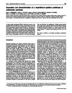
Figure 1. Protocol to construct expression vectors containing celJV or/and celG.
Cloning of cellulase genes Primer pairs of celJV-E (5′–ggaattcgaaagtactttgtggcgcgg–3′, underlined is EcoRI site) and celJV-X (5′– tctcgaggtgagagcttgcgcattcct–3′, underlined is XhoI site) were designed according to ACI11355 in GenBank to amplify Klebsiella sp. strain IV-3 1,4-endoglucosidase gene. The primers pairs of celG-BS (5′–gagatctgagctcgcccctgcgtgctttagtg–3′, single-underlined is BglII site and double-underlined is SacI site) and celG-SH (5′– tgtcgacaagcttgcgctgatcctgtttcgcc–3′, single-underlined is SalI site and double-underlined is HindIII site) were designed according to ACI07560 in GenBank to amplify Klebsiella sp. strain IV-3 glucoside hydrolase family 8 gene. The amplified genes were named as celJV and celG, respectively. They were inserted into pMD19-T simple, and the recombinant plasmids were named as pT-JV and pT-G (Figure 1). Expression of recombinant enzymes in E. coli According to the protocol (Figure 1), a number of vectors
expressing celJV or/and celG were constructed, including those on the basis of secretory vector pET-22b(+) for p22-JV (celJV), p22-G (celG), and p22-JV-G (celJV and celG), those based on intracellular vector pETDuet-1 for pDin-JV (celJV) and pDin-JV-G (celJV and celG), and those based on pDout, which combined the pelB leader of pET-22b(+) with pETDuet-1, for pDout-JV (celJV) and pDout-JVG (celJV and celG). After verified by sequencing, they were introduced into E. coli BL21(DE3). The engineered E. coli were inoculated into 2 × TY medium (16 g.l-1 tryptone, 10 g.l-1 yeast extract, 5 g.l-1 NaCl, pH 7.2) supplemented with 100 μg.ml-1 ampicillin and cultured at 180 r.min 1 , 37°C. When A600 reached 0.6-0.8, isopropyl β-D-1thiogalactopyranoside (IPTG) with a final concentration of 1 mM was added and then continued to incubate for 12 h. Twenty ml cell cultures were collected by centrifugation (4°C, 5000 ×g, 10 min), resuspended in a sodium phosphate buffer (pH 7.4) and kept for further analysis. For SDS-PAGE analysis, lysozyme with a final concentration of 0.3 mg.ml-1 was added to the resuspension. After incubating for 30 min at room temperature, the cell pellets was then put into ultrasonic for lyses. The supernatant and precipitate proteins were
2060
Afr. J. Microbiol. Res.
harvested respectively by centrifugation (4°C, 12,000 ×g, 15 min) and then subjected for 12.5% SDS-PAGE analysis. Analysis of degradation capability and enzymatic activity The resuspensions were also directly smashed using ultrasonic disruptor and then centrifugated (4°C, 12,000 ×g, 15 min) to obtain the supernatant as crude enzyme solution. After determined the protein content using Coomassie brilliant blue method, 20 μl solution with protein concentration of 5 μg.μl-1 was dipped onto CMC-Na plates (the proliferation medium supplemented with 15 g.l 1 decarburization agar strip, pH7.0). After incubation for 24 h at 37°C, the plates were put at 60°C for 1 h, then stained with 0.2% (w/v) Congo red for 15 min and destained with 1 M NaCl solution (Ng et al., 2009). Two ml solution with protein concentration of 1 μg.μl-1 was transferred into 8 ml Mandel’s inorganic salt solution containing FINE pieces of 1 × 4 cm decarbonization quantitative filter paper (Xinhua Paper Co., Ltd, Hangzhou, China). After culturing for three days at 30°C and 150 r.min -1, the filter papers were weighted. The specific activity was also determined by adding 100 μg protein extract into 4 ml CMC-Na liquid medium and cultured for 12 h at 180 r.min -1, 37°C. The activity was evaluated based on the amount of reducing sugar produced in the medium. The amount of reducing sugar was determined by DNS method (Lin et al., 2011). One unit (U) is defined as the amount of enzyme needed to produce 1 µg of reducing sugar per hour.
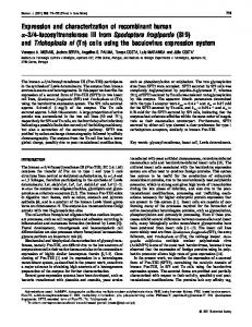
RESULTS AND DISCUSSION Screening for cellulolytic strains To isolate cellulolytic strains with high degrading activity, 48 colonies from samples I-VII that cleared the proliferation medium within 4 days were transferred onto the Congo red plates. Fifteen of them with larger degrading zones were selected. Most of the colonies did not have hypha, indicating that they are bacteria. As Cellulomonas sp. is one of the most intensively studied genera to produce cellulases (Kuhad et al., 2010), it was used as a bench mark in the study. After screened again on Congo red plates, 7 strains that showed a comparable or higher cellulose degrading capability than C. fimi were screened out. Their degrading zones diameter and the diameters ratio of degrading zones and colonies were shown in Figure 2. IV-3 was the strain that had highest cellulose degrading capability. Phylogenetic analysis of the isolated cellulolytic bacteria The genomic DNAs of the isolated bacteria were amplified using 3 pairs of primers. 27F/1522R was found optimal to amplify IV-3, V-1, VI-1, VII-3 and VII-7, while 27F/1492R to VII-6 and VII-8. Sequencing revealed that IV-3, VII-3 and VII-7 is 1532 bp, V-1 and VI-1 is 1530 bp, while VII-6 and VII-8 is 1504 bp. BLAST alignment showed that the isolated bacteria belong to either Klebsiella or Acinetobacter. After downloading the existing 16S rRNA gene sequences of 19
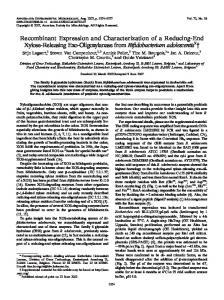
Klebsiella, 19 Acinetobacter, and the known outgroups of 5 Enterobacteriacea, a phylogenetic tree along with the isolated strains was built (Figure 3). V-1 and VI-1 were classified into Acinetobacter oleivorans as they clustered with A. oleivorans DR1 with bootstrap values higher than 50%, while VII-3, VII-6 and VII-8 were classified into K. pneumonia as they clustered with K. pneumoniae sctccT53, K. pneumonia 342 and K. pneumoniae HUBIV-004, respectively, all having bootstrap values higher or equal to 50%. However, IV-3 was only roughly classified into Klebsiella sp. as it located between K. pneumoniae or K. variicola with the bootstrap values less than 50%. VII-7 branched out alone and clustered with some strains of the Enterobacteriaceae family. It could be roughly classified into E. bacterium. The phylogenetic location of IV-3 between K. pneumoniae and K. variicola is consistent with the longlasting controversy in classification of the two species. It was proposed that K. variicola was a new species in Klebsiella sp., and genetically isolated from K. pneumonia (Rosenblueth et al., 2004). Some strains that initially identified as K. pneumonia were actually K. variicola (Brisse and van Duijkeren, 2005; Rosenblueth et al., 2004). K. pneumonia could be classified into four groups as KpI, KpII-A, KpII-B and KpIII (Alves et al., 2006). K. variicola possibly belong to KpIII (Rosenblueth et al., 2004), and is a member of K. pneumonia complex (Søgaard et al., 2007). No effective methods have been proposed to differentiate them in practice so far (Westbrook et al., 2000), though several indicators such as ribitol fermentation capability, a test system containing 18 biochemical indicators (Alves et al., 2006; Hansen et al., 1998). We can also not identify IV-3 accurately based on 16S rRNA gene sequences in this study. Further research is still needed to identify IV-3 accurately. Cloning of the cellulase genes from Klebsiella sp. strain IV-3 A 1116 bp fragment named celJV were amplified using celJV-E/celJV-X, while a 1023 bp fragment named celG was amplified using primer pairs of celG-BS/celG-SH. Sequencing revealed that there are 13 bases difference but no amino acid difference between celJV and the existing 1,4-endoglucosidase gene of K. pneumoniae 342 (Accession No. ACI11355). Similarly, there are 9 bases difference while only one amino acid difference between celG and the existing glucoside hydrolase family 8 gene of K. pneumoniae 342 (accession No. ACI07560). Therefore, celJV and celG were considered as the 1,4endoglucosidase gene and glucoside hydrolase family 8 gene, respectively. Compared with C. fimi, the cellulase genes from Klebsiella sp. strain IV-3 has superiority in two aspects. First, they showed a higher degrading ability, and the gene function is stronger. Second, both genes are approximately 1 kb in length, while C. fimi cenB encoding
Li et al.
2061
Figure 2. Screening for cellulolytic strains that had a comparable or higher cellulose degrading capability than Cellulomonas fimi. Thirty colonies from each strain were randomly chosen to estimate the cellulose degrading capability according to the diameters (mm) of degrading zones (Φzone) (A) and the ratio of Φzone to diameters of colony (Φcolony) (B).
endoglucanase and cbhB encoding a second exocellobiohydrolase is over 3 kb with a high GC content (Meinke et al., 1991; Shen et al., 1995). So they are shorter than C. fimi and it is easier for DNA manipulation. Expressing cellulase genes of Klebsiella sp. strain IV3 in E. coli A series of expression vectors were transformed into E. coli BL21(DE3). SDS-PAGE analysis showed that the recombinant protein(s) could be expressed as soluble form (in supernatant) in pETDuet-1 (Figure 4A), but mainly as inclusion bodies (in precipitate) in pET-22b(+) (data not shown) and in pDout, which combined the pelB leader of pET-22b(+) with pETDuet-1 (Figure 4B). As a signal peptide, pelB leader helps protein translocate into
bacterial periplasm (Lei et al., 1987; Ng et al., 2013). However, the situation is not happened in the study, which was possibly owing to the multiple disulfide bonds or highly hydrophobic domains of the proteins (Gao et al., 2013; Lei et al., 1987; Ng et al., 2013). Furthermore, the efficiency of pDin-JV expressing recombinant CelJV was higher than that of pDin-JV-G expressing CelJV/CelG (Figure 4A). Cellulose degradation capability of the recombinant enzyme(s) from the engineered E. coli expressing celJV or/and celG The recombinant enzyme(s) first evaluated the cellulose degradation capability on Congo red plates (Figure 5A). Intracellular pDin-JV-G that expressed two genes had
2062
Afr. J. Microbiol. Res.
Figure 3. Phylogenetic tree of the isolated strains based on 16S rRNA gene information. Italics on right are the Latin Names of registered species. A, Acinetobacter; K, Klebsiella; M, Moraxella; E, Enterobacteriaceae in E. Bacterium and Escherichia in E. coli. The roman figures after the Latin names are the number of strains. Bracket showed the registered number in GenBank. M. bovoculi, M. lacunata, E. bacterium and E. coli are outgroups. Figures on the branches are the Bootstrap values, and they are believable if higher than 50%.
Li et al.
2063
Figure 4. 12.5% SDS-PAGE analysis of the engineered E. coli. expressing celJV or/and celG. sup: supernatant. ppt: precipitate. Marker: Protein molecular weight standards, from top to bottom is 97.2, 66.4, 44.3, 29, 20.1 and 14.3 kDa. Arrows indicated the recombinant celJV bands without/with the signal peptide expressed in pETDuet-1 (43.2 kDa) and pDout (44.6 kDa), respectively.
Figure 5. The cellulose degrading capability of the recombinant enzyme(s) from the engineered E. coli. expressing celJV or/and celG. A and data B of cellulose degrading capability of the recombinant enzyme(s) from the engineered E. coli. strains evaluated on CMC-Na Congo red plates. For the analysis, the cell pellets were smashed and then centrifugated to obtain the crude enzyme solution. Twenty μl of solution with protein concentration of 5 μg.μl -1 was dipped onto CMC-Na plates.
largest degrading zone (10.6 mm) (Figure 5B), which corresponded to 2.6-fold of Klebsiella sp. strain IV-3. Intracellular pDin-JV that expressed single gene celJV
had the second largest degrading zone (7.5 mm), which corresponded to 1.9-fold of IV-3. Secretory pDout-JV-G that expressed two genes had no degrading zone at all,
2064
Afr. J. Microbiol. Res.
Figure 6. The enzymatic activity of the recombinant enzyme(s) from the engineered E. coli. expressing celJV or/and celG. A. The filter papers decomposition capability of the engineered E. coli strains. B. The cellulose specific activity of the recombinant enzyme(s) from the engineered E. coli strains, which was evaluated by the amount of reducing sugar produced in CMC-Na liquid Mandel’s solution. One unit is defined as the amount of enzyme needed to produce 1 µg of reducing sugar per hour.
while the rest of vectors had comparable degrading ability to IV-3. Even though pDin-JV-G expressed CelJV/CelG with a lower efficiency, it still had a higher degrading capability than pDin-JV, indicating that the concurrence of CelJV and CelG is more efficient to cellulose degradation than CelJV alone, which suggested that enzymatic synergism is helpful for cellulose degradation. The enzymatic activity of the recombinant enzyme(s) from the engineered E. coli expressing celJV or/and celG As the recombinant enzyme(s) from pETDuet-1 derived vectors had higher cellulose degrading capability than that of pET-22b(+) on Congo red plates, their degradation capability on decarbonization filter papers were also evaluated (Figure 6A). pDin-JV-G decreased the most
amount of filter paper (11.1%), which corresponded to 2.1-fold of Klebsiella sp. strain IV-3 followed by pDin-JV (7.1%), which corresponded to 1.3-fold of IV-3. pDout-JV decreased the same amount of filter paper as IV-3. pDout-JV-G decreased the least amount of filter paper (1.4%), which showed no difference as the negative control of pETDuet-1. However, the filter paper weights of pETDuet-1 and pDout-JV-G were also reduced to some extent (Figure 6A). This suggested that autolysis of filter strips occurred in Mandel’s culture medium, which may interfere with the determination. Furthermore, it was not easy to completely wash away the impurities such as bacteria and ion precipitations attached on the filter papers after shake-culturing for 3 days, which may also affect the determination. That is why the large statistical deviations still existed although we have performed the experiment for several times. Therefore, we think that Congo red CMC-Na degradation is a more simple and
Li et al.
effective method to estimate the degradation capability of cellulolytic bacteria or recombinant enzyme(s). The specific activity of the recombinant cellulase(s) expressed in pETDuet-1 and its derived vectors were also determined (Figure 6B). pDin-JV-G expressing two cellulases simultaneously had the highest specific activity (30.2 U.mg-1 protein), which corresponded to 28-fold of Klebsiella sp. strain IV-3. No much difference was found -1 between pDin-JV (22.2 U.mg protein) and pDout-JV -1 (18.3 U.mg protein) that expressed single gene respectively, while pDout-JV-G had the lowest specific -1 activity (2.5 U.mg protein). The specific activity of the recombinant enzymes in engineered E. coli corresponded well with their cellulose degrading capability. The recombinant enzyme(s) expressed in engineered E. coli in the study showed a higher degradation activity to soluble cellulose (CMC-Na) than to crystal cellulose (filter paper). This result is consistent with that ethanol producing recombinant E. coli FBR5 degraded the filter paper with a much lower activity (34 U.ml-1) than that of CMC-Na cellulase (1011 U.ml-1) and xylanase (1149 U.ml1 ) (Saha et al., 2011). Therefore, how to improve the degradation capability of cellulose on crystalline is still a big problem and need further researches. Conclusion In this paper, we have isolated seven nature strains that have a comparable or higher cellulose degrading capability than Cellulomonas fimi. Phylogenetic analysis indicated that they are all bacteria and belong to either Acinetobacter or Klebsiella. After cloning the cellulase genes encoding 1,4-endoglucosidase (celJV) and glucoside hydrolase family 8 (celG) from Klebsiella sp. strain IV-3 that had the highest cellulose degrading capability, they were inserted separately or simultaneously into several types of vectors. Our data indicates that intracellular pETDuet-1 was the appropriate vector for expressing these cellulase genes, while the enzymatic synergism is helpful for cellulose degradation. Conflict of interests The authors did not declare any conflict of interest. ACKNOWLEDGEMENTS This work was supported by the grants of the State Key Laboratory of Medicinal Chemical Biology (201503017), the Key Program of the Natural Science Foundation of Tianjin (12JCZDJC22900; 14JCZDJC34100), Tianjin International Science and Technology International Cooperation Project (09ZCGHHZ00500), and the Funds for National Basic Science Personnel Training (J1103503).
2065
REFERENCES Adachi W, Sakihama Y, Shimizu S, Sunami T, Fukazawa T, Suzuki M, Yatsunami R, Nakamura S, Takénaka A (2004). Crystal structure of family GH-8 chitosanase with subclass II specificity from Bacillus sp. K17. J. Mol. Biol. 343:785-795. Alves MS, Dias RC, de Castro AC, Riley LW, Moreira BM (2006). Identification of clinical isolates of indole-positive and indole-negative Klebsiella spp. J. Clin. Microbiol. 44: 3640–3646. Anand AAP, Vennison SJ, Sankar SG, Prabhu DI, Vasan PT, Raghuraman T, Geoffrey CJ, Vendan SE (2010). Isolation and characterization of bacteria from the gut of Bombyx mori that degrade cellulose, xylan, pectin and starch and their impact on digestion. J. Insect Sci. 10:1-20. Brisse S, van Duijkeren E (2005). Identification and antimicrobial susceptibility of 100 Klebsiella animal clinical isolates. Vet. Microbiol. 105: 307–312. Gao B, Zhangsun D, Wu Y, Lin B, Zhu X, Luo S (2013). Expression, renaturation and biological activity of recombinant conotoxin GeXIVAWT. Appl. Microbiol. Biotechnol. 97:1223–1230. Gilbert HJ (2010). The biochemistry and structural biology of plant cell wall deconstruction. Plant Physiol. 153:444–455. Grice EA, Snitkin ES, Yockey LJ, Bermudez DM, NISC Comparative Sequencing Program, Liechty KW, Segre JA (2010). Longitudinal shift in diabetic wound microbiota correlates with prolonged skin defense response. Proc. Natl. Acad. Sci. USA 107:14799-14804. Hansen DS, Gottschau A, Kolmos HJ (1998). Epidemiology of Klebsiella bacteraemia: a case control study using Escherichia coli bacteraemia as control. J. Hosp. Infect. 38:119-132. Johnson JL (1994). Similarity analysis of rRNAs. In: Gerhardt P, Murray RGE, Wood WA, Krieg NR (eds) Methods for General and Molecular Bacteriology: American Society for Microbiology, Washington, DC. pp. 683-700. Kuhad RC, Gupta R, Khasa YP (2010). Bioethanol production from lignocellulosic biomass: an overview. In: Lal B (ed) Wealth from Waste, Teri Press, New Delhi. pp. 53-106. Lei SP, Lin HC, Wang SS, Callaway J, Wilcox G (1987). Characterization of the Erwinia carotovora pelB gene and its product pectate lyase. J. Bacteriol. 169:4379-4383. Lin CW, Wu CH, Tran DT, Shih MC, Li WH, Wu CF (2011). Mixed culture fermentation from lignocellulosic materials using thermophilic lignocellulose-degrading anaerobes. Process Biochem. 46:489-493. Liu SL, Chen WZ, Liu G, Xing M (2011). Enhanced secreting expression and improved properties of a recombinant alkaline endoglucanase cloned in Escherichia coli. J. Ind. Microbiol. Biotechnol. 38:855-861. Lynd LR, Weimer PJ, van Zyl WH, Pretorius IS (2002). Microbial cellulose utilization: fundamentals and biotechnology. Microbiol. Mol. Biol. Rev. 66:506-577. Maki M, Leung KT, Qin W (2009). The prospects of cellulase-producing bacteria for the bioconversion of lignocellulosic biomass. Int. J. Biol. Sci. 5:500-516. Meier A, Kirschner P, Bange F, Vogel U, Bö1tger EC (1994). Genetic alterations in streptomycin-resistant Mycobacterium tuberculosis: mapping of mutations conferring resistance. Antimicrob. Agents Chemother. 38:228-233. Meinke A, Braun C, Gilkes NR, Kilburn DG, Miller Jr RC, Warren RA (1991). Unusual sequence organization in CenB, an inverting endoglucanase from Cellulomonas fimi. J. Bacteriol. 173: 308–314. Ng IS, Chi X, Wu X, Bao Z, Lu Y, Chang JS, Ling X (2013). Cloning and expression of Cel8A from Klebsiella pneumoniae in Escherichia coli and comparison to cel gene of Cellulomonas uda. Biochem. Eng. J. 78:53-58. Ng IS, Li CW, Yeh YF, Chen PT, Chir JL, Ma CH, Yu SM, Ho TD, Tong CG (2009). A novel endo-glucanase from the thermophilic bacterium Geobacillus sp. 70PC53 with high activity and stability over a broad range of temperatures. Extremophiles 13:425-435. Ogura J, Toyoda A, Kurosawa T, Chong AL, Chohnan S, Masaki T (2006). Purification, characterization, and gene analysis of cellulase (Cel8A) from Lysobacter sp. IB-9374. Biosci. Biotechnol. Biochem. 70:2420-2428. Percival Zhang YH, Himmel ME, Mielenz JR (2006). Outlook for cellulase improvement: screening and selection strategies. Biotechnol.
2066
Afr. J. Microbiol. Res.
Adv. 24:452-481. Rosenblueth M, Martínez L, Silva J, Martínez-Romero E (2004). Klebsiella variicola, a novel species with clinical and plant-associated isolates. Syst. Appl. Microbiol. 27:27-35. Saha BC, Nichols NN, Qureshi N, Cotta MA (2011). Comparison of separate hydrolysis and fermentation and simultaneous saccharification and fermentation processes for ethanol production from wheat straw by recombinant Escherichia coli strain FBR5. Appl. Microbiol. Biotechnol. 92:865-874. Shen H, Gilkes NR, Kilburn DG, Miller Jr RC, Warren RA (1995). Cellobiohydrolase B, a second exo-cellobiohydrolase from the cellulolytic bacterium Cellulomonas fimi. Biochem. J. 311:67-74. Søgaard M, Hansen DS, Fiandaca MJ, Stender H, Schønheyder HC (2007). Peptide nucleic acid fluorescence in situ hybridization for rapid detection of Klebsiella pneumoniae from positive blood cultures. J. Med. Microbiol. 56:914-917. Sukharnikov LO, Cantwell BJ, Podar M, Zhulin IB (2011). Cellulases: ambiguous nonhomologous enzymes in a genomic perspective. Trends Biotechnol. 29:473-479.
Van Dyk JS, Pletschke BI (2012). A review of lignocellulose bioconversion using enzymatic hydrolysis and synergistic cooperation between enzymes-factors affecting enzymes, conversion and synergy. Biotechnol. Adv. 30:1458-1480. Vuong TV, Wilson DB (2009). Processivity, synergism, and substrate specificity of Thermobifida fusca Cel6B. Appl. Environ. Microbiol. 75:6655-6661. Waghmare PR, Kshirsagar SD, Saratale RG, Govindwar SP, Saratale GD (2014). Production and characterization of cellulolytic enzymes by isolated Klebsiella sp. PRW-1 using agricultural waste biomass. Emir. J. Food Agric. 26:44-59. Westbrook GL, O’Hara CM, Roman SB, Miller JM (2000). Incidence and identification of Klebsiella planticola in clinical isolates with emphasis on newborns. J. Clin. Microbiol. 38:1495-1497.