limb mesenchyme, skeletogenesis, cellular condensation. SUMMARY. Expression and functional involvement of N-cadherin in embryonic limb chondrogenesis.
177
Development 120, 177-187 (1994) Printed in Great Britain The Company of Biologists Limited 1994
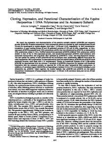
Expression and functional involvement of N-cadherin in embryonic limb chondrogenesis Steven A. Oberlender and Rocky S. Tuan* Department of Orthopaedic Surgery and Department of Biochemistry and Molecular Biology, Thomas Jefferson University, Philadelphia, PA, 19107, USA *Author for correspondence
SUMMARY Cell adhesion molecules have been shown to be important mediators of morphogenesis and pattern formation. In this study, we have shown that N-cadherin is expressed in a specific spatiotemporal manner in the developing limb bud during chondrogenesis in vivo and in cultured limb mesenchyme in vitro. The time period of maximal expression of N-cadherin corresponds to the period of active cellular condensation, an event believed to be a necessary prerequisite for chondrogenic differentiation. To directly assess the functional involvement of N-cadherin in cellular condensation, we have examined the effects of perturbing Ncadherin activity on both cell aggregation and chondrogenesis using NCD-2, a rat monoclonal antibody directed against the binding region of N-cadherin. Non-immune rat IgG was used as a control. Our results show that functional N-cadherin is necessary for chondrogenesis to proceed both in vivo and in vitro. Limb mesenchymal cells exhibited characteristic Ca2+-dependent cell aggregation in suspension, which was inhibited in the presence of exogenous NCD-2. In micromass cultures of limb mesenchymal cells,
NCD-2 inhibited overt chondrogenesis in a dose-dependent manner. Furthermore, NCD-2 inhibition of chondrogenesis in micromass cultures was time-dependent, suggesting that N-cadherin is crucially involved during the latter half of the first 24 hours of culture, a time period most likely corresponding to active cellular condensation. NCD-2 also significantly influenced limb development when injected into embryonic limb buds in vivo. In addition to significant inhibition of chondrogenesis and developmental delays, gross developmental deformities and perturbation of overall pattern formation were also observed. Taken together, these results demonstrate that N-cadherin is functionally required in mediating the cell-cell interactions among mesenchymal cells important for chondrogenesis in micromass culture in vitro and in the intact limb bud in vivo.
INTRODUCTION
solubilized cadherins (Ozawa et al., 1989; Peifer et al., 1992). The three catenins, α, β, and γ, are closely related to vinculin, armadillo, and plakoglobin, respectively. The expression of a cell adhesion molecule during development usually coincides with substantial morphogenetic events. Chondrogenesis in the embryonic chick limb bud represents an interesting model system, in view of the early cellular condensation phase (Fell and Canti, 1934; Thorogood and Hinchliffe, 1975; Ede, 1983; Newman et al., 1985). At around stage 23/24 of development (Hamburger and Hamilton, 1951), mesenchymal cells of the limb bud core come into close apposition with one another (i.e. condensation) and subsequently differentiate into the first cartilaginous structure in the limb. This is accompanied by the synthesis and secretion of collagen type II and the protein core of the large chondroitin-rich proteoglycan known as aggrecan. The close apposition of the mesenchymal cells could result from or facilitate the formation of cell-cell interactions necessary for chondrogenesis. Interestingly, Widelitz et al. (1993) recently reported that N-CAM, a neural Ca2+-independent cell adhesion molecule, was
Cell-cell adhesion and the events that subsequently take place between interacting cells, play important functional roles in biological processes (Grunwald, 1991). Cell adhesion molecules are recognized as being important mediators of both embryonic developmental and morphogenetic events, as well as stabilizers of adult tissue structures. The two main families of cell adhesion molecules are the Ca2+-dependent cadherin superfamily, and the Ca2+-independent immunoglobulin superfamily (CAMs; Edelman and Crossin, 1991; Takeichi, 1990, 1991; Geiger and Ayalon, 1992). Members of both of these families of cell adhesion molecules mediate cell-cell adhesion in a homophilic manner. The classical cadherins, such as Ncadherin (Hatta et al., 1988) and E-cadherin (Nagafuchi et al., 1987), are transmembrane glycoproteins of about 120×103 Mr that require interaction with the cytoskeleton in order to mediate extracellular binding. Such interactions are likely to involve the catenins, a group of at least three proteins (α, β, and γ) which both co-localize and co-immunoprecipitate with
Key words: cell-cell interaction, morphogenesis, extracellular matrix, limb mesenchyme, skeletogenesis, cellular condensation
178
S. A. Oberlender and R. S. Tuan
expressed in condensing aggregates of chick limb mesenchymal cells both in vivo, and cultured as micromass in vitro. In addition, these investigators demonstrated that inhibiting NCAM mediated cell-cell adhesion with specific antibodies against N-CAM, resulted in the inhibition of overt chondrogenesis in vitro. In considering how other cell adhesion processes may be functionally involved in limb bud mesenchymal condensation and chondrogenesis, it is noteworthy that San Antonio and Tuan (1986) previously demonstrated that exogenous Ca2+ significantly stimulates chondrogenesis in limb mesenchymal micromass cultures. This effect acts predominantly during the first 24 hours of culture, when mesenchymal cells are actively condensing (San Antonio and Tuan, 1986; Evans and Tuan, 1988; Tuan, 1991). Additionally, Bee and Von der Mark (1990) have also described a Ca2+-dependent cell adhesion phenomenon in isolated limb mesenchymal cells. These observations suggest that Ca2+ may affect or influence the cell-cell interactions in mesenchymal condensation, perhaps involving cadherins. In this study, we have investigated the possible involvement of cadherins in chondrogenesis, particularly during mesenchymal condensation. Specifically, we have focused on neural cadherin (N-cadherin) because of its transient expression in various mesenchymal populations during embryonic development (Hatta and Takeichi, 1986; Hatta et al., 1987). To test this postulate, we have first identified N-cadherin expression in limb mesenchyme, and secondly, perturbed N-cadherin mediated cell-cell adhesion in embryonic limbs in vivo and in cultured limb mesenchyme in vitro by employing specific monoclonal antibodies that recognize an epitope in the binding region of N-cadherin. Our results show that N-cadherin is expressed in populations of mesenchymal cells that are undergoing active cellular condensation, and is absent from differentiated cartilage. In addition, perturbation of N-cadherin binding function significantly inhibits both limb mesenchymal Ca2+-dependent aggregation, and chondrogenesis in vitro and in vivo. MATERIALS AND METHODS Chick embryos and shell-less embryo culture Fertilized White Leghorn chicken eggs (Truslow Farms, Chestertown, MD) were incubated at 99°F in a humidified egg incubator for the desired period of time. For in vivo perturbation of N-cadherin, shellless, cultured chick embryos were used as previously described by Tuan (1980). Briefly, after 3 days of incubation in ovo, chick embryos were placed without the eggshell into ringstands lined with plastic kitchen wrap, covered with a Petri dish lid, and maintained in a humidified incubator at 37.5°C with constant air flow. This method permitted easy access to the limb bud and continuous observation of development. Limb mesenchymal cell isolation and differential protease sensitivity This isolation procedure was adapted from that described by Ahrens et al. (1977) and modified by San Antonio and Tuan (1986). Briefly, limb buds were dissected from chick embryos at stage 23/24, cut, and enzymatically dissociated in Ca2+-, Mg2+-free saline G (CMFSG) containing 0.1% trypsin (Type II, Sigma) and 0.1% collagenase (Worthington). Isolated cells were resuspended in CMFSG + 10% fetal calf serum (FCS), counted with a hemocytometer in the presence of trypan
blue (over 95% dye exclusion), adjusted to the appropriate plating density (routinely between 10-12×106 cells/ml), plated in a 100 µl drop per well on 6 well tissue culture dishes (Corning), allowed to attach, and then covered with 3 ml of culture medium (CM) containing Ham’s F-12, 10% FCS, 0.2% chick embryo extract, and 1% penicillin-streptomycin (CM). These cells were allowed to recover in culture for 15-18 hours, and then collectively dissociated with 0.01% trypsin in the presence of either 1.5 mM CaCl2 (TC) or 1 mM EGTA (TE) as described by Takeichi (1977). Isolated cells were resuspended in HCMFSG and counted. In certain experiments, the HCMFSG was supplemented with additional reagents as described below. Analysis of cell aggregation TC- (or TE-) treated cells were resuspended in HCMFSG (±2 mM CaCl2), and supplemented with either control rat IgG (Sigma) or purified NCD-2 antibodies. To initiate cell aggregation, 2-2.5×10⁵ cells were placed into the well of a 24-well plate previously coated with 1% bovine serum albumin (BSA), covered with 1 ml of HCMFSG with or without the addition of antibodies and 2 mM CaCl2, and then incubated at 37°C on a gyrating shaker at 80-100 rpm. An aliquot from each well was removed at various time points and counted with a Coulter Counter (Model ZM) equipped with a 100 µm aperture. The degree of aggregation was determined by dividing the number of particles counted at each time point by the number of particles at time zero. Micromass cultures A 10 µl drop of TC cells at 8-15×106 cells/ml was plated per well on a 24 well plate and allowed to attach for 1.5-2 hours at 37°C and 5% CO2, after which 1 ml of CM was added to each well. For most experiments, the medium was replaced with fresh medium every 24 hours. The effects of various agents were tested by direct additions to the culture medium, and cells cultured for the desired period of time. For some experiments, FCS was heated at 56°C for 30 minutes prior to use to inactivate the complement components. Chondrogenesis was quantified based on sulfate incorporation ([35S]Na2SO4, 2.5 µCi/ml, ICN) and staining with Alcian blue 8-GX (Sigma) at pH 1.0 (San Antonio and Tuan, 1986). N-cadherin monoclonal antibodies The NCD-2 rat hybridoma, a kind gift of Dr M. Takeichi, produces a monoclonal antibody that specifically recognizes the extracellular binding region of N-cadherin and effectively neutralizes its normal binding functions (Hatta and Takeichi, 1986). IgG antibodies were isolated from the NCD-2 culture supernatant by (NH4)2SO4 precipitation and bulk phase DEAE-Sephadex ion-exchange chromatography (Harlow and Lane, 1988), diluted to known concentrations, and stored at −20°C until use. Immunohistochemical localization of N-cadherin Chick embryonic limb buds Limb buds were excised from embryos at various stages of development (stages 18-30), fixed in 4% paraformaldehyde-PBS at 4°C for 1-2 hours, rinsed in Tris-buffered saline with 2 mM CaCl2 (TBS/Ca) and then frozen-embedded in Tissue-Tek OCT compound (Miles). Cryosections (8 µm) were placed onto Neoprene coated slides for immunohistochemical staining (Lagunowich and Grunwald, 1989). Briefly, after blocking with 5% normal goat serum (NGS) and washing, the sections were incubated with undiluted NCD-2 culture medium for 30 minutes at room temperature. Controls were incubated in either unconditioned culture medium or TBS/Ca. After incubation, the slides were washed in TBS/Ca, blocked a second time in 5% NGS, and then incubated with fluorescein-conjugated goat anti-rat antibody (Zymed) at a dilution of 1:1000 for 30 minutes at room temperature. The sections were viewed with an Olympus BH-2 microscope equipped with epifluorescence and Nomarski differential interference optics.
N-cadherin and chondrogenesis Micromass limb mesenchyme cultures Micromass cultures plated at 11×106 cells/ml on 8-chamber glass Lab-Tek slides (Nunc) were processed for immunohistochemical staining as described above with the following modifications: blocking with 10% NGS, incubation with purified NCD-2 IgG (5 mg/ml) at 50 µg/ml, and secondary antibody at a dilution of 1:4000. Immunoblot analysis of N-cadherin The micromass cell culture samples were solubilized in 60 mM Tris (pH 6.8) and 2% SDS and protein concentration determined (micro BCA kit, Pierce Chemical). Samples were adjusted to equal protein concentrations in a reducing sample buffer and analyzed by electrophoresis in a 7.5% polyacrylamide SDS gel. The proteins were electrophoretically transferred to nitrocellulose and incubated with purified NCD-2 antibodies (50 µg/ml) followed by biotin-conjugated goat anti-rat antibody (Zymed) at a dilution of 1:500. After washing, the blots were incubated with streptavidin-alkaline phosphatase and developed histochemically with Nitro blue tetrazolium (NBT) and 5bromo-4-chloro-3-indolyl phosphate (BCIP). Injection of limb bud with NCD-2 antibodies in vivo The limb buds of shell-less chick embryos at stages 22-24 were injected with antibodies. To visualize the efficacy of injection, the vital dye, Nile Blue sulfate, was added to solutions of both antibodies used, control rat IgG and NCD-2 (10 mg protein/ml). Because the embryos were always situated on top of the yolk with mostly only its right side exposed, and it was undesirable to disrupt any vital embryonic vasculature, only one of the limbs, either the fore- or hindlimb was injected with NCD-2, and the other limb with rat IgG at the same concentration. The injections were carried out using a pulled 10 µl capillary tube attached to a microsyringe (Drummond), mounted on a micromanipulator. The volume injected into the limbs was typically between 0.5-1.0 µl (i.e. 5-10 µg of antibodies), and each embryo always received the same volume in each of its limbs. After the injections were completed, the embryos were placed back into the incubator and allowed to develop, uninterrupted. Whole-mount Alcian blue staining of chick embryos Two days after injection, the embryos were removed and fixed in PBS-buffered 10% formalin and subjected to whole-mount Alcian blue staining (Kimmel and Trammell, 1981). Briefly, the embryos were stained in acid-alcohol Alcian blue for 17 hours, cleared and macerated in 2% KOH, hardened in 50% glycerin, and stored in 100% glycerin. The limbs were then removed from the embryos and viewed and photographed using a Wild stereo microscope. All comparisons were made between the injected limb and the contralateral, uninjected limb. In addition, the rat IgG injected limbs served as both a control for possible effects due to the injection buffer or the presence of rat IgG, as compared to the contralateral, uninjected limb.
RESULTS Immunohistochemical localization of N-cadherin expression in limb bud in situ and in micromass cultures in vitro Limb buds from various stages of chick embryos, from prechondrogenic to cartilaginous stages, were examined for the presence of N-cadherin by indirect immunofluoresence. Ncadherin was detected as early as stage 17/18, shortly after limb bud formation and was found to localize in a sparsely scattered pattern around the center of the limb bud (data not shown). At stage 24/25, when cellular condensation was clearly evident, N-cadherin was detected at high levels in the central, aggregating region of the limb bud (Fig. 1A,B). As the limb
179
continued to develop, the condensed, central region began to lose N-cadherin protein expression, while the peripheral mesenchyme began to express N-cadherin. By stage 29/30, mature cartilage was visible and clearly displayed no immunoreactivity for N-cadherin, while the surrounding dense mesenchyme continued to show a high level of N-cadherin expression (Fig. 1C,D). It is noteworthy that some of these N-cadherin-positive cells would eventually contribute to the expanding cartilaginous core by appositional growth. In micromass cultures in vitro, mesenchymal cells aggregate eventually to form discrete, cartilaginous nodules, separated by fibroblasts and myocytes (Ahrens et al., 1977). Immunohistochemistry showed that by 12 hours in culture, N-cadherin was expressed in aggregating (or condensing) regions within the culture, and the surrounding cells displayed no evident signal (data not shown). Expression of N-cadherin in the condensing mesenchyme intensified as a function of culture time and was maximal at around 18 hours (Fig. 1E,F), with the pattern of expression remaining essentially unchanged. During these early stages of in vitro culture, cells were seen to aggregate actively, and the aggregates would eventually give rise to differentiated, cartilaginous nodules, which did not express Ncadherin. The expression and distribution pattern of Ncadherin in micromass cultures in vitro was thus reminiscent of that in the developing limb bud in situ. NCD-2 interferes with Ca2+-dependent cell aggregation To test the hypothesis that Ca2+ stimulates chondrogenesis by promoting mesenchymal cellular aggregation and condensation, we carried out cell adhesion studies employing the experimental design first described by Takeichi (1977). Limb bud mesenchymal cells were first allowed to recover from initial dissociation and isolation for 15-18 hours in culture, and were then dissociated by mild treatment with trypsin in the presence of 1.5 mM Ca2+ (TC) or 1 mM EGTA (TE). It has been shown (Takeichi, 1977; Urushihara et al., 1979) that TE treatment cleaves all cell surface cadherins and renders them non-functional, whereas TC treatment leaves cadherins intact and functional. The TC and TE dissociated cells were subsequently placed in suspension cultures and the kinetics of cell aggregation was analyzed. As shown in Fig. 2, TC cells readily aggregated in the presence of Ca2+ and control rat IgG antibodies, but displayed minimal aggregation in the absence of Ca2+ (Note: the baseline aggregation was most likely due to artifactual adhesion caused by cellular debris and DNA derived from necrotic cells and/or mediated by Ca2+-independent mechanisms). Characteristic of cells capable of Ca2+-dependent cell adhesion, TE treatment substantially lowered their ability to aggregate, the level being similar with or without 2 mM Ca2+ (data not shown). This adhesion profile strongly suggests that the limb mesenchymal cells express cell surface cadherins and that they are functional in mediating cell-cell interactions in a Ca2+-dependent manner. This was confirmed by immunoblot analysis (Fig. 2, inset), showing intact N-cadherin protein in TC cells immediately following proteolytic isolation, but not in TE cells. To conclusively determine whether N-cadherin was indeed mediating this aggregation event, we tested the effect of NCD-2 (see Materials and methods) on the aggregation of TC cells in suspension. As illustrated in Fig. 2, when NCD-2 was added at the same concentration as the control
180
S. A. Oberlender and R. S. Tuan
Fig. 1. Immunohistochemical detection and localization of N-cadherin in the chick embryonic limb bud in situ and in micromass culture of limb mesenchyme in vitro. (A,C,E) Immunofluorescent staining of N-cadherin in limb bud cryosections (A,C) and in fixed micromass cultures (E). (B,D,F) Corresponding views of the sections using Nomarski differential interference optics. (A,B) Stage 24/25 limb sectioned transversely, showing prominent expression of N-cadherin in the condensing mesenchymal (M) core region (arrows). The epithelium (E) demarcates the edge of the limb bud. (C,D) Stage 29/30 limb sectioned longitudinally, revealing mature cartilage (C) which was negative for N-cadherin expression, and dense mesenchyme (M) which was positive for N-cadherin expression. Scale bar, 100 µm for A-D. (E,F) Micromass culture 18 hours into incubation, displaying intense N-cadherin staining in a condensing aggregate of mesenchymal cells (arrow). The immediate surrounding mesenchyme was slightly positive and would most likely be incorporated into the cartilaginous core, while the mesenchyme located further away remained negative for N-cadherin expression. Scale bar, 50 µm for E and F.
antibodies in the presence of the same concentration of Ca2+, aggregation of the TC cells was significantly perturbed. It should be noted that NCD-2 inhibition of TC cell aggregation was less than complete, i.e. to the same level as the Ca2+ deficient group. One explanation is that Ca2+-independent cell adhesion molecules were present and functional on the surfaces of these cells; another possibility is that the NCD-2 antibodies did not completely obliterate N-cadherin action, perhaps because of sub-optimal stoichiometry. NCD-2 inhibits chondrogenesis in vitro To test whether NCD-2 interference of cell aggregation or adhesion could affect subsequent chondrogenic differentiation,
NCD-2 was added at various concentrations to the culture medium of TC cells plated in micromass cultures for the first 24 hours of incubation, corresponding to the time period in which Ca2+ exerted its maximal effect (San Antonio and Tuan, 1986; Tuan, 1991). After 72 hours of incubation, the cultures treated with non-immune control rat IgG antibodies demonstrated roughly the same number of Alcian blue stained cartilaginous nodules, regardless of the concentration of antibody (Fig. 3A). However, increasing concentrations of NCD-2 significantly perturbed nodule formation in a dose-dependent manner in these micromass cultures (Fig. 3A). This profile of NCD-2 dose-dependent inhibition was also observed when chondrogenesis was measured in terms of [35S]sulfate incor-
N-cadherin and chondrogenesis
formation (Fig. 3E), while NCD-2-treated cultures were significantly inhibited in their ability to undergo cellular condensation (Fig. 3D) and subsequent chondrogenesis (Fig. 3F). Complement-mediated toxicity was unlikely to be involved in the observed effects of NCD-2, since replacing FCS with heatinactivated FCS resulted in similar inhibition of chondrogenesis (data not shown).
120
% Single cells
100
*
80
*
60
-Ca
*
+Ca, NCD-2
* 40
+Ca, Rat IgG 20
0 0
20
40
60
80
181
100
Time (min) Fig. 2. NCD-2 inhibition of Ca2+-dependent aggregation of TC cells in suspension culture. Recovered limb mesenchymal cells were dissociated by the TC method (see Materials and methods) and placed into suspension cultures either with (+Ca) or without (−Ca) 2 mM CaCl2 in the medium. The cultures with 2 mM Ca2+ were supplemented with either 300 µg/ml control rat IgG antibodies (Rat IgG) or 300 µg/ml NCD-2 antibodies (NCD-2). TC cells only aggregated to a minimal extent in the absence of Ca2+ (−Ca) as determined by the percentage of single cells remaining in culture at different time points. In the presence of 2 mM Ca2+ and control antibodies (+Ca, Rat IgG), the TC cells aggregated in a typical Ca2+dependent manner. However, in the presence of 2 mM Ca2+ and NCD-2 antibodies (+Ca, NCD-2), the TC cells were significantly perturbed in their ability to aggregate. Values represent the mean ± s.d. of 3 cultures. * Denotes statistical significance (P