patients withB- or T-cell leukemia (n = 26) at various stages of maturation with probes to two additional TCR genes, TCRG and TCRA (encoding the TCR y and a ...
Proc. Natl. Acad. Sci. USA Vol. 83, pp. 8759-8763, November 1986 Medical Sciences
Immunoglobulin and T-cell receptor gene rearrangement and expression in human lymphoid leukemia cells at different stages of maturation (clonallty/lineage/T-lymphocyte differentiation)
MICHAEL P. DAVEY*t, KATHLEEN F. BONGIOVANNI*, WILHELM KAULFERSCH*, THOMAS QUERTERMOUSt, J. G. SEIDMANt, MICHAEL S. HERSHFIELD§, JOANNE KURTZBERG§, BARTON F. HAYNES§, MARK M. DAVIS$, AND THOMAS A. WALDMANN* *Metabolism Branch, National Cancer Institute, National Institutes of Health, Bethesda, MD 20892; tDepartment of Genetics, Harvard Medical School, Boston, MA 02115; §Departments of Medicine and Pediatrics, Duke University Medical Center, Durham, NC 27710; and sDepartment of Medical Microbiology, Stanford University School of Medicine, Stanford, CA 94305
Contributed by Thomas A. Waldmann, July 31, 1986
tiation of a pluripotent stem cell into a mature T cell, a process of DNA rearrangement juxtaposes a Ds with a Jp segment and then a Vq region with this Dl6-J/ junction to assemble the complete variable region gene and permit transcription of mRNA for the complete TCRB peptide. The DNA recombinations generate changes in the location of restriction endonuclease sites that can be used in Southern blot analysis to distinguish the rearranged from the germ-line form ofthis gene. The TCRA gene complex consists of at least 13 families of Va genes and perhaps 50 Ja gene segments in a tandem array present in a greater than 60-kilobase (kb) region on the 5' side of a single Ca gene. Detecting rearrangement of the TCRA locus using Ca probes has been difficult because the restriction enzymes used have sites of action between Ca and the 5' Ja genes and DNA recombinations would not give a new band on Southern blot analysis. A third gene, TCRG, has been identified in T cells that has many properties in common with TCRA and TCRB genes including assembly from gene segments resembling V, J, and C regions, rearrangement, and expression in T cells (2-4). In addition to multiple variable region genes there are at least two J and two C region segments (Fig. 1C) (5, 6). Over the last five years it has been possible to determine clonality, lineage, and stage of maturation of lymphoid malignancies by analyzing the arrangement ofgenes encoding IG (7-9) and TCRB (10). Furthermore, an analysis of IG gene rearrangements in precursor B-cell leukemia cells has revealed a hierarchy of IG gene rearrangements in which heavy chain genes precede light chain genes and K light chain genes precede X light chain genes (7, 11). However, while this type of analysis has permitted the assignment of a population of patients with the same type of leukemia to a specific lineage, ambiguity can still exist in individual cases at the gene level. For example, approximately 30% of patients with acute lymphocytic leukemia (ALL) that have cells that lack T-cell markers and surface IG rearrange both IG and TCRB genes (12). In an attempt to clarify such cases of ambiguous lineage, we have analyzed the arrangement of IG and TCR genes in mature and precursor B- and T-cell leukemia cells at various stages of maturation. IG, TCRG, and TCRB gene rearrangements were studied as well as expression of TCRG, TCRB, and TCRA mRNAs. Leukemia cells from several of the patients exhibited a more immature phenotype than those in previous studies. The spectrum of leukemias studied provid-
ABSTRACT The use of probes to genes (IG and TCRB) encoding immunoglobulins (IG) and the ,3 chain of the T-cell antigen receptor (TCRB), respectively, have become a sensitive means to assess clonality and lineage in lymphoid malignancies. It has become apparent that some individual cases show rearrangements of both IG and TCRB genes. In an attempt to more accurately define cell lineage we have analyzed cells from patients with B- or T-cell leukemia (n = 26) at various stages of maturation with probes to two additional TCR genes, TCRG and TCRA (encoding the TCR y and a chains, respectively), as well as the IG heavy chain joining region (IGHJ) and TCRB genes. On Southern blot analysis, the mature T-cell leukemia cells studied had rearranged TCRG and TCRB while IGHJ remained as in the germ line. The mature B-cell leukemia cells studied had rearranged IGHJ with germ-line TCRG and TCRB. These data suggest that, in the majority of more mature leukemias, cells have rearranged IG or TCR genes but not both. In contrast, cells from five of nine precursor B-cell leukemia patients and cell lines from one of four precursor T-cell leukemia patients had rearranged both IGHJ and TCR genes. TCRG and TCRB mRNAs were expressed in the cells of precursor T- but not B-cell leukemia patients studied. The spectrum of leukemia cells studied within the T-cell series permitted an assessment of the order of TCR gene rearrangements. Two of 13 patients had cells with germ-line TCRG and TCRB, 2 patients had cells with rearranged TCRG alone, and the remainder had cells with rearranged TCRG and TCRB. TCRG and TCRB mRNAs were expressed in precursor T-cell leukemia cells, whereas TCRB and TCRA were expressed in mature T-cell leukemia cells. These results parallel observations from mouse studies on gene expression and support the view of a hierarchy of TCR gene rearrangements in Tlymphocyte ontogeny. TCRG genes are rearranged first, subsequently TCRB genes are rearranged, followed by TCRA gene activation.
The T-cell antigen receptor (TCR) is a 90-kDa heterodimer consisting of 40- to 50-kDa a and A3 subunits (TCRA and TCRB, respectively). Similar to the immunoglobulin (IG) genes, the genes encoding the subunits undergo somatic rearrangements in the course of lymphocyte ontogeny (see ref. 1 for review). The human TCRB gene complex in its germ-line form is composed of discontinuous gene subsegments consisting of multiple variable regions (V3) with duplicate sets of diversity (DO), joining (J), and constant (C3) gene segments (Fig. 1B). At some point during the differen-
Abbreviations: ALL, acute lymphocytic leukemia; ATL, adult T-cell leukemia; C, constant; CLL, chronic lymphocytic leukemia; D, diversity; J, joining; TCR, T-cell receptor; V, variable; a, a subunit of TCR; ,B, /3 subunit of TCR; y, y subunit of TCR; LC, leukemia
The publication costs of this article were defrayed in part by page charge
payment. This article must therefore be hereby marked "advertisement"
cells; kb, kilobase(s). tTo whom reprint requests should be addressed.
in accordance with 18 U.S.C. §1734 solely to indicate this fact.
8759
Proc. Nad. Acad. Sci. USA 83 (1986)
Medical Sciences: Davey et al.
8760
A 16 kb-
Bam HI GL 17
B
Eco RI GL 10 --
24 kb-
W' =
24
JH
Eco RI
M
JHPRIBE
JH PROBE
I
Vp Dp1
'
Eco RI GL 17
C Bam HI GL 17 15 _ kb --= 12.5 kb-
3.4 kb- m
1.5 kb- ai
_Z1 5
,
.
Eco RI EcoRl BarnHI [co RI BamjlH BarnHI Vv
Ji8
I
Ix vl cCi r Ci
12.5 EcoRI EcoRI BarnHI S
Cy PROBE 1kb - ~-Jy PROBE -
Bam HI
Barn HI
Eco RI
-.
J2Ss
C2
-C* o o
G
ed data consistent with a hierarchy of TCR gene rearrangement and expression comparable to the IG gene rearrangement observed in leukemias of the B-cell series. A preliminary report of this work has been presented. 11
MATERIALS AND METHODS Cells. DNA and RNA were extracted from cell lines or from cell suspensions of peripheral blood mononuclear cells from patients grouped according to the following characteristics. Leukemia cells 1-4 (LCs 1-4), from patients 1-4, respectively, with chronic lymphocytic leukemia (CLL), expressed surface IG and had clonal rearrangements of both heavy and light chain IG genes. These patients were considered to have mature B-cell leukemias. LCs 5-9, from patients 5-9, respectively, with ALL, had leukemia cells that reacted with the monoclonal antibody B4, lacked surface IG, failed to react with any T-cell specific monoclonal antibodies, and showed rearrangement of the IGHJ gene. These patients were considered to have precursor B-cell leukemias. LCs 10-13 and 18-21 represent precursor B- and T-cell lines,
ItThis work was presented in part to the national meeting of the American Society for Clinical Investigation, May 2-5, 1986, Washington, D.C., abstr. 656A.
111111
J( 1
MA
V 1' 111
CP 1 D2 JP2
2
0:3-C, PROBE--El 1kb
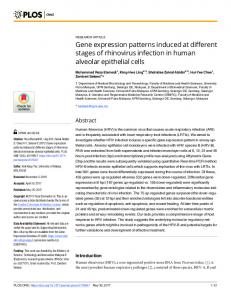
FIG. 1. Restriction maps and examples of Southern blots for the IG and TCR gene probes used in this study. (A) The JH probe hybridizes to a 16-kb EcoRI fragment in the germ-line (lane GL) configuration, here demonstrated by a T-cell line. In this example of the precursor B-cell leukemia line LC 10 (lane 10) at least a D-Jjuxtaposition has occurred on both chromosomes resulting in the loss of the 16-kb germ-line band and the appearance of two new rearranged bands (arrows). (B) Both Cp genes (C01 and Cp2) are found on a germ-line 24-kb BamHI fragment. T-cell depleted peripheral blood mononuclear cells (lane GL) from patient 17 with ATL shows the germ-line band while the leukemia cells (LC 17, lane 17) show evidence of rearrangements on both chromosomes resulting in two new rearranged bands (arrows). (C) The T-cell Cy probe hybridizes to 15-(C01) and 12.5-(Cl2) kb BamHI fragments and the Jy probe hybridizes to 1.5-(Jyl) and 3.4-(Jy2) kb EcoPJ fragments in the germ-line (lanes GL) configuration, again shown for T-cell depleted peripheral blood mononuclear cells from patient 17. Here LC 17 (lanes 17) shows a deletion of both sets of Ji and Cl genes with a rearrangement of both relevant chromosomes that alters the restriction fragments bearing the J2 and C2 genes.
respectively, whose phenotypes and IG gene patterns have been described (8). There were cells from four patients (LCs 14-17) with adult T-cell leukemia (ATL) based on described criteria (13). These patients were considered to have mature T-cell leukemias. LCs 22-26, considered to represent either stem- or precursor T-cell leukemias (14), expressed the 3A1 antigen (CD-7) (15, 16) but lacked T3, T4, T6, and T8 antigens. LCs 22-26 were negative for BA1, B1, or B2 expression and, with the exception of LC 23, did not express the MO1 antigen. LCs 22 and 26 expressed the T11 antigen (CD-2). In addition, when placed in culture, LCs 22, 24, and 26 had the ability to spontaneously differentiate along myeloid, erythroid, and megakaryocytoid lineages, respectively (ref. 17; J.K., M.S.H., and B.F.H., unpublished data). Flow Cytometric Analysis. The source of the monoclonal antibodies and the methods used for the phenotypic determinations were performed as described (13). Southern Blot Analysis. DNA extraction, gel electrophoresis, transfer, and hybridization were performed as described (8, 9). The probes used were as follows: JH, a 2.4-kb germ-line Sau3A fragment that recognizes the IGHJ locus (Fig. LA) (18); CB, a 700-base-pair EcoRI fragment containing a mouse Cp region that recognizes both human Cp genes (Fig. 1B) (13); Ta^, a 1.1-kb fragment containing V, J, and C sequences (19) (a kind gift from Tak Mak); TC,,, a 300-base-pair Pst I-BamHI fragment that recognizes both Cy genes (Fig. 1C) (4); and TJy,
Medical Sciences:
Proc. Natl. Acad. Sci. USA 83 (1986)
Davey et al.
a 1.0-kb EcoRI-HindIII fragment that recognizes both JY genes (Fig. 1C) (6). RNA Gel Blot Analysis. RNA was extracted by the guanidinium isothiocyanate technique (20). Total (20 jig) and poly(A)-selected (10 gg) mRNA was denatured in 50% (vol/vol) formamide at 650C and electrophoresed through 1% agarose with 6.6% (vol/vol) formaldehyde in Mops buffer (20). The RNA was transferred to a nylon matrix (Zeta-Probe; Bio-Rad). Nylon blots were used for repeated hybridizations by washing in 0.1% SSC/0.1% NaDodSO4 at 950C for 20 min (SSC = 0.15 M NaCl/0.015 M sodium citrate, pH 7.0).
RESULTS IG and TCR Gene Rearrangements in B- and Precursor B-Cell Leukemia Cells. The normal germ-line arrangements of IGHJ, TCRG, and TCRB genes for the various 32P-labeled probes and restriction enzymes were defined by examining DNA from circulating leukocytes from healthy persons. The JH probe detects a germ-line 16-kb EcoRI fragment (18) (Fig. LA). The Cp probe hybridizes to both C3 gene segments on a single 24-kb BamHI fragment (13) (Fig. 1B). The Cy genes consist of two closely related gene segments found on 15-(C1) and 12.5-(C2) kb BamHI fragments (6) (Fig. 1C). The Jy,probe hybridizes with both Jy genes present on 3.4-(J2) and 1.5-(J1) kb EcoRI fragments (Fig. 1C) (6). The four patients with B-cell CLL studied had germ-line TCRG and TCRB patterns (Table 1). However, in studies including our own with larger sample sizes (12, 13), about 10% of mature B-cell leukemia cells were shown to have TCRB rearrangements. IGHJ was rearranged in all cases. Leukemia cells or cell lines from patients with precursor B-cell leukemia had rearranged IGHJ genes in all cases, however, TCRG or TCRB genes were also rearranged in five of nine cases studied. One contained a rearrangement of TCRG alone, four had rearranged both TCRG and TCRB, whereas the remaining four had retained their TCRG and TCRB genes in the germ-line configurations (Table 1). There were no cells studied with an isolated TCRB rearrangement. IG and TCR Gene Rearrangements in T-, Precursor T-Cell, and Stem-Cell Leukemia Cells. Leukemia cells from the four patients with ATL (Table 2) had a germ-line IGHJ pattern. In all cases there was rearrangement of TCRG and TCRB genes. Likewise, all precursor T-cell leukemia cell lines rearranged Table 1. B-cell leukemia cells Gene configuration*
LC 1 2 3 4
RA RA RA RA
5
RA RA RA RA RA
6 7 8 9
mRNA expressiont
TCRB TCRG TCRG TCRB TCRA Chronic lymphocytic leukemia
IG
NA GL GL GL NA GL GL NA GL GL GL NA Precursor B-cell ALL RA RA NA NA RA RA GL GL NA GL RA NA RA NA RA Precursor B-cell lines
NA NA NA NA
NA NA NA NA
NA NA NA NA NA
NA NA NA NA NA
RA RA RA 10 (Reh) GL GL 11 (Nalm 6) RA 1.6 12 (Nall 1) RA GL GL 1.6,1.3 GL GL 13 (Nalm 1) RA RA, rearranged; GL, germline; NA, not available. *Gene patterns were determined with JH, CS, C., or JY probes. tA minus (-) sign indicates absence of specific mRNA; the number indicates the size of the mRNA species detected in kb.
8761
Table 2. Stem and T-cell leukemia cells mRNA expression Gene configuration IG TCRB TCRG TCRG TCRB TCRA LC Adult T-cell leukemia 1.6 1.7 GL RA RA 1.3,1.0 14 1.6 1.3 RA RA GL 15 1.6 1.3,1.0 GL RA RA 16 1.6 1.3 RA GL RA 17 Precursor T-cell lines 1.3 RA RA 18 (Molt 4) GL 1.3 RA 1.7 RA 19 (CEM) GL 1.3 1.7 RA RA 20 (HSB-2) RA 1.3 RA RA 1.7 21 (8402T) GL Stem and precursor T-cell leukemia cells 1.7 1.3,1.0 1.6,1.3 GL RA RA 22 1.7 1.0 GL GL RA 23 GL 24 GL GL 1.6 1.7 RA 25 GL GL NA NA NA GL GL GL 26 Data are expressed as in Table 1.
TCRG and TCRB genes (Table 2). As reported (8) the T-cell line HSB-2 has rearranged IGHJ. A germline IGHJ pattern was present in all stem and precursor T-cell leukemia cells (Table 2). Cells from two patients (LCs 24 and 26) were germ-line for TCRG and TCRB, cells from two patients (LCs 23 and 25) had rearranged TCRG alone while cells from one patient (LC 22) had rearranged TCRG and TCRB. Again, there were no examples of an isolated TCRB rearrangement. DNA from LCs 23-26, digested with EcoRI and HindIII and hybridized with the Cp probe, likewise showed a germ-line pattern (data not shown). Expression of TCR Genes in B- and T-Cell Leukemia Cells. Because of the difficulty in accurately determining TCRA gene rearrangement, we have attempted to identify activation of the TCRA gene complex by looking for mRNA expression of this gene. Mature TCRA mRNA encoding the complete TCRA peptide is 1.6 kb, however, smaller (1.3 kb), presumably immature transcripts have been described (19). Mature TCRB mRNA is 1.3 kb while an immature 1.0 kb mRNA transcript from D-J junctions that does not involve the rearrangement of the V,3 gene can be detected (21, 22). In murine thymocytes and cytotoxic T-lymphocyte clones mRNA of =1.7 kb containing Cy sequences has been detected (2, 32). TCRG and TCRB mRNA was not detected in any of the precursor B-cell lines studied (Table 1). TCRA was expressed in LCs 11 and 12, where TCRG and TCRB genes were in germ-line form. Mature T-cell leukemia cells (LCs 14-17) all expressed TCRB and TCRA while TCRG expression was detected in one case (Table 2). None of the precursor T-cell lines (LCs 18-21) expressed TCRA. However, all expressed TCRB and with one exception (LC 18) expressed TCRG as well (Table 2). Expression in stem or precursor T-cell leukemia cells (LCs 22-26) was more variable. LC 24, which showed a germ-line pattern for TCRG and TCRB genes, lacked expression of any T-cell receptor specific mRNA. Two cases (LCs 23 and 25) that expressed TCRG had different patterns of TCRB and TCRA expression. LC 23 had a germ-line TCRB pattern on Southern analysis and expressed the immature TCRB message (1.0 kb) that lacks V,3 sequences. LC 25 was likewise germ line for TCRB but expressed TCRA mRNA. LC 22 rearranged TCRG and TCRB genes and expressed mRNA for all three T-cell receptor genes.As a quantitative control for the presence of mRNA all samples showed a band of equal intensity when hybridized
precursor
8762
Medical Sciences: Davey et al.
with a cDNA probe for actin [pHFA-1, a kind gift of Larry Kedes (23), data not shown].
DISCUSSION In an attempt to define lineage in lymphoid malignancies we have analyzed IG, TCRG, and TCRB gene rearrangements in lymphoid leukemia cells at different stages of maturation. The T-cell malignancies expressing a mature phenotype studied here (e.g., ATL, Table 2) as well as the majority reported (13) show a germ-line pattern of IGHJ and in all cases a rearrangement of TCRG and TCRB genes. IG light chain gene rearrangements have not been noted in T-cell leukemia cells to date (9, 24). All mature B-cell leukemia cells analyzed here (e.g., CLL, Table 1) showed rearranged IGHJ and germ-line T-cell receptor genes. However, previous studies have shown 09o% of cells from B-cell CLL patients, had rearranged TCRB (13, 24). Furthermore, all mature B-cell leukemia cells show IG light chain gene rearrangements. These data suggest that when IG light and heavy chain gene as well as T-cell receptor probes are utilized the more mature leukemia cells show an unambiguous pattern of IG or TCR gene rearrangements. Patients with leukemias of immature lymphocytes have cells that show a greater tendency to rearrange both IG and TCR genes. Cells from four of five patients with precursor B-cell leukemias, one of four precursor B-cell leukemia cell lines (Table 1), and one of four precursor T-cell leukemia cell lines (Table 2) manifested such dual rearrangements. Previous studies have shown (10, 13, 24) that 10% of patients with T-cell ALL and 30% of patients with non-B-, non-T-cell ALL (12) had cells with rearranged IG and TCRB genes. The greater frequency of dual rearrangements in leukemias of the precursor B-cell series is in agreement with previous studies (24). Since bands reflecting rearrangement are presumably not lost as B cells mature, the immature leukemia cells may present patterns that are not on the common pathway of normal B-cell maturation. One possible explanation for the high incidence of TCRG and TCRB gene rearrangement in precursor B-cell leukemias is that they lack the signals present in more mature cells that terminate gene rearrangements. In this view, activation of both B- and T-cell genes within the same cell may be explained in part by the use ofthe same recombinational mechanism by both IG and TCR genes (25-27). Recombinases responsible for IG and TCR gene rearrangements may remain active until an effective "stop signal" (e.g., mature IG or TCR molecules) is produced. Such a situation exists in B cells where additional V to D-J rearrangements are usually prevented once an effective V-D-J rearrangement leading to the production of an IG molecule occurs (28, 29). Thus if a precursor B cell has a productive heavy chain mRNA produced as a result of an early recombinational event, recombinases may be suppressed or open chromatin sites closed, and no further rearrangements (e.g., ofT-cell genes) can occur. A transforming event after this stage would lead to an unambiguous B-cell malignancy. However, if a productive mRNA leading to the expression of IG is not produced, recombinases may remain active, genes of both the T- and B-cell series may be activated, and a transforming event at this stage could lead to the clonal expansion of a population of cells with dual rearrangements. All leukemias in which TCRB is rearranged likewise rearranged TCRG, therefore, studies of TCRG rearrangement will not resolve the question of lineage in ambiguous cases of B- and T-cell malignancies. However, determination of TCRG and TCRB mRNA expression may be helpful. For example, the precursor B-cell precursor line Reh (LC 10) showed rearrangement of IGHJ, TCRG, and TCRB, but lacked expression of TCRG and TCRB mRNA. In contrast, the precursor T-cell cell line HSB-2 (LC 20) likewise rearranged all three genes and expressed TCRG and TCRB mRNA. One drawback in relying on mRNA for classification
Proc. Natl. Acad. Sci. USA 83
(1986)
is that contamination of an RNA preparation with RNA from a minority normal T-cell population with high levels of expression may yield misleading results. TCRA mRNA expression was not helpful in discriminating between cases of ambiguous lineage. As expected, it was present in mature T-cell leukemia cells (Table 2), but could also be detected in two precursor B-cell leukemia cell lines (LCs 11 and 12), and in LC 25 representing an early precursor T-cell leukemia. In addition, the lymphoblastoid B-cell partners to HSB-2 and 8402T expressed a 1.3-kb TCRA message (data not shown). The presence of TCRA mRNA expression in lymphoid cells must be interpreted with caution as situations exist where expression does not correlate with rearrangement. Various size Ja-Ca transcripts lacking translation initiation codons can be found in B cells (30). Probes to the Ja region have been developed (31) that will allow a more exact association between rearrangement and expression. The use of DNA probes to detect clonality has several valuable clinical uses. Besides its obvious value as an adjunct to cancer diagnosis, it can be used to monitor the effect of therapy and as an early indicator of relapse (13). Analysis of leukemia cells can likewise lead to a better understanding of events that normally occur in the course of lymphocyte ontogeny. For example, by examining the spectrum of leukemias of the T-cell series described here, from stem cells or very early T-cell precursors bearing only the T-cell antigen defined by the 3A1 monoclonal antibody to mature T-cell leukemias, we provide evidence for a hierarchy of rearrangement of TCR genes. Two of 13 LCs had germ-line TCRG and TCRB (LCs 24 and 26), LCs 23 and 25 rearranged TCRG alone, and the remainder rearranged both TCRG and TCRB. With one exception (LC 18), precursor T-cell leukemia lines expressed both TCRG and TCRB mRNA while mature T-cell leukemia cells expressed TCRB and TCRA mRNA (LC 14 also expressed TCRG mRNA). Of the nine precursor B-cell leukemia cells studied, one rearranged TCRG alone (LC 8) while four rearranged both TCRG and TCRB. TCRA mRNA detected in the setting of germ-line TCRB genes (as in several B-cell lines) may represent truncated messages and not V-J rearrangements. The present data on human T-cell leukemia lineage parallels the observations in thymocytes of fetal mice (32) and observations with leukemia T-cell lines (31) and supports the view that the TCRG gene is rearranged first, followed by the TCRB gene. This in turn is followed by TCRA gene activation. Other investigators have likewise found that activation of TCRB genes precedes TCRA genes in thymic ontogeny (33-35). Thus as in B cells there is an apparent PLURIPOTENT STEM CELL
PRE THYMO-
CYTE
STAGE OF PERIPHERAL INTRATHYMIC DEVELOPMENT* T CELL III 11
3A1
11,T T4,B
Til
T~rT T
Tor TB A1+or -
ItDNA Rearrangement
___.
Ty
TP
___
mRNA
Expression ___
--
___ __
Ty
TP Ta
FIG. 2. TCR gene rearrangements and expression in relation to the coordinate sequence of cell surface antigen expression. Ty, TCRG; To, TCRB; T., TCRA. *The phenotypes of the leukemia T cells studied were determined and classified according to the convention of Reinherz et al. (36), however, we have also included 3A1, an early T-cell lineage differentiation antigen (37, 38). t3A1 expression is variable in normal peripheral T cells. It is expressed on OKT8 cells but is lacking from a subset of OKT4 cells, Sezary cells (16, 39), and most ATL cells.
Medical Sciences:
Davey et al.
hierarchy of receptor gene rearrangement. Accompanying this is a coordinate sequence of cell surface antigen expression in these leukemia cells (Fig. 2). The earliest recognizable T-cell precursors that rearrange only the TCRG gene express the 3A1 antigen alone. Cells subsequently rearrange the TCRB gene and begin to express the antigen identified by the T11 monoclonal antibody. The mature T-cell leukemia cells express T3, T4, or T8 peptides, with rare exception no longer express 3A1, begin to express TCRA gene mRNA, and, for the leukemias studied here, no longer express measurable quantities of TCRG message (except LC 14). These correlations of cell surface antigen expression and T-cell receptor gene rearrangements and expression provide further evidence that the lymphocytic leukemia cells are cells at distinct stages of T-cell development.
Proc. Natl. Acad. Sci. USA 83 (1986)
15.
16.
17.
18. 19. 20. 21.
This work was supported in parts by Grants CA28936, K0400695, AI-19938, and AI18436 from the National Institutes of Health. J.K. is a fellow of the Leukemia Society of America and the American Cancer Society. 1.- Kronenberg, M., Siu, G., Hood, L. E. & Shastri, N. (1986) Annu. Rev. Immunol. 4, 529-591. 2. Saito, H., Kranz, D. M., Takagaki, Y., Hayday, A. C., Eisen, H. N. & Tonegawa, S. (1984) Nature (London) 309, 757-762. 3. Saito, H., Kranz, D. M., Takagaki, Y., Hayday, A. C., Eisen, H. N. & Tonegawa, S. (1984) Nature (London) 312, 36-40. 4. Murre, C., Waldmann, R. A., Morton, C. C., Bongiovanni, K. F., Waldmann, T. A., Shows, T. B. & Seidman, J. G. (1985) Nature (London) 316, 549-552. 5. Lefranc, M. P. & Rabbitts, T. H. (1985) Nature (London) 316, 464-466. 6. Quertermous, T., Murre, C., Dialynas, D., Duby, A., Strominger, J., Waldmann, T. A. & Seidman, J. G. (1986) Science 231, 252-255. 7. Korsmeyer, S. J., Hieter, P. A., Ravetch, J. V., Poplack, D. G., Waldmann, T. A. & Leder, P. (1981) Proc. Natl. Acad. Sci. USA 78, 7096-7100. 8. Korsmeyer, S. J., Arnold, A., Bakhshi, A., Ravetch, J. V., Siebenlist, U., Hieter, P. A., Sharrow, S. O., LeBien, T. W., Hersey, J. H., Poplack, D. G., Leder, P. & Waldmann, T. A. (1983) J. Clin. Invest. 71, 301-313. 9. Arnold, A., Cossman, J., Bakhshi, A., Jaffe, E. S., Waldmann, T. A. & Korsmeyer, S. J. (1983) N. Engl. J. Med. 309, 1593-1599. 10. Minden, M. D., Toyonaga, B., Ha, K., Yanagi, Y., Chin, B., Gelfand, E. & Mak, T. (1985) Proc. NatI. Acad. Sci. USA 82, 1224-1227. 11. Hieter, P. A., Korsmeyer, S. J., Waldmann, T. A. & Leder, P. (1981) Nature (London) 290, 368-372. 12. Tawa, A., Hozumi, N., Minden, M., Mak, T. W. & Gelfand, E. W. (1985) N. Engl. J. Med. 313, 1033-1037. 13. Waldmann, T. A., Davis, M. M., Bongiovanni, K. F. & Korsmeyer, S. J. (1985) N. Engl. J. Med. 313, 776-783. 14. Hershfield, M. S., Kurtzberg, J., Harden, E., Moore, J. O.,
22. 23. 24. 25. 26.
27. 28. 29. 30.
31.
32. 33. 34.
35.
8763
Whang-Peng, J. & Haynes, B. F. (1984) Proc. Natl. Acad. Sci. USA 81, 253-257. Haynes, B. F., Mann, D. L., Hamler, M. E., Schroer, J. A., Shelhamer, J. H., Eisenbarth, G. S., Strominger, J. L., Thomas, C. A., Mostowski, H. S. & Fauci, A. S. (1980) Proc. Natl. Acad. Sci. USA 77, 2914-2918. Haynes, B. F., Metzgar, R. S., Minna, J. D. & Bunn, P. A. (1981) N. Engl. J. Med. 304, 1319-1323. Kurtzberg, J., Bigner, S. H. & Hershfield, M. S. (1985) J. Exp. Med. 162, 1561-1578. Ravetch, J. V., Siebenlist, U., Korsmeyer, S. J., Waldmann, T. A. & Leder, P. (1981) Cell 27, 583-591. Yanagi, Y., Chan, A., Chin, B., Minden, M. & Mak, T. W. (1985) Proc. Natl. Acad. Sci. USA 82, 3430-3434. Maniatis, T., Fritsch, E. & Sambrook, J. (1982) Molecular Cloning: A Laboratory Manual (Cold Spring Harbor Laboratory, Cold Spring Harbor, NY), pp. 187-209. Clark, S. P., Yoshikai, Y., Taylor, S., Siu, G., Hood, L. & Mak, T. W. (1984) Nature (London) 311, 387-389. Yoshikai, Y., Anatoniou, D., Clark, S. P., Yanagi, Y., Sangster, R., van den Elsen, P., Terhorst, C. & Mak, T. W. (1984) Nature (London) 312, 521-524. Gunning, P., Ponte, P., Okayama, H., Engel, J., Blau, H. & Kedes, L. (1983) Mol. Cell. Biol. 3, 787-795. Pelicci, P.-G., Knowles, D. M. & Dalla-Favera, R. (1985) J. Exp. Med. 162, 1015-1024. Davis, M. M. (1985) Annu. Rev. Immunol. 3, 537-560. Chien, Y.-H., Gascoigne, N. R. J., Kavaler, J., Lee, N. E. & Davis, M. M. (1984) Nature (London) 309, 322-326. Yancopoulos, G. D., Blackwell, T. K., Suh, H., Hood, L. & Alt, F. W. (1986) Cell 44, 251-259. Weaver, D., Costantini, F., Imanishi-Kari, T. & Baltimore, D. (1985) Cell 42, 117-127. Reth, M. G., Ammirati, P., Jackson, S. & Alt, F. W. (1985) Nature (London) 317, 353-355. Calman, A. F. & Peterlin, B. M. (1986) Fed. Proc. Fed. Am. Soc. Exp. Biol. 45, 377 (abstr.). Sangster, R. N., Minowada, J., Sucia-Foca, N., Minden, M. & Mak, T. W. (1986) J. Exp. Med. 163, 1491-1508. Raulet, D. H., Garman, R. D., Saito, H. & Tonegawa, S. (1985) Nature (London) 314, 103-107. Royer, H. R., Acuto, O., Fabbi, M., Tizard, R., Ramachandran, K., Smart, J. E. & Reinherz, E. L. (1984) Cell 39, 261-266. Collins, M. K. L., Tanigawa, G., Kissonerghis, A.-M., Ritter, M., Price, K. M., Tonegawa, S. & Owen, M. J. (1985) Proc. Natl. Acad. Sci. USA 82, 4503-4507. Royerj H. R., Ramarli, D., Acuto, O., Campen, T. J. & Reinherz, E. L. (1985) Proc. NatI. Acad. Sci. USA 82,
5510-5514. 36. Reinherz, E. L., Kung, P. C., Goldstein, G., Levey, R. H. & Schlossman, S. F. (1980) Proc. Natl. Acad. Sci. USA 77, 1588-1592. 37. Haynes, B. F. (1984) Clin. Res. 32, 500-507. 38. Lobach, D. F., Hensley, L. L., Ho, W. & Haynes, B. F. (1985) J. Immunol. 135, 1752-1759. 39. Matutes, E., Robinson, D., O'Brien, M., Haynes, B. F., Zola, H. & Catovosky, D. (1983) Leukemia Res. 7, 787-801.