accumulating homoserine 0-phosphate when supplied with homoserine. In broken-cell ... synthesize their own threonine, we describe here the introduction ... A number of bacterial genes have been expressed successfully ... 50 (HI form) to remove a trace contaminant which was not .... Oxo acids were detected by forming.
Biochem. J. (1992) 281, 865-870 (Printed in Great Britain)
865
Expression of Escherichia coli homoserine kinase in mouse 3T3 cells William D. REES, Susan M. HAY and Harry J. FLINT Rowett Research Institute, Greenburn Road, Bucksburn, Aberdeen AB2 9SB, Scotland, U.K.
The Escherichia coli gene for homoserine kinase (thrB) has been cloned into a simian-virus-40-based eukaryotic expression vector which also includes a neomycin-resistance gene. Mouse 3T3 cells transfected with this plasmid were selected for resistance and screened for homoserine kinase activity. It has thus been possible to isolate clones which are capable of accumulating homoserine 0-phosphate when supplied with homoserine. In broken-cell preparations the kinetic constants for the production of homoserine 0-phosphate were similar to those of the wild-type E. coli enzyme. These experiments demonstrate that E. coli homoserine kinase can be expressed in an animal cell and that it can successfully phosphorylate L-homoserine in the intact cell utilizing endogenous ATP.
INTRODUCTION Developments in transgenic technology now allow the transfer of genes across species boundaries and the expression of prokaryotic genes in eukaryotic cells. One possible application of these methods is the introduction of pathways for the biosynthesis of essential amino acids into farm animals (Rees et al., 1990). The most important of these are the family of amino acids derived from aspartic acid, namely lysine, threonine and methionine. None of the enzyme steps leading from aspartate to the synthesis of these amino acids in prokaryotes or lower eukaryotes is presently known to occur in mammalian cells. As a first step towards producing mammalian cells that can synthesize their own threonine, we describe here the introduction of bacterial homoserine kinase activity into mammalian cells. Homoserine is the branch point for threonine synthesis in Escherichia coli and its conversion into threonine involves only two enzyme steps: homoserine kinase and threonine synthase. In E. coli the enzyme homoserine kinase phosphorylates L-homoserine using ATP as the phosphate donor to produce homoserine 0-phosphate. The enzyme is composed of two identical 29 kDa subunits (Burr et al., 1976). The gene coding for homoserine kinase (designated thrB) has been identified in the E. coli genome and is located within an operon which specifies three of the four threonine-biosynthetic genes. This operon has been cloned and sequenced (Cosart et al., 1981; Parsot et al., 1983). A number of bacterial genes have been expressed successfully in animal cells. These include phosphotransferase (Mulligan & Berg, 1980; Southern & Berg, 1982), kinase (Goff & Berg, 1979; Schumperli et al., 1982), dehydrogenase (Hartman & Mulligan, 1988), acetyltransferase (Gorman et al., 1982) and decarboxylase activities (Hartman & Mulligan, 1988). In order to achieve expression of prokaryotic genes in eukaryotic systems, it is necessary to incorporate a eukaryotic promoter and mRNAprocessing signals (Mulligan & Berg, 1980). The most readily available and versatile eukaryotic expression vectors are based on simian virus 40 (SV40). In the present paper we describe the subcloning of the coding region for the E. coli homoserine kinase gene into an SV40-based expression vector and the successful expression of the enzyme in mouse 3T3 cells. Our results demonstrate that the enzyme is active in this cell line and is
capable of phosphorylating homoserine with endogenous ATP to produce an intracellular pool of homoserine 0-phosphate. MATERUILS AND METHODS General The plasmid pIP3 (Parsot et al., 1983) was generously given by Dr. I. Saint-Girons, Institut Pasteur, Paris, France. The plasmid pSVL was from Pharmacia, and the plasmid pMClneoPA was from Stratagene. Enzymes were supplied by Pharmacia, Boehringer or Northumbria Biologicals. L-[U-'4C]Homoserine (40 mCi/mmol) was supplied by Amersham International and was further purified by ion-exchange chromatography on Dowex50 (HI form) to remove a trace contaminant which was not retained by this column. Recombinant DNA methods These were as described by Maniatis et al. (1982). DNA fragments were end-filled by following the protocol of Ware et al. (1989). A partially purified preparation of E. coli homoserine kinase, free from threonine synthase activity, was isolated by DEAE-Sephadex chromatography of an E. coli extract to give a final specific activity of 3 ,mol/min per mg of protein and stored as a slurry in (NH4)2SO4 (Szczesiul & Wampler, 1976). Mouse 3T3 cells were cultured in Dulbecco's modified Eagle's medium containing 12% (v/v) foetal-calf serum. The cells were transfected with DNA by the method of Parker & Stark (1979) using a calcium phosphate precipitate.
Preparation of enzyme extracts Mouse 3T3 cells were grown to confluence in 90 mm-diameter Petri dishes. The cells were washed twice with ice-cold phosphatebuffered saline (PBS) and then scraped into 0.5 ml of PBS with a rubber policeman. The cells were transferred to a Microfuge tube, the dish washed with a further 0.5 ml of PBS, and the cells harvested by centrifugation for 1 min in a Microfuge. The supernatant was removed, and the cells were resuspended in 200 ll of breaking buffer (Szczesiul & Wampler, 1976) and lysed by sonication for 15 s at 60 W with a 0.1 cm probe. The sonicated extracts were then centrifuged for 15 min at 100000 g and the supernatant used for the assay.
Abbreviations used: PCA, perchloric acid; SV40, simian virus 40; PBS, phosphate buffered saline (10 mM-sodium phosphate/0.15 M-NaCl, pH 7.4); G418, Geneticin, 0-2-amino-2,7-dideoxy-D-glycero-a-D-glucoheptopyranosyl(I --4)-0-3-deoxy-C4-methyl-3-(methylamino)-fl-L-arabinopyranosylD-streptamine.
Vol. 281
W. D. Rees, S. M. Hay and H. J. Flint
866
Homoserine kinase assay The activity of homoserine kinase was determined by a radiochemical assay in which L-homoserine 0-phosphate is precipitated as its barium salt. L-[U-'4C]Homoserine (5 x 10⁴ c.p.m.) was incubated with 20 4u1 of enzyme extract in a final volume of 25 1d containing a final concentration of 10 mmATP. After incubation at 37 °C the reaction was stopped by the addition of 0.2 ml of 0.15 M-Ba(OH)2. After mixing, 0.2 ml of 0.15 M-Na2CO3 was added and the precipitate was collected by centrifugation (13 000 g for 1 min). The supernatant was removed and the pellet washed with 1 ml of distilled water. The precipitate was then redissolved in 0.2 ml of 0.2 M-HCI and the radioactivity determined by liquid-scintillation counting. In initial screening experiments the label was used without dilution with unlabelled homoserine, giving a substrate concentration of 4.7 /M. As this is well below the Km for the enzyme the rates obtained are only a relative measure of the activity between different preparations and do not represent the maximal velocity. For the comparison of activities in different clones (see Fig. 2 below) additional unlabelled L-homoserine was added to give a final concentration of 2.0 mM. Under these conditions the rate of conversion remained linear until 15 % of the homoserine had been converted into homoserine 0-phosphate, and the rate reflects the maximal activity present.
Analysis of products For the analysis of intracellular products cells were cultured to confluence in 35 mm-diameter dishes. The medium was removed and 0.4 ,uCi of L-[U-'4C]homoserine was added in 0.5 ml of PBS. The incubation was continued for the required time with regular agitation to prevent the centre of the dish from drying out. The dish was then washed ten times with ice-cold PBS and extracted with 0.2 ml of 2 M-perchloric acid (PCA). The PCA extract was transferred to a Microfuge tube and the dish washed with a further 0.2 ml of PCA. The extract was centrifuged to remove protein and the supernatant neutralized by the addition of KOH. The precipitate was removed by centrifugation and the supernatant used for further analysis. The distribution of radioactivity among the intracellular products was analysed by separation on Dowex- 1 (formate form) columns using a linear gradient of sodium formate (0-2 M) (Kuo et al., 1964). Known standards were added to the sample, and radioactivity was determined by liquid-scintillation counting. Oxo acids were detected by forming their dinitrophenylhydrazine derivatives, and amino compounds were detected by their reaction with ninhydrin. Paper electrophoresis was by the method of Szczesiul & Wampler (1976). Samples together with a Bromophenol Blue marker were applied to Whatman 3MM paper soaked in 56 mM-acetic acid, and the electrophoresis was run at 500 V for 1-2 h until the Bromophenol Blue, which runs more slowly than the homoserine 0-phosphate, had run approximately half the length of the paper. The paper was dried and cut into strips. The distribution of radioactivity was determined by cutting the strips into 1 cm sections, which were placed in scintillation vials, eluted with 0.5 ml of distilled water and 5 ml scintillant added. The location of a homoserine standard was determined by spraying a strip
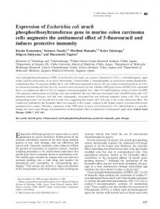
50 (HI form) column (0.5 ml bed volume) and eluting with 1 ml of distilled water. The effluent from the column was then concentrated by evaporation with a stream of nitrogen. A sample (5 x 10⁴ c.p.m.) of this product was then incubated with 20 1ll of 3T3 cell extract and, after a suitable time, the [14C]homoserine 0phosphate was precipitated by the addition of 0.1 ml of 0.15 MBa(OH)2 and 0.1 ml of 0.15 M-Na2CO3. The mixture was centrifuged in a Microfuge (13 000 g for 2 min) and the radioactivity released into the supernatant was measured. RESULTS Transient expression The approach used to insert the thrB coding sequences into the plasmid pSVL, giving pSVThrB, is shown in Fig. 1. The eukaryotic expression vector pSVL (Lusky & Botchan, 1981) has an SV40 late promoter, the VP1 intron sequence and a polyadenylation sequence. Coding sequences inserted into the multiple cloning site of this plasmid have been reported (Templeton & Eckhart, 1984) to be expressed in cultured eukaryotic cells. From the sequence of the thr operon (Cosart et al., 1981; Parsot et al., 1983) a number of possible restriction sites were available for isolating the thrB gene from pIP3. Since pSVL will express protein from the first AUG encountered in the inserted sequence, we chose the Hinfl site in order to eliminate an out-offrame AUG start codon present only 4 bp upstream of the authentic start codon. A 1.047 kbp Hinfl-EcoRI fragment of the plasmid pIP3 was prepared by digestion with both restriction endonucleases, followed by isolation of the fragment. The
Hinfl
EcoRI
47
47
"AUG thrA
thrB 1047 bp
P
thrC7t' +
47 thrB
VPI intro
SV40 late
SV40 poly(A) site pSVthrB 5936bp
promoter
s
pR322 on
ampR
SV40/thrB TK promoter
pSVthrBneo 7069bp
with ninhydrin. Measurement of L-homoserine 0-phosphate hydrolysis L-[U-'4C]Homoserine was incubated with ATP and E. coli homoserine kinase under conditions similar to those described for the enzyme assay. After incubation for 1 h the L-[U- 4C]homoserine 0-phosphate produced was separated from remaining homoserine by passing the material through a Dowex-
pIP3
''AUG
+
neo
pBR322 ori Fig. 1. Diagrammatic representation of the preparation of the plasmids pSVThrB and pSVthrBneo The diagrams are not drawn to scale. Abbreviations: VPI, SV40 VPI intron and processing signals; ampR, ampicillin-resistance gene; neo, neomycin-resistance gene; ori, origin. amp
1992
*~ ._
Homoserine kinase in mouse cells a
Size
(kb)
2.9
b
...,,..;.
c
..
..........
Fig. 2. Northern blot of mRNAs isolated from transient-expression experiments RNA isolated from mouse 3T3 cells transfected with the plasmid pSVThrB were hybridized with a probe prepared from the Hinfl/EcoRI fragment of the plasmid pIP3. Each lane was loaded with 2.5 ,ug of total RNA. Lanes a and c are control cells transfected with pIP3 (25 /tg/dish), and lane b is from cells transfected with pSVThrB (25 ,ug/dish). The arrows indicate the positions of the 28 and 18 S eukaryotic ribosomal RNAs and the 16 and 23 S prokaryotic ribosomal RNAs relative to a 5.5 kb EcoRI fragment of pIP3 (arrow on the right).
overhanging ends of the fragment were end-filled with dNTPs and the Klenow fragment of DNA polymerase I, and the fragment was ligated into SmaI-digested alkaline-phosphatasetreated pSVL. This ligated DNA was used to transform E. coli strain DH5a to ampicillin resistance. The orientation of inserts was determined by digestion with ScaI and AvaI. Approx. 50% were in the correct orientation with respect to the SV40 promoter. Suitable clones were screened by transfection of mouse 3T3 cells, followed by assay 48-72 h later for homoserine kinase activity. Preliminary experiments showed that [14C]homoserine was converted into a barium-precipitable product (assumed to be homoserine 0-phosphate, as discussed below) by extracts of the transfected cells. Optimal expression of enzyme activity was obtained when the cells were transfected with 2-5,ug of DNA/50 mm dish. Greater amounts of DNA lead to reduced growth, although we do not believe that this is due to expression of homoserine kinase activity, since cells transfected with pSVL constructs with inactive inserts show similar reductions in the growth rate (results not shown). The homoserine kinase activity detected was in the range of 0.02-0.05 nmol/h per mg of protein using tracer homoserine. Total RNA was extracted from transformed and nontransformed cells and analysed by Northern blotting and hybridization with a 32P-labelled probe prepared from the Hinfl-EcoRI fragment of pIP3. As shown in Fig. 2, RNA species complementary to the probe were detected specifically in cells transiently expressing homoserine kinase activity after transVol. 281
867
formation with pSVthrB and not in cells transfected with pIP3, where no expression is found. The two major transcription products (estimated to be larger than 4.4 kbp on the basis of three separate experiments using rRNAs as standards) were considerably larger than the sizes that would be predicted for the mature mRNA. Lower-molecular-mass species were, however, also detected.
Stable transformants In order to allow the selection of stable transformants, a further construct, pSVthrBneo, was prepared from pSVThrB as follows. An XhoI-SalI fragment of the plasmid pMClneoPA containing a neomycin-resistance cartridge coupled to the herpes simplex thymidine kinase promoter was ligated into pSVThrB at a SalI site beyond the eukaryotic expression sequence. Ampicillinresistant E. coli clones which contained an insert were characterized by restriction mapping. Stably transfected 3T3 cell lines were raised either by cotransfection with 2.5 ,ug of pSVthrB and 2.5 ,ug of pMClneoPA or with 2.5 ,ug of pSVthrBneo/50 mm dish. Cells were cultured in the presence of 400 ,tg of Geneticin (G-418)/ml for 21-28 days until colonies of resistant cells became apparent. There was no apparent loss of transformation frequency by the inclusion of the homoserine kinase gene. Control 3T3 cells, transformed with pMClneoPA to produce G-418 resistance, gave similar numbers of colonies with a transformation rate of approx. 40 colonies/dish. Suitable candidate colonies of different sizes were cloned and used for assay. Lysates of the cloned cells were produced and these were assayed for homoserine kinase activity using homoserine at 2 mm final concentration, and the averaged results for three experiments are shown in Fig. 3. The co-transformed cell lines showed a greater difference in the activity expressed, with some clones being inactive and the pSVthrBneo clones showing a higher average level of expression (Fig. 3). The soluble RNA fraction (Hesketh et al., 1991) was prepared from clone MC/B- 1 I and from G-418-resistant control cells, and the RNA was separated on a denaturing formamide gel. A Northern blot of this membrane was then analysed by hybridization with a 32P-labelled probe prepared as described previously. The results of this experiment (Fig. 4) showed an mRNA species complementary to the probe which was present in the MC/B-1I clone and not present in the G-418-resistant controls. Clone MC/B-10, which showed a much lower level of enzyme activity (Fig. 3), showed some evidence for a similar complementary RNA, although this was close to the limit of detection in these experiments (results not shown). The size of the RNA species (smaller than the 2.0 kbp 18 S ribosomal subunit) was much smaller than that seen in the transientexpression experiments. Clone Bneo-6 was used to investigate further the metabolism of homoserine and the kinetics of homoserine metabolism.
Metabolism of I'4Clhomoserine in intact cells The above results suggest that homoserine kinase is active in broken-cell preparations from transformed cells. In order to investigate whether the enzyme is also active in vivo, intact cells were incubated with [14C]homoserine, and the labelled intermediates were isolated and characterized. ThrB-transfected 3T3 cells or G-418-resistant controls were incubated with tracer [14C]homoserine for 2 h. After this time the cells were extensively washed to remove extracellular radioactivity, and PCA extracts of the intracellular products were prepared. In thrB-transfected cells the extract contained 10.6-12.5 % of the total radioactivity added to the dish, compared with 1.5-1.8 % which was retained in the control neomycinresistant cells (ranges for two experiments). The accumulation of
868
I eF'
.,;
W. D. Rees, S. M. Hay and H. J. Flint HS 8 6 4 2
I*~, ql0*0 % 50
5
10
15
20
25
10
15
20
25
10 15 20 Fraction no.
25
E 10ci
(6
Clone number
Fig. 3. Homoserine kinase activity of a series of G-418-resstant 3T3 cell
(b) neo
86-
.? .>
4-
cc
._
Cells were either co-transfected with both pSVthrB and pMC I neoPA (MC/B clones) or with psvthrBneo (Bneo clones). The data represent the averages of three separate determinations using a substrate concentration of 2 mM.
2-
m
x
0
5
w-
10
-
(c) pSVthrBneo
864
-
2-
:::.
0
:..:
--
.:.::.
28 S
.::.
*..:
2-OB
10
0
5
:::; :;
::
..::
....
*... :::: ..::
*:
.:
....::.
18 S
;.;
....
...
...........
.... :. :. :::
*:
......
*:.
:::.
............ ....: !..
:. .:
:..::
.'.;:
:.:..:.::.
a
b
Fig. 4. Northern blot of soluble mRNAs isolated from G-418-resnt 3T3 cell clones Each lane was loaded with 25 jug of total soluble RNA derived from G-418-resistant control cells (a) and clone MC/B 11 (b), which expresses homoserine kinase activity. The resulting blot was hybridized with a probe derived from the Hinfl/EcoRI fragment of pIP3. The arrows indicate the location of the 28 and 18 S ribosomal RNAs.
radioactivity was linear up to 2 h, but thereafter the rate declined, possibly owing to loss of cell viability in the simple medium used. There was no increase in the intracellular radioactivity of the G-418-resistant controls. In order to establish the fate of [14Cjhomoserine, extracts from these experiments were separated on a Dowex-l (formate form) column and fractionated by elution with a gradient of sodium formate (Fig. 5). Fig. 5(a) shows the product of the reaction with semi-purified E. coli homoserine kinase and ATP. Figs. 5(b) and 5(c) show the products from similar extracts of the intracellular products of control and thrB-transfected 3T3 cells. Unchanged tracer homoserine is not retained by this column and passes out in the initial fractions; 96% of the radioactivity present in the control neomycin-resistant cells was eluted with the homoserine standard. It can be seen that the new product formed by thrBtransfected 3T3 cells (5c) has a similar retention time to homoserine 0-phosphate produced by the bacterial enzyme (5a), and this material accounted for between 95 and 100% of the total radioactivity applied to the column. The G-418-resistant
Fig. 5. Ion-exchange chromatography of the products of homoserine metaboism in varieus mouse 3T3 cell lines Samples of the neutralized PCA extracts of 3T3 cells which had been previously incubated with radiolabelled homoserine were chromatographed on a Dowex-1 (formate form) column using a gradient of sodium formate. The distribution of radioactivity is shown for (a) a semi-purified preparation of E. coli homoserine kinase (HSK), (b) control G-418-resistant cells carrying the pMClneoPA plasmid (neo) and (c) cells carrying the pSVthrBneo plasmid (pSVthrBneo). The arrows indicate the position of the homoserine (HS) and 2-oxobutyrate (2-OB) standards.
controls (5b) show no products in this region of the column effluent. Additional evidence that the new product is indeed homoserine 0-phosphate is provided by the observation that the material in the neutralized extract is 86% precipitable as a barium salt. This column is also able to resolve oxo acids, and the location of a 2-oxobutyrate standard is shown. When rat liver preparations which oxidize homoserine were used in a similar experiment, there was a significant accumulation of radioactivity in the 2-oxobutyrate pool, as this is the first oxidation product of homoserine in rat liver. There was no evidence for the production of oxo-acid products in our cell extracts, and we therefore conclude that there is little or no oxidation of homoserine occurring even in the control 3T3 cells. In order to confirm further the identity of the new product the samples were analysed by high voltage electrophoresis (Fig. 6). Samples of the PCA extract of cells were electrophoresed on Whatman 3MM paper as described by Szczesiul & Wampler (1976). The location of homoserine 0-phosphate was determined by preparing a standard by the action of semi-purified homoserine kinase from E. coli (Fig. 6a). It can be seen that the extract from cells carrying pSVThrB (Fig. 6c) has a peak corresponding to a new product. This peak accounts for 85 % of the radioactivity applied to the electrophoretogram and has a similar RF to the product from the bacterial enzyme. There is also one minor unidentified peak accounting for approx. 10% of the total 1992
Homoserine kinase in mouse cells
869
HS
5-
1515-
1 I
10 -
I
(a) HSK
4cn_ xa 0
5_1
2-
5
0
10
15
20
E 15 I
-0.6 -0.4 -0.2
(b) neo
._
_/I
ccitx
0
i-
_40
-5
15
0
10 .
10
15
2(0
(c) pSVthrBneo
5
I
_
+
I
_
0
-5
0
5 10 Migration (cm)
15
20
Fig. 6. Electrophoretic analysis of the products of homoserine metabolism in various mouse 3T3-cell lines Samples of the neutralized PCA extracts of 3T3 cells which had previously been incubated with radiolabelled homoserine were electrophoresed on Whatman 3MM paper at 500 V. The distribution of radioactivity is shown for (a) a semi-purified preparation of E. coli homoserine kinase (HSK), (b) control G-418-resistant cells carrying the pMClneoPA plasmid (neo) and (c) cells carrying the pSVthrBneo plasmid (pSVthrBneo). The arrow indicates the position of the homoserine (HS) standard.
radioactivity present on the electrophoretogram. There is no evidence for any product in the G-418-resistant control cells, all of the activity being present in the same region as the homoserine standard (Fig. 6b). These electrophoretic results then confirm the results from ion-exchange chromatography and show a new product identical with that produced by homoserine kinase. Fate of homoserine 0-phosphate in broken cells ['4C]Homoserine 0-phosphate was prepared as described in the Materials and methods section, and incubated with crude 3T3-cell Ilysates at 37 °C for 30 min in order to establish whether it was subject to further metabolism. In this time 23-40 % of added tracer was hydrolysed and no longer precipitable as its barium salt. In lysates subjected to brief centrifugation (10000 g for 2 min) 18-25 % of the added homoserine 0-phosphate was lost in a 30 min incubation. Analysis of the products of homoserine 0-phosphate breakdown by ion-exchange chromatography showed the non-precipitable material released to run as a single peak which was indistinguishable from the homoserine standard. The breakdown of homoserine 0-phosphate is thought to be due to the release of non-specific phosphatases by the disruption of lysosomes. This activity was not detected in homogenates produced in iso-osmotic buffers by mechanical homogenization (results not shown), when lysosomes Vol. 281
0.2
0.4
0.6
0.8
-1
"..
5
-
.
0
[S] (mM) Fig. 7. Kinetic parameters for the interaction of homoserine with homoserine kinase expressed in mouse 3T3 cells The data represent the average results for three separate experiments, and the line was fitted by linear regression. The calculated parameters are: Km, 0.45 mM; V..., 0.348 nmol/min per mg of protein.
.D 10 -
;.0 5._
0
are less likely to be disrupted. The homoserine 0-phosphate phosphatase activity was greatly reduced when the ultrasonic lysates were centrifuged at 100000 g for 15 min, when less than 5 % of the added [14C]homoserine 0-phosphate was hydrolysed in 30 min. There was no loss of soluble homoserine kinase activity with high-speed centrifugation, and this procedure was adopted for homoserine kinase assays on transformed cells. We do not believe that this non-specific phosphatase can significantly reduce cytoplasmic homoserine 0-phosphate accumulation in intact 3T3 cells, since at least 85 % of the intracellular homoserine is recovered as homoserine 0-phosphate in the experiments described in Figs. 5 and 6.
Kinetics of homoserine kinase from mammalian cells By restricting conversion to less than 10 % of the [14C]homoserine added, we were able to determine the initial rates of kinase activity with different concentrations of homoserine and from these data determine the Km with respect to homoserine. The enzyme found in mouse-cell homogenates gave a value of 0.45 mm (Fig. 7). This is similar to the values of 0.2 mM reported for the purified E. coli enzyme by Burr et al. (1976) and 0.3 mM by Theze et al. (1974). The enzyme purified from Brevibacterium flavus also yielded a similar Ki: 0.77 mM (Miyajima & Shiio, 1972). This suggests that the bacterial enzyme expressed in the mammalian cell has undergone a normal folding of the polypeptide chains and that the enzyme exhibits its usual catalytic activity. Since we cannot measure the kinetic parameters of homoserine kinase in the intact cell, it is not possible to show that it is fully active in the environment of the mammalian cell, but these experiments point to an intracellular accumulation of homoserine 0-phosphate when cells carrying pSVThrB are incubated with L-homoserine and to the fact that homoserine 0-phosphate accumulates in the cells.
DISCUSSION Our data show that functional homoserine kinase is expressed when the coding region for E. coli homoserine kinase is placed under the control of the strong SV40 late promoter and transfected into mammalian cells. Furthermore, the enzyme is active intracellularly in 3T3 cells, suggesting that it is being supplied with sufficient cofactors to function in vivo. Our data suggest that the enzyme is located in the cytosol, since the activity remains in the cytosolic fraction when membrane components are sedimented by high-speed centrifugation. The similar Km for the native E. coli homoserine kinase and the enzyme produced by mammalian cells suggests that the protein has folded to produce
W. D. Rees, S. M. Hay and H. J. Flint
870 an active catalytic site, although without some means of determining the amount of enzyme protein present, we are unable to confirm that the catalytic-centre activity is unchanged. Analysis of the proteins contained in the control and thrBtransfected cells by SDS/PAGE shows no differences between the two cell lines, and there is no evidence that there is a large production of protein with only a very limited catalytic capacity. Assuming that the enzyme is folded correctly, the specific activity of homoserine kinase found in extracts of the clone Bneo6 (0.34 nmol/min per mg of protein) when compared with a value of 58 ,umol/min per mg of protein reported by Burr et al. (1976) for the purified enzyme indicates that the homoserine kinase enzyme accounts for less than 0.001 % of the total protein in the transformed cells. Previous reports have described the expression of bacterial phosphotransferase, dehydrogenase, acetyltransferase and decarboxylase activities in a variety of animal cell types. Both galactokinase and thymidine kinase from E. coli have been expressed without apparent ill effect in animal cells (Goff & Berg, 1979; Schumperli et al., 1982). We have found no evidence for disturbances of the growth of these cells expressing the bacterial homoserine kinase. Phosphorylation of serine residues in proteins is important for the control of many vital mammalian cell functions, but it would appear that there is no interference from this kinase activity. This is not surprising, since there is no evidence that the thrB gene product has protein kinase activity. Baker (1988) has noted a similarity in the sequence of the human retinoic acid receptor and E. coli homoserine kinase. The lack of effect on growth seen in the present experiments suggests that there is no effect of homoserine kinase acting as a DNA-binding protein, although since the retinoic acid receptor is a DNAbinding protein, the cytosolic location of homoserine kinase may prevent it from binding to DNA. Other attempts to express bacterial genes in mammalian cells have used a similar approach of inserting eukaryotic promoters and message-processing sequences around the bacterial coding region. The activities reported by other authors for similar prokaryotic genes under the control of SV40 promoters are in the order of 0.1-5 nmol/min per mg of protein in cell extracts (Mulligan & Berg, 1981 ; Schumperli et al., 1982). The homoserine kinase activity of 0.34 nmol/min per mg of protein is therefore within the range of that seen for other enzyme systems. A crucial point in the transfer of new metabolic capacities to animal cells is that the introduced enzyme is able to function and build up a sufficiently large pool for onward metabolism. Homoserine is oxidized by the enzyme cystathionine y-lyase (Deme & Chatagner, 1972) and it is therefore important that the bacterial enzyme can compete with the oxidative enzyme and produce its product in the intact animal cell. Computer simulations (Rees et al., 1990) assuming the in vitro kinetic constants of the oxidative activity found in rat liver suggest that, provided homoserine kinase can be expressed at a sufficiently high level, homoserine phosphate will be formed with only a small percentage of the incoming homoserine being lost to oxidation. The capacity of 3T3 cells to oxidize homoserine is much lower than that found in rat liver, and in this case a significant pool of homoserine accumulates in the cells with only minimal losses to oxidation. Since the liver is the main site of homoserine oxidation, it will be interesting to introduce these plasmid constructs into primary hepatocyte cultures to investigate the functioning of the new pathway in an oxidative tissue. Some breakdown of homoserine 0-phosphate is observed in sonicated cell extracts, but we believe that this is due to the release of lysosomal phosphatases in the sonicated preparations that we are using. When membrane-bound phosphatase is removed by high-speed centrifugation, this homoserine 0-phosReceived 24 June 1991; accepted 5 August 1991
phate phosphatase activity is removed too, leaving the homoserine kinase activi4~in the supernatant. Analysis of the products would suggest that the only labelled product formed from [14C]homoserine 0-phosphate is homoserine, indicating that the reaction is indeed a dephosphorylation. In homogenates produced from rat liver by the use of milder mechanical homogenization procedures, there is no evidence for hydrolysis. It would appear, therefore, that, in the intact cell, the homoserine kinase and phosphatase activities are sequestered from one another. The major transcripts from pSVthrB in transient-expression experiments were apparently almost as large as the plasmid itself, although other small transcripts were also present. This might be due to a failure to terminate transcription correctly or to some form of secondary structure preventing the functioning of the polyadenylation site. Alternatively there may be transcription from cryptic promoters in the pBR322 portion of the plasmid (Langner et al., 1986). Since enzyme activity is detected, there must be some form of transcribable message produced, although which of the species detected is active is not clear from these experiments. However, in the stable clones a much smaller message is present at a much lower abundance, and this, we believe, may be the functional mRNA species. These experiments demonstrate that E. coli homoserine kinase can be expressed in an animal cell and that it can successfully phosphorylate L-homoserine in the intact cell using endogenous ATP. We thank Dr. I. Saint-Girons for generously providing the plasmid pIP3. We also thank Dr. J. E. Hesketh and Mrs. G. P. Campbell for their help and assistance in the analysis of mRNA. REFERENCES Baker, M. E. (1988) Biochem. J. 255, 748-749 Burr, B., Walker, J., Truffa-Bachi, P. & Cohen, G. N. (1976) Eur. J. Biochem. 62, 519-526 Cosart, P., Katinka, M. & Yaniv, M. (1981) Nucleic Acids Res. 9, 339-347
Deme, D. & Chatagner, F. (1972) Biochim. Biophys. Acta 258, 643-654 Goff, S. P. & Berg, P. (1979) J. Mol. Biol. 133, 359-383 Gorman, C. M., Moffat, L. F. & Howard, B. H. (1982) Mol. Cell. Biol. 2, 1044-1051 Hartman, S. C. & Mulligan, R. C. (1988) Proc. Natl. Acad. Sci. U.S.A. 85, 8047-8051 Hesketh, J. E., Campbell, G. P. & Whitelaw, P. F. (1991) Biochem. J. 274, 607-609 Kuo, M. H., Saunders, P. P. & Broquist, H. P. (1964) J. Biol. Chem. 239, 508-515
Langner, K.-D., Weyer, U. & Doerfier, W. (1986) Proc. Natl. Acad. Sci. U.S.A. 83, 1598-1602 Lusky, M. & Botchan, M. (1981) Nature (London) 293, 79-81 Maniatis, T., Fritsch, E. F. & Sambrook, J. (1982) Molecular Cloning: A Laboratory Manual, Cold Spring Harbor Laboratory, Cold Spring Harbor, NY Miyajima, R. & Shiio, I. (1972) J. Biochem. (Tokyo) 71, 219-226 Mulligan, R. C. & Berg, P. (1980) Science 209, 1422-1427 Mulligan, R. C. & Berg, P. (1981) Proc. Natl. Acad. Sci. U.S.A. 78, 2072-2076 Parker, B. A. & Stark, G. R. (1979) J. Virol. 31, 360-369 Parsot, C., Cosart, P., Saint-Girons, I. & Cohen, G. N. (1983) Nucleic Acids Res. 11, 7331-7345 Rees, W. D., Fuller, M. F. & Flint, H. J. (1990) Bio/Technology 8, 629-633
Schumperli, D., Howard, B. H. & Rosenberg, M. (1982) Proc. Natl. Acad. Sci. U.S.A. 79, 257-261 Southern, P. J. & Berg, P. (1982) J. Mol. App. Genet. 1, 327-341 Szczesiul, M. & Wampler, D. E. (1976) Biochemistry 15, 2236-2244 Templeton, D. & Eckhart, W. (1984) Mol. Cell. Biol. 4, 817-821 Theze, J., Kleidman, L. & Saint-Girons, I. (1974) J. Bacteriol. 118, 577-581
Ware, C. E., Bauchop, T. & Gregg, K. (1989) J. Gen. Microbiol. 135, 921-930
1992