Y13-259 (Furth et al., 1982), as shown in Figure 2. The con- struction was verified by sequencing the plasmid DNA in the region of the translational initiation site.
The EMBO Journal vol.5 no.6 pp.1351-1358, 1986
Expression of p21 proteins in Escherichia coli and stereochemistry of the nucleotide-binding site
J.Tucker, G.Sczakiel, J.Feuerstein, J.John, R.S.Goody and A.Wittinghofer Max-Planck-Institut fiir Medizinische Forschung, Abteilung Biophysik, Jahnstrasse 29, 69 Heidelberg, FRG Communicated by K.C.Holmes
v-Ha-ras encoded p21 protein (p21V), the cellular c-Ha-ras encoded protein (p21C) and its T24 mutant form P21T were produced in Escherichia coli under the control of the tac promoter. Large amounts of the authentic proteins in a soluble form can be extracted and purified without the use of denaturants or detergents. All three proteins are highly active in GDP binding, GTPase and, for p2lv, autokinase activity. Inhibition of [3H]GDP binding to p21C by regio- and stereospecific phosphorothioate analogs of GDP and GTP was investigated to obtain a measure of the relative affinities of the three diphosphate and five triphosphate analogs of guanosine. p21 has a preference for the Sp isomers of GDPaS and GTPaS. It has low specificity for the Sp isomer of GTP,BS. Together with the data for GDP,BS and GTP-yS these results are compared with those obtained for elongation factor (EF)Tu and transducin. This has enabled us to probe the structural relatedness of these proteins. We conclude that p21 seems to be more closely related to EF-Tu than to transducin. Key words: p21 protein/expression/stereochemistry/nucleotide-binding site
Introduction The ras oncogene family, which includes the Ki-, Ha- and Nras proto-oncogenes, encodes a closely related group of proteins of -21 000 daltons mol. wt, termed p21 proteins, which are GTP-binding proteins with low GTPase activity (for a review, see Bishop, 1985; Gibbs et al., 1985). They are believed to play an essential role in growth and/or development. A number of structural comparisons and structure predictions have been published in an attempt to explain the structural implications of the amino acid replacements at positions 12, 13 and 59,61, which lead to tumorigenic activation of the protein (Gay and Walker, 1983; Wierenga and Hol, 1983; Leberman and Egner, 1984; Halliday, 1983; Pincus and Brandt-Rauf, 1985; Murakami, 1985). Since secondary structure predictions at the very best are only 60 % accurate and are sometimes contradictory to each other, the actual determination of the three-dimensional structure is essential. Models for the tertiary structure of the nucleotide-binding site of p21 have been built on the basis of partial sequence homologies between elongation factor (EF)-Tu and p21 proteins and using the tertiary structure of the GDP-binding domain of trypsinized EF-Tu from Escherichia coli determined by X-ray crystallography (McCormick et al., 1985; Jurnak, 1985). Since crystals suitable for X-ray crystallography have not yet been obtained, we have looked for other means to obtain structural information from the p21 proteins. Thiophosphate analogs of © IRL Press Limited,
Oxford, England
nucleoside di- and triphosphates have been used to study the active site geometry and metal ion coordination in the nucleotidebinding site of many proteins (for a review, see Eckstein, 1985). We have used this method to study the active site of p21 proteins and compared it with results obtained earlier with EF-Tu (Wittinghofer et al., 1982; Leupold et al., 1983); and recently with transducin (Yamanaka et al., 1985). The source of the p21 protein was a bacterial expression system based on the tac promoter which produces high amounts of authentic and soluble p2l1 proteins which are highly active in GTPase and nucleotide binding.
Results Construction of expression vectors To express p21 in E. coli we used the expression vector pKMtacl (deBoer et al., 1983) following the scheme outlined in Figure 1. The 880-bp HindIII fragment of Ha-MuSV was inserted and the correct coding sequence for the p21 protein from v-Ha-ras (= p2lv) was regenerated by inserting a synthetic linker consisting of two 19-mers. For expression of the ras construction the lac repressor overproducing strain RRIAM15 was used. The final clone ptacrasv was identified by immunoblotting the crude extract of the induced culture with anti-ras monoclonal antibody Y13-259 (Furth et al., 1982), as shown in Figure 2. The construction was verified by sequencing the plasmid DNA in the region of the translational initiation site. The normal and the T24 mutant Ha-ras proteins were also produced by replacing the 880-bp Hindu fragment of ptacrasv with the HindHI-Sall fragment of pSKcHras and pSKT24 (Gross et al., 1985) as shown in Figure 1. These constructs are also transformed into RRIAM15. They are stably maintained in this strain and the cells can be easily grown, even in large fermenters. In medium containing high concentrations of ampicillin (100 itg/ml) the tac promoter was repressed and very little p21 protein could be identified by SDS-polyacrylamide gel electrophoresis in the crude lysate (see Figure 2). Expression of p21 proteins could be induced with 50-100 AiM isopropyl-f-D-thiogalactopyranoside (IPTG) to produce amounts of proteins much greater than 20% of the soluble protein for all three constructions. On SDS - PAGE the viral and the T24 mutant form of ras protein have an apparent mol. wt of 23 500 daltons. The cellular form of p21 (p2 IC) has a slightly higher mobility, which has been observed before by other authors (Fasano et al., 1984; McGrath et al., 1984). A small amount of a 65 000-dalton protein is induced, which also reacts with monoclonal antibody Y13-259 (Furth et al., 1982). The nature of this protein is presently unknown. Purification of p21 proteins Lysis of the cell paste and purification by a two-column procedure could be performed essentially by the same method which we have used for the isolation of bacterial EF-Tus and EF-Ts (Leberman et al., 1980), except that after DEAE-Sepharose, AcA54 was used for gel filtration chromatography. No detergent was 1351
J.Tucker et al.
5' P-AATTCTATGACAGAATACA GATACTGTCTTAGTTCGA-P synthetic 19 mers
Fig. 1. Construction of the ptacrasv expression vector. Plasmid pRASI (R.Mueller, unpublished) containing the 2200-bp EcoRI-BamHI fragment of HaMuSV (Ellis et al., 1980) was excised with HindIII, which generates a 880-bp fragment containing the p21 coding region except four codons at the N terminus and 300 bp of 3'-non-coding region (Yasuda et al., 1984). This fragment was ligated into the pKMtacl expression vector (deBoer et al., 1983) cleaved with HindIll. The correct orientation of the p21 coding region behind the tac promoter was verified by Pvu-EcoRI double digests. This pre-ras plasmid was partially digested with HindIlI and EcoRI, the digestion mixture was separated on an agarose gel. The band corresponding to the desired 5300-bp fragment was electroeluted and ligated to the mixture of phosphorylated 19-mers with the indicated sequences, which restored the p21 coding region and the proper distance between ATG start codon and the Shine-Dalgarno sequence (Shine and Dalgarno, 1974). The ligation mixture was transformed into RRIAM15. The resulting clones were screened by preparing crude lysates from minicultures induced with 50 /M IPTG and analyzing them by SDS-PAGE and immunoblotting. H, E, P, Pv and B denote the restriction enzymes HindIlI, EcoRI, PstI, PvuII and BamHI, respectively.
used in the isolation procedure. For analysis of the column effluents the simple GDP exchange assay with nitrocellulose filters was used (Leberman et al., 1980). Figure 3 shows the polyacrylamide gel electrophoresis of crude lysate and the two column effluents for the purification of p21C. The viral and T24 mutant human p21 proteins were purified similarly and lanes 4 and 5 show these two purified proteins. 100 g of cell paste could thus be processed to obtain 320 ± 30 mg of the three proteins as shown in Table I, which gives examples for one preparation of each of the three different p21 proteins. Table I and the polyacrylamide electrophoresis data also show that >20 % of the total soluble protein in the cellular extract is isolated as p21 protein with > 95 % purity. Protein sequencing has been performed
1352
for the p2 IV protein and the results show that the N terminus of the protein is not blocked, that methionine is the first amino acid and that a unique and expected sequence is obtained as far as arginine in position 12. On SDS -polyacrylamide gels the two transforming, but not the cellular protein occasionally appeared as two bands, both of which reacted with p21 antibody (see Figures 2 and 3). We are presently investigating whether posttranslational modification is responsible for this phenomenon.
Characterization ofpurified proteins The u.v. spectrum of the purified proteins is identical to that reported for the fusion protein described by Poe et al. (1985). We also find that the purified p21 proteins contain one equivalent
p21 Nucleotide-binding site
1
2
3
4
5
i.:
r--, t
,!.
* 68 * *
4b *
*
29 *
41~:
4u_ *
W
20
13
*
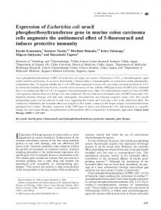
Fig. 2. Induction of p2lv in bacterial cultures. Cells of an overnight culture of ptacrasv in RPIAM15 in LB medium containing 1000g/ml of ampicillin in the presence (induced) or absence (uninduced) of 1 mM IPTG were collected. They were washed and a cleared lysate of soluble protein in 100 lI of buffer A was prepared as described in Materials and methods. 25 tld of this solution were treated with SDS sample buffer and separated on a 15% acrylamide gel as described by Laemmli (1970). The lanes were either stained with Coomassie Blue (lanes 1-3) or used for immunoblotting (lanes 4-5) as described in Materials and methods. The lanes from left to right are: 1, uninduced; 2, induced; 3, mol. wt markers with mol. wts in kd as indicated; 4, uninduced; 5, induced.
of bound nucleotide as one would expect for the high binding constants (of the order of 108 M-1) reported for the binding of GDP and GTP to p21 (Hattori et al., 1985). Contrary to what
has been found by Poe et al. (1985), we find that besides GDP (ribo) also deoxy-GDP and a small amount of GTP remain bound to the proteins, which together add to 1 mol of nucleotide per mol of protein (data not shown). 80 % of bound nucleotide is GDP only when the elution from the second column (AcA54) is done in the presence of GDP. The p21 proteins are highly active in GDP binding, GTPase activity and the viral protein is also highly active in autophosphorylating activity (see Table II). The GDP binding activity is higher than reported by other authors and is the theoretical maximum 46 500 pmol bound GDP per mg of protein if one makes the (reasonable) assumption that 1 mol protein binds 1 mol GDP. The GTPase activity for the purified p21c is 0.02 min-', as high as the value reported for the p21 fusion protein (Temeles et al., 1985). Table II also shows that the transforming proteins p21V and P21T have a 5- to 8-fold lower GTPase activity, which has been observed by many authors (McGrath et al., 1984; Sweet et al., 1984; Gibbs et al., 1984; -
-
Manne et al., 1985). The proteins have also been used for proton and phosphorus magnetic resonance measurements (n.m.r.), where they have been shown to be highly structured, native proteins (Roesch et al., 1986; Roesch et al., unpublished). The pro-
teins also can be crystallized although the crystals obtained not yet suitable for X-ray crystallography.
are
Stereochemistry of the nucleotide-binding site Phosphorothioate analogs of GDP and GTP contain sulfur in place of a non-bridging oxygen atom in one of the phosphorus atoms. Substitution of sulfur for oxygen produces a chiral center at the a-phosphorus of GDP and at the a- and (3-phosphorus atoms of GTP. The absolute stereochemistry of the diastereomers is known (see Eckstein, 1985, for references) and they are designated Sp and Rp according to their configuration. Thus there are five regioand stereospecific thio-GTP analogs: (Sp)-GTPaS, (Rp)-GTPaS, (Sp)-GTP,BS, (Rp)-GTPflS, GTPRyS, and three thio-GDP analogs: (Sp)-GDPaS, (Rp)-GDPaS and GDP,BS, which can be used to probe the active site stereochemistry of p21. Figure 4 illustrates the different phosphate oxygens of GTP which are replaced by 1353
J.Tucker et al.
668 _ _
45
_
J ' ' _2 9
-
n
F20
.1 3
Fig. 3. SDS-PAGE of fractions from the purification of p21C and purified p21V and P21T. The lanes from left to right contain: mol. wt makers as indicated on the right, crude lysate, DEAE-Sepharose pool, AcA54 pool, P21T, p21V mol. wt markers.
sulfur in the thio-GTP analogs. Pro-R and pro-S indicate the oxygen ligands on a- or $-phosphorus, whose substitution by sulfur leads to the corresponding (Rp) and (Sp) isomers, respectively. The diastereomers of GDP(a-S) have structures corresponding to those of GTP(ca-S). We have measured the relative affinity of the phosphorothioates of GDP and GTP by exchanging GDP bound to p21 with these analogs. Figure 5 shows that the exchange reaction between bound GDP and free radioactive [3H]GDP, which is also the basis of the GDP binding activity measurements, is a very slow reaction at room temperature and takes > 6 h before equilibrium is reached. Addition of excess EDTA speeds up the reaction considerably so that equilibrium conditions are reached after 15 min. It appears that the metal ion in the p21.GDP.Me2+ complex is readily available to solvent. Thus the dissociation of bound GDP, which is probably rate-limiting in the exchange reaction, is increased dramatically after metal ion removal, which has also been observed for EF-Tu (Eccleston, 1981). Inhibition of [3H]GDP binding was measured with increasing concentrations of thiophosphate analogs. The exchange reaction was performed in the presence of EDTA, but after attainment of equilibrium excess magnesium was added. After incubating for another 30 min the reaction mixture was filtered through nitrocellulose filters and washed. Figure 6 shows as an example 1354
Table I. Summary of protein yields for the purification of c-Ha-ras encoded p2lc, its T24 mutant forms, p2lT and the v-Ha-ras encoded protein p2lv, starting with 100 g cell paste
p21c Crude DEAE ACA54
(mg)
(mg)
P21T
P21v
1591 915 350
1319 870 318
1486 889
(mg)
325
Table II. Biochemical activities of the three purified p21 proteins
Activity
Protein
P21c
P21v
P21T
GDP-binding (U/mg)
46 500
GTPase (min-1) Autokinase (min1)
0.02 -
45 100 0.005 3.8 x 10⁻⁴
45 600 0.0025 -
the inhibition of GDP binding by (Sp)-GDPczS and (Rp)-GDPcaS. p21C shows a selectivity for the (Sp)-isomer of GDPaS and the relative affinities of the analogs versus GDP can be estimated from the concentrations of analog which cause 50% inhibition
p21 Nucleotide-binding site
~~~0
0
0
Table m. Relative affinities of phosphorothioate analogs of GDP and GTP to p21, and to EF-Tu (Wittinghofer et al., 1982; Leupold et al., 1983) and transducin (Yamanaka et al., 1985) as taken from the literature
/
Protein
Analog
p
Guo
P
P
GDP[caS] Sp
00 pro-S
Rp Sp/Rp
0
0
pro-R pro-R
pro-S
Fig. 4. Schematic drawing showing the oxygens of the (x-, ,B- and -yphosphates of GTP. The ca- and $-phosphorus atoms are pro-chiral centers, so that substitution of one of the non-bridging oxygens (designated pro-R) by sulfur leads to a chiral center with the Rp absolute configuration, and substitution of the other (pro-S) to the Sp configuration. The diastereomers of GDP(ci-S) have the same configuration as those of GTP(ci-S). Substitution of one of the three -y-phosphate oxygens does not create a chiral center.
GDP[OS] GTP[aS] Sp Rp Sp/Rp GTP[3S] Sp Rp Sp/Rp
GTP[yS] GTP
p21C
EF-Tu
1.3 0.2 6.5 0.36 0.6 0.05 12.0 0.016 0.019 0.84 0.29 1.5
0.23 0.0083 27.0 0.041 0.29a O.015a 19.3a 0.0103a 0.0108a 0.95a 0.36a O.Ola
Transducin 4.0 0.6 6.7 0.4 10
0.83 < 10-2
> 83 > 10
The relative affinities of analogs is the inverse of the ratio of concentration [analog]/[GDP] which causes 50% inhibition of GDP binding, as demonstrated for the GTPcxS analogs in Figure 4 (not shown for the other analogs). The affinity of p21C for GDP is defined as one. aThese values were obtained by measuring the inhibition of the GTPase reaction with varying concentrations of GTP analogs. The 50% inhibition value is thus not a measure of relative affinity versus GDP. bMiller and Weissbach (1970).
24
Table IV. Turnover number for the guanosine triphosphatase reaction between p21C and GTP and phosphorothioate analogs of GTP, determined with 0.1 mM protein and 1 mM nucleotide as described in Materials and methods
22
60
120
180
Time [mini
220
Fig. 5. Kinetics of the GDP exchange reaction. 25 ,aM p21 was incubated with 10 zM [3H]GDP in the presence or absence of 0.6 mM EDTA in a final volume of 1 ml. At the indicated times, 50 M1 aliquots were removed and the amount of [3H]GDP bound to protein was detenrmined as described in Materials and methods.
of GDP binding. Thus 8 ^tM (Sp)-GDPavS and 54,u.M (Rp)-
GDPaxS cause 50% inhibition, and the ratio of affinities of the (Sp)- and (Rp)- isomers (Sp/Rp) is 6.5. The 50% inhibition values are used to obtain the relative affinities for all the thiophosphate analogs as shown in Table III. If we take the binding constant for p21.GDP as unity [its absolute value is 1.0 x 10⁸ M-1 as determined by Hattori et al. (1985)], we find relative binding constants for (Sp)-GDPaS of 1.3 and 0.2 for the (Rp)-isomer, which means that (Sp)-GDP(aeS) binds better than GDP. Using the same method, we find that the affinity of GTP is higher than GDP by a factor of 1.4, which agrees well with what has been found by Finkel et al. (1984), Hattori et al. (1985) and Manne et al. (1984), who have reported that the two nucleotides bind with similar affinities. (Sp)-GTP(ctS) has a relative affinity of 0.6, i.e. it binds more weakly than either GDP or GTP. The relative affinities for the GTPaeS diastereomers (Sp/Rp) are of the same order but the relative strength is 12.6 as compared with 6.5 for the GDPaS diastereomers. Both GTP,BS isomers have
Substrate
Turnover number (min1)
GTP
(Sp)-GTPOS (Rp)-GTPOS
0.02 0.005 0.004 0.0005 0.002
GTPyS
-
(Sp)-GTPuS (Rp)-GTPciS
relatively low affinities for p21 and the association constant is lower by a factor of 52-62, but there is a slight preference for one diastereomer, (Rp), which is favored over the (Sp)-isomer by a factor of 1.2. The substrate activity of the GTP analogs was also tested and the results are shown in Table IV. The diastereomers of GTP(c-S) are cleaved at similar rates, and this is considerably lower than that for GTP. The diastereomers of GTP(f-S) are cleaved even more slowly, but with a clear preference for the Rp isomers. Discussion In this report we describe the construction of an E. coli expression system which can be used to produce high amounts of three different p21 proteins in soluble form. Several reports have appeared describing the use of different expression vectors to produce p21 proteins in E. coli. Except in one case (Gross et al., 1985), they all lead to the expression of fusion proteins or insoluble forms of the protein which need considerable amounts of chaotropic salts or detergents to be solubilized. From the results presented here and those of Gross et al. (1985) it is apparent that the native protein is highly soluble. The use of denaturing 1355
J.Tucker et al.
100 90
80 70
E 60
D-
o 50
NZ40 30 20
-
10 0
100
200
300
400
500
Nucleotide conc. [,uM] Fig. 6. Inhibition of [3H]GDP binding to p21C by GDPcaS analogs. 2.2 1iM p21C was incubated with 9 AM [3H]GDP and varying concentration of (Rp)GPDaxS (.) and (Sp)GDPaiS (x) with excess EDTA. After 150 min at 0°C excess Mg2+ was added and the bound [3H]GDP was determined as described in Materials and methods.
agents or detergents may in fact perturb the native protein conformation and may change its biochemical and biological characteristics. The protein described here is highly soluble even at the concentrations (-- 30 mg/mi) used for n.m.r. measurements (Roesch et al., 1986). It is produced in E. coli in such high amounts that the purification of gram amounts of protein for structural studies is relatively easy. In earlier work we investigated the stereochemistry of the GDPand GTP-binding site of bacterial EF-Tu by using phosphorothioates of GDP and GTP together with e.p.r. and n.m.r. measurements (Wittinghofer et al., 1982; Leupold et al., 1983; Kalbitzer et al., 1984). This enabled us to propose a model for interactions at the nucleotide-binding site which was later confirmed by the available X-ray data (LaCoeur et al., 1985). The X-ray coordinates were then used to build a model of p21 (McCormick et al., 1985; Jurnak, 1985). p21, as can be seen from the data in Table HI, has a preference for the (Sp)-isomer of GDPaS and GTPaS and this is also true of EF-Tu (Leupold et al., 1983; data shown in Table III). In case of EF-Tu, it was shown that there is no interaction between the (x-phosphate group and the metal ion bound with the nucleotide, which leads to the conclusion that the specificity for the Sp diastereomers of GDPCtS and GTPaS is due to an energetically important interaction of the protein with the pro-R oxygen of this phosphate group and it is likely that this is also true for p21. We expect an unambiguous clarification of this feature from our current e.p.r. experiments with p21. There is very little specificity for the binding of the diastereomers of GTPflS, again in agreement with the results obtained with EF-Tu, and in contrast to the high Sp/Rp ratio seen for transducin (Yamanaka et al., 1985; data shown in Table III). In the case of p21, it was possible to measure the rate of cleavage of the GTP(f-S) diastereomers directly, and this showed a clear preference for the Rp isomer. A common feature for all three proteins, and indeed for many others studied, is that the replacement of either oxygen of the f-phosphate group by sulfur leads to a large reduction in affinity, suggesting that both oxygens are involved in important interactions, in general one being bound to the metal ion and one to the protein. EF-Tu shows a reversal of specificity (Rp with Mg2 +, Sp with Cd2 +) for the diastereomers of GTP,BS when Mg2 + is replaced by Cd2 +,
1356
which is a strong indication that there is an interaction between the pro-S oxygen of the f-phosphate group and the metal ion (Leupold et al., 1983). We have recently confirmed this interpretation by e.p.r. spectroscopy (Kalbitzer et al., in preparation). Again, our current e.p.r. experiments should give a definitive answer to this question for p21, but the preference for the Rp diastereomer of GTP(,B-S) in the cleavage reaction suggests that here there is also an interaction of the metal ion with the pro-S oxygen of the :-phosphate group. In contrast with EF-Tu, there is no metal ion dependence of the specificity of transducin for the diastereomers of GTPf3S (Yamanaka et al., 1985). As suggseted by the authors, this indicates either that there is no interaction of the metal ion with the f-phosphate group or that a very strong interaction with the protein forces the metal to bind to the same residue (sulfur) regardless of the nature of the metal ion. Assuming the latter interpretation to be correct, this leads to the initially unexpected conclusion that the pro-S oxygen (see Figure 4) is bound to the metal ion, as it is the case for EF-Tu, despite the seemingly contrasting properties with respect to the specificity for the diastereomers. From the results presented and discussed here together with the cited work on EF-Tu and transducin, we can conclude that there are similarities in the active site stereochemistry between p21, EF-Tu and transducin with respect to both the a- and f-phosphate groups. The similarity between the interactions of the fl-phosphate groups of GTP with EF-Tu and with p21 is probably greater than that between the interactions of either of these proteins with transducin, although this cannot be maintained with certainty until the e.p.r. results for p21 are available. It should be pointed out that many other nucleoside triphosphatases and kinases (e.g. myosin and adenylate kinase; Hofmann and Goody, 1980; Connolly et al., 1982) have been shown to have an interaction between the metal ion and the pro-R oxygen atom of the fl-phosphate group, suggesting a different geometry around this phosphate when compared with the GTP-binding proteins discussed here. Both GDPflS and GTP-yS, for which diastereomers are not formed by the replacement of oxygen by sulfur, are fairly good analogs for GDP and GTP. In this respect, p21 is more similar to transducin than to EF-Tu, since GDP,BS is a poor analog of GDP for ET-Fu (see Table III). This only applies to EF-Tu in
p21 Nucleotide-binding site
the presence of Mg2+ since, in the presence of excess EDTA, GDP3S is a good analog (Wittinghofer et al., 1982). This has led us to suggest that in the EF-Tu.MgGDP complex all three
non-bridging ,B-phosphate oxygens are involved in important interactions, one with metal ion and two with the protein. An extension of the same arguments leads to the conclusion that this does not apply to p21 or transducin, i.e. that in these complexes perhaps only one of the ,3-phosphate oxygens is bound to protein. Both EF-Tu and transducin are guanosine triphosphatases, and the interconversion between the GTP and GDP bound states is coupled to their translocation, either to and from the ribosome (Kaziro, 1978), or to and from the cyclic-GMP phosphodiesterase in the membrane, which amplifies the signal of transducin (Stryer, 1983). In the case of ras proteins it is postulated, and it has been shown for the yeast ras proteins, that they stimulate membranebound adenylate cyclase (Toda et al., 1985; DeFeo-Jones et al., 1985). It is thus of interest to look for similarities and differences between the proteins. On the basis of the results on p21 presented here and the published work on EF-Tu and transducin, there are some obvious similarities but also quantitative differences. Amongst the similarities are the very low GTPase activities of the isolated proteins (i.e. when they are not interacting with the other components of their in vivo systems), the high binding affinities for guanosine nucleotides and certain stereochemical similarities of their interactions with nucleotide and metal ions discussed above. A quantitative comparison suggests that there is perhaps more similarity between p21 and EF-Tu than between p21 and transducin or EF-Tu and transducin, although in one respect, i.e. the interaction of the (-phosphate of GDP with metal ion and protein, p21 and transducin are similar to each other but significantly different from EF-Tu, as discussed above. The models presented for the structure of the nucleotide-binding site of p21 (McCormick et al., 1985; Jurnak, 1985) might be good aproximations of the real three-dimensional structure, although the results discussed here show that there are obviously differences in detail. In considering the similarities of the three proteins, the question of the function of p21 arises, since it has been shown that the RAS1 and RAS2 gene products of yeast, which regulate adenylate cyclase activity, are highly homologous to mammalian ras gene products, at least in their N-terminal 180 amino acids (Dhar et al., 1984; Powers et al., 1984). Thus it has been postulated that these proteins have an analogous function to that which G-proteins and transducin have in the amplification of extracellular signals. The human Ha-ras oncogene product, however, is not a component of adenylate cyclase (Beckner et al., 1985) and it does not seem to be specifically ADPribosylated. In addition, in a recent report by Birchmeier et al. (1985) it was shown that human Ha-ras p21 proteins injected into oocytes induce maturation but do not change the cyclic AMP concentration. These results and our analysis of the relatedness of the active site geometries suggest that the mammalian ras proteins are perhaps functionally different from the G-proteins and transducin. Materials and methods [3H]GDP (400 GBq/mmol) was obtained from Amersham Buchier and was diluted specific activity with unlabelled GDP (Pharma Waldhof). Phosphorothioate analogs of GDP and GTP were synthesized according to Goody and Leberman (1979) and Roesch et al. (1984) except that glycerol kinase was used to remove traces of (Rp)-GTPI3S from (Sp)-GTP(S as described by Connolly et al. (1982). DEAE-Sepharose CL-6B was from Pharmacia and AcA54 from LKB. IPTG was from Serva Heidelberg. Nitrocellulose filters, type BA85, to the desired
0.45 AM were from Schleicher and Schuell. H.p.l.c. grade acetonitrile was obtained from Baker. All other reagents were of the highest purity available. Plasmids and strains Plasmid prasI contains the 2200-bp Eco-BamHI fragment of Ha-MuSV cloned between EcoRI and BamHI of pBR322 (R.Mueller, unpublished). pKM-tacl (deBoer et al., 1983) and the two 19-mers (see Figure 1) were a kind gift of A.Ullrich. RRIAM15 [leu,pro,thi,lacAM15,r-,m-,F'lacIQZ,AM15,pro'] is a lac repressor overproducing strain derived from HB1O1 (Boer and Roulland-Dussoix, 1969), which was obtained from B.Mueller-Hill. Plasmids pSKcHras and pSKT24 (Gross et al., 1985) were obtained from O.Fasano. Cloning methods Restriction endonucleases, alkaline phosphatase, T4 ligase and polynucleotide kinase were commercial products of Boehringer Manhheim or BRL and were used following the Laboratory Manual of Maniatis et al. (1982). Transformation was done according to the method of Hanahan (1983) with frozen cells. The sequence of the final ptacrasv construction was verified by sequencing according to the method of Maxam and Gilbert (1977). Cell-free extract and immunoblotting A 1 ml miniculture of ptacrasv in RRIAM15 was grown in LB medium containing 30 jg/ml of ampcillin. 10 Al of this culture was used as an inoculum for a fresh 1 ml culture in LB medium with 100 Ig/ml of ampicillin in the presence and absence of 100 ytM IPTG. After growing the cells overnight at 37°C, they were collected by centrifugation for 10 s in an Eppendorf centrifuge, washed with 10 mM Tris-HCI pH 7.4 and lysed basically by scaling down the procedure of Leberman et al. (1980). The crude cell extract was run on a 15 % SDS-PAGE essentially as described by Laemmli (1970). One part of the gel was stained with Coomassie Blue, the other was blotted onto nitrocellulose filters and reacted with the anti-p21 antibody Y13-259 (Furth et al., 1982) and with a second anti-rat antibody as described by Yamamoto et al. (1985). Cell culture 4 1 of an overnight culture of RRIAM15 containing the appropriate plasmid in LB-medium containing 50 yg of ampicillin/ml were used as an inoculum for 80 1 of rich medium (in g/l:Difco casamino acids, 10; Difco yeast extract 7.5; KH2PO4.H20, 5; K2HPO4, 5; MgSO4.7H20, 1; glycerol, 18; ampicillin 0.1). Growth was carried out at 37°C to an A260 of 1.5 and then the culture was induced with 50 AM IPTG for 8 h at 37°C. The cells were harvested by centrifugation to give a 2000 g-2400 g cell paste, which was stored at -70°C. Isolation procedure 105 g of frozen cell paste was homogenized with 500 ml of buffer A [64 mM Tris, 50 mM HCI, 1 mM NaN3, 0.5 mM dithioerythritol (DTE), pH 7.6 at 4°C]. Phenylmethylsulfonayl fluoride (PMSF) to 0.01 mM, benzamidine to 0.01 mM, 1 ml 0.5 M EDTA pH 7.6 and 200 mg of lysozyme were added and the suspension re-homogenized. After 30 min at 4°C 5 ml of 1 M MgCl2, 2.5 ml of 4% sodium deoxycholate and 20 mg of DNase I were added. After a further 30 min of incubation at 4°C the viscosity of the suspension had decreased and it was then centrifuged at 12 000 r.p.m. for 30 min in a Sorvall GSA rotor at 4°C. The supernatant, 540 ml, was applied to a column of DEAE-Sepharose CL-6B (5 x 27 cm) equilibrated in buffer B (buffer A containing 10 mM MgCI2). The column was washed with 500 ml of buffer B and then developed with a 5000 ml linear gradient of 0-0.4 M NaCl in buffer B. The flow-rate was 3.6 ml/min and 20-ml fractions were collected. Fractions containing p21 (as detected by the GDP exchange assay and by SDS-PAGE) were pooled and brought to 60% saturation with ammonium sulfate. After 20 min the precipitate was recovered by centrifugation, dissolved in 15 ml of buffer B, clarified by centrifugation and applied to a system of two consecutive columns of AcA54 (2.5 x 200 cm) in buffer C (buffer B containing 0.1 mM GDP and 200 mM NaCI). The column was developed with buffer C at a flow-rate of 1.2 ml/min and 9-ml fractions were collected. The second major peak of GDP exchange activity was located and fractions free from impurities (as detected by SDS gel electrophoresis) were pooled and brought to 70% saturation with ammonium sulfate. The precipitated protein was collected by centrifugation, dissolved in 20 ml buffer B and dialyzed against buffer B containing 10 mM DTE overnight. All purification procedures were carried out between 0 and 4°C. H.p.l.c. A C- 18 reversed-phase column (0.4 x 25 cm) filled with Shandon Hypersil 5 A was obtained from Abimed. The system consisted of a Beckman 280B pump and a u.v. III-detector (252 nm) from Latek. The column was run at ambient temperature with a flow-rate of 2.5 nl/min with phosphate buffer (50 mM, pH 6.5) containing 0.2 mM tertiary butylammoniumbromide, 3 % (v:v) acetonitrile and 0.2 mM NaN3. In this system the order of elution is GMP, GDP and GTP (retention times 2.14, 3.67 and 6.02 min, respectively). The amount and identity of nucleotide bound to p21 could be determined by applying the protein sample directly to this column and eluting with the buffer. The column was calibrated with solutions of appropriate nucleotides of known concentrations.
1357
J.Tucker et al.
[3H]GDP exchange reaction For monitoring the column effluents during the isolation procedure the [3H]GDP exchange assay as described previously for EF-Tu was used (Leberman et al., 1980). Maximum exchange of protein bound GDP against [3H]GDP (200-300 c.p.m./pmol) was achieved by incubation of 0.2-2 yM p21 and 8-10 AM [3H]GDP in buffer A for 30 min at room temperature in the presence of 0.6 mM EDTA. The incubation mixture is filtered through nitrocellulose filters and the filter-bound radioactivity determined. Inhibition studies 2.2 1tM p21C, 9 jAM [3H]GDP and varying concentrations of analog with 0.6 mM EDTA were incubated for 2.5 h at 0°C in a total volume of 50 Al. 1 ytl of 0.5 M MgCl2 was added and the mixture was incubated for 30 min. The probes were filtered and the filter-bound radioactivity determined. Determination of protein concentration Protein concentration was assayed routinely the method of Bradford (1976) and in some cases by the method of Lowry et al. (1950) using bovine serum albumin (BSA) for calibration. Both methods gave the same results. The values obtained agreed well with the results obtained from the [3H]GDP exchange assay assuming a 1:1 complex between p21 and GDP (Poe et al., 1985). The amount of GDP bound was also determined by h.p.l.c. using a calibration curve obtained with a GDP solution of known concentration. Assuming a 1:1 complex between nucleotide and protein, this led to the same value for the protein concentration as that obtained by the method of Bradford (1976). We could thus determine a molar absorption coefficient for the complex of E280 = 18 450 M/cm at ambient temperature. The value for E280 is higher than the one published by Poe et al. (1985). GTPase assay GTPase was assayed by using the calibrated h.p.l.c. system as described above. 5 A1 of the reaction mixture (0.1 mM p21 and 1 mM nucleotide in buffer B) were injected onto the h.p.l.c. column after incubation for various times at 37°C and analyzing for the decrease of guanosine triphosphate or the increase in guanosine diphosphate concentration.
Acknowledgements We thank Dr T.Graf for monoclonal antibody Y13-259, O.Fasano for plasmids pSKcHras and pSKT24 and Dr A.Ullrich for the pKM-tacI expression vector and for the two 19-mers. We thank Dr E.Schiltz for the protein sequence data, M.Isakov for technical assistance and Dr K.C.Holmes for his continued encouragement and support.
References Beckner,S.K., Hattori,S. and Shih,T.Y. (1985) Nature, 317, 71-72. Birchmeier,C., Broek,D. and Wigler,M. (1985) Cell, 43, 615-621. Bishop,J.M. (1985) Cell, 42, 23-38. Boyer,A.W. and Roulland-Dussoix,D. (1969) J. Mol. Biol., 41, 459-472. Bradford,M.M. (1976) Anal. Biochem., 72, 248-254. Connolly,B., Romaniuk,P.J. and Eckstein,F. (1982) Biochemistry, 21, 1983-1989. deBoer,H.A., Comstock,L.J. and Vassen,M. (1983) Proc. Natl. Acad. Sci. USA, 80, 21-25. DeFeo-Jones,D., Tatchell,K., Robinson,L.C., Sigal,I.S., Vass,W.C., Lowy,D.R. and Scolnick,E.M. (1985) Science, 228, 179-184. Dhar,R., Nieto,A., Koller,R., DeFeo-Jones,D. and Scolnick,E.M. (1984) Nucleic Acids Res., 12, 3611-3618. Eccleston,J. (1981) Biochemistry, 20, 6265 -6272. Eckstein,F. (1985) Annu. Rev. Biochem., 54, 367-402. Ellis,R.W., DeFeo,D., Maryak,J.M., Young,H.A., Shih,T.Y., Chang,E.H., Lowy,D.J. and Scolnik,E.M. (1980) J. Virol., 36, 408-420. Fasano,O., Aldrich,T., Tamanoi,F., Taparowsky,E., Furth,M. and Wigler,M. (1984) Proc. Natl. Acad. Sci. USA, 81, 4008-4012. Finkel,T., Der,C.J. and Cooper,G.M. (1984) Cell, 34, 151-158. Furth,M.E., Davis,L.J., Fleurdelys,B. and Scolnik,E.M. (1982) J. Virol., 43, 294-304. Gay,N.J. and Walker,J.E. (1983) Nature, 301, 262-264. Gibbs,J., Sigal,I.S., Poe,M. and Scolnick,E.M. (1984) Proc.Natl. Acad. Sci. USA, 81, 5704-5708. Gibbs,J., Sigal,I.S. and Scolnick,E.M. (1985) Trends Biochem. Sci., 10,
350-353. Goody,R.S. and Leberman,R. (1979) FEBS Lett., 102, 269-272. Gross,M., Sweet,R.W., Yokoyama,G.S.S., Fasano,O., Goldfarb,M., Wigler,M. and Rosenberg,M. (1985) Mol. Cell. Biol., 5, 1015-1024. Halliday,K.R. (1983) J. Cyclic Nucl. Res., 9, 435-448. Hanahan,D. (1983) J. Mol. Biol., 166, 557-580.
1358
Hattoni,S., Ulsh,L., Halliday,K. and Shih,Y. (1985) Mol. Cell. Biol., 5, 1449-1455. Hofmnann,W. and Goody,R.S. (1980) J. Muscle Res. and Cell Motil., 1, 101 - 115. Jurnak,F. (1985) Science, 230, 32-36. Kalbitzer,H.-R., Goody,R.S. and Wittinghofer,A. (1984) Eur. J. Biochem., 1411, 591 -597. Kaziro,Y. (1978) Biochim. Biophys. Acta, 505, 95-127. LeCour,T.F.M., Nyberg,J., Thirup,S. and Clark,B.F.C. (1985) EMBO J., 4, 2385-2388. Laemmli,U.K. (1970) Nature, 227, 680-685. Leberman,R. and Egner,U. (1984) EMBO J., 3, 339-341. Leberman,R., Antonsson,B., Giovanelli,R., Guariguata,R., Schumann,R. and Wittinghofer,A. (1980) Anal. Biochem., 104, 29-36. Leupold,C., Goody,R.S., Roesch,P. and Kalbitzer,H.-R. (1983) Eur. J. Biochem., 124, 237-241. Lowry,O.H., Rosebrough,N.J., Farr,A.L. and Randall,R.J. (1950) J. Biol. Chem., 193, 265-275. Maniatis,T., Fritsch,E.F. and Sambrook,J. (1982) Molecular Cloning. A Laboratory Manual. Cold Spring Harbor Laboratory Press, NY. Manne,V., Yamazaki,S. and Kung,H.-F. (1984) Proc. Natl. Acad. Sci. USA, 81, 6953-6957. Manne,V., Bekesi,E. and Kung,H.-F. (1985) Proc. Natl. Acad. Sci. USA, 82, 376-380. Maxam,E.M. and Gilbert,W. (1977) Proc. Natl. Acad. Sci. USA, 74, 560-564. McCormick,F., Clark,B.F.C., LaCour,T.F.M., Kjelgaard,M., Norskov-Lauritsen,L. and Nyberg,J. (1985) Science, 230, 78-82. McGrath,J.P., Capon,D.J., Goeddel,D.V. and Levinson,A.D. (1984) Nature, 310, 640-644. Miller,D.L. and Weissbach,H. (1970) Arch. Biochem. Biophys., 141, 26-37. Murakami,M. (1985) J. Theor. Biol., 114, 193-198. Pincus,M.R. and Brandt-Rauf,P.W. (1985) Proc. Natl. Acad. Sci. USA, 82, 3596-3600. Poe,M., Scolnik,E.M. and Steiz,R.B. (1985) J. Biol. Chem., 260, 3906-3909. Powers,S., Kataoka,T., Fasano,O., Goldfarb,M., Strathern,J., Broach,J. and Wigler,M. (1984) Cell, 36, 607-612. Roesch,P., Goody,R.S. and Isakov,M. (1984) Phosphor Su, 21, 9-16. Roesch,P., Wittinghofer,A., Tucker,J., Sczakiel,G., Leberman,R. and Schlichting,I. (1986) Biochem. Biophys. Res. Commun., in press. Shine,J. and Dalgarno,L. (1974) Proc. Natl. Acad. Sci. USA, 71, 1342- 1346. Stryer,L. (1983) Cold Spring Harbor Symp. Quant. Biol., 48, 841-852. Sweet,R.W., Yokoyama,S., Kamatu,T., Feramisco,J.R., Rosenberg,M. and Gross,M. (1984) Nature, 311, 273. Toda,T., Uno,I., Ishikawa,T., Powers,S., Kataoka,T., Broek,D., Cameron,S., Broach,J., Matsumoto,K. and Wigler,M. (1985) Cell, 40, 27-36. Wierenga,R.K. and Hol,W.G.J. (1983) Nature, 302, 842-845. Wittinghofer,A., Goody,R.S., Roesch,P. and Kalbitzer,H.R. (1982) Eur. J. Biochem., 124, 109-115. Yamamoto,K., Koch,N., Steinmetz,M. and Hammerling,D.J. (1985) J. Immunol., 134, 3461-3467. Yamanaka,G., Eckstein,F. and Stryer,L. (1985) Biochemistry, 24, 8094-8101. Yasuda,S., Furuichi,M. and Soeda,E. (1984) Nucleic Acids Res., 12, 5583 -5588. Received on 10 March 1986; revised on I April 1986