rom the Department of Obstetrics and Gynecology,* School of. Medicine, and the ... ase activity and how levels of telomerase activity are regulated over the ...
American Journal of Pathology, Vol. 153, No. 6, December 1998 Copyright © American Society for Investigative Pathology
Expression of Telomerase Activity in Human Endometrium Is Localized to Epithelial Glandular Cells and Regulated in a Menstrual PhaseDependent Manner Correlated with Cell Proliferation
Masaaki Tanaka,* Satoru Kyo,* Masahiro Takakura,* Taro Kanaya,* Tetsuya Sagawa,* Kaname Yamashita,† Yasunori Okada,§ Eiso Hiyama,‡ and Masaki Inoue* rom the Department of Obstetrics and Gynecology,* School of Medicine, and the Department of Surgery,† Cancer Research Institute, Kanazawa University, Kanazawa, Department of Pathology, School of Medicine, Keio University, Tokyo,§ and the Department of General Medicine,‡ Hiroshima University, Hiroshima, Japan
Telomerase activity is observed in most malignant tumors and germ cells , whereas normal somatic cells usually do not express it. Human endometrium is composed of glandular and stromal components and exhibits dramatic changes in proliferative activity during the menstrual cycle , which is exquisitely regulated by estrogen function. We previously reported that normal human endometrium expresses telomerase activity. However , it remains unclear which of the above components are the major sources of telomerase activity and how levels of telomerase activity are regulated over the menstrual cycle. Quantitative analysis of telomerase activity revealed that it changes dramatically over the course of the menstrual cycle and is strictly regulated in a menstrual-phase-dependent manner. Maximal activity equivalent to that in endometrial cancer was present in late proliferative phase , and minimal activity in late secretory phase. Postmenopausal endometrium and endometrium treated with anti-estrogen drugs exhibited decreased telomerase activity. Testing isolated epithelial glandular cells and stromal cells , we found that telomerase activity was localized to epithelial glandular cells. In situ RNA hybridization analysis also revealed epithelial-specific expression of human telomerase RNA. In vitro analysis of cultured epithelial cells demonstrated that telomerase activity is correlated with epithelial proliferation but not affected by estrogen treatment. These findings suggest that expression of telomerase activity is specific to epithelial cells and linked to cell proliferative status. The involvement of estrogen in telomerase regulation remains to be elucidated. (Am J Pathol 1998, 153:1985–1991)
Telomeres are the distal ends of human chromosomes composed of tandem repeats of the sequence TTAGGG.1 Possible functions of telomeres include stabilization of chromosome ends and prevention of their degradation, end-to-end fusions, rearrangements, and chromosome loss. Human telomeres inevitably undergo progressive shortening with cell division through replication-dependent sequence loss at DNA termini.2 Telomerase is a specialized ribonucleoprotein polymerase containing an integral RNA with a short template element that directs the synthesis of telomeric repeats at chromosome ends to prevent the shortening of human telomeres.3,4 The function of telomerase is thus thought to include the maintenance of telomeres, which results in the attainment of cellular immortality and oncogenesis.5,6 A variety of cell lines and malignant tumors are known to express telomerase activity,7–10 whereas most normal somatic cells do not express this activity, suggesting that telomerase activation may be a critical step in cell immortalization and oncogenesis. However, normal somatic cells such as hematopoietic cells, epidermal keratinocytes, and cervical epithelial cells, all of which have high regenerative potential, are also known to express telomerase activity, yet the functional significance of this activity and its regulatory factors remains to be determined.11–13 Human endometrium undergoes a complex pattern of proliferation, secretion, and breakdown over an approximately 28-day period. The proliferative activity of endometrium increases through the proliferative phase, and maximal activity is achieved in late proliferative phase, which corresponds to the pre-ovulatory peak of estrogen activity. In contrast, endometrial proliferative activity decreases through the secretory phase, with minimal activity in late secretory phase, which corresponds to the post-ovulatory peak of progesterone and subsequent decrease in both estrogen and progesterone activities. Thus, human endometrium is highly regenerative tissue,
Accepted for publication August 31, 1998. Address reprint requests to Dr. Satoro Kyo, Department of Obstetrics and Gynecology, Kanazawa University School of Medicine, 13–1, Takaramachi, Kanazawa, Ishikawa 920-0934, Japan. E-mail: satoruky med,kanazawa-u.ac.jp.
1985
1986 Tanaka et al AJP December 1998, Vol. 153, No. 6
and its proliferative activity dramatically changes through the menstrual cycle. We previously reported that normal endometrium expresses telomerase activity.14 As human endometrium is composed of a variety of cell types, such as glandular epithelial cells and stromal cells, it is of interest to determine which cells are the major source of telomerase activity. As dramatic changes in proliferative activity are observed during the menstrual cycle, human endometrium might be an ideal model for examination of the correlation between telomerase activity and cell proliferation. It would also be of interest to determine whether estrogen regulates telomerase activity in human endometrium. These questions prompted us to characterize the telomerase activity in endometrium. In the present study, we measured levels of telomerase activity in endometria in various menstrual phases using a quantitative method and determined the localization of telomerase activity using fractionated endometrial cells and in situ hybridization of human telomerase RNA. Our findings suggest that expression of telomerase activity is confined to epithelial glandular components of endometrium and is correlated with cell proliferation status. They also suggest that signaling pathways that facilitate epithelial proliferation might be involved in telomerase regulation.
Materials and Methods Tissue Samples Fifty-two normal human endometrial samples and nineteen endometrial cancer samples were obtained after hysterectomy performed at Kanazawa University Hospital. The normal endometrial samples included 36 samples from women with regular menstrual cycles, 9 samples from postmenopausal women, and 7 samples from women undergoing hormonal therapy. The collected endometrial tissues were sampled for histopathological diagnosis, and the remaining portions of the samples were frozen at 280°C until used for telomerase assay. The phase of the menstrual cycle was determined by histological examination of the tissues and also confirmed by determination of the day of the menstrual cycle.
Telomerase Assay To determine telomerase activity, the telomeric repeat amplification protocol (TRAP) assay was performed as previously described.7 Samples were suspended in phosphate-buffered saline (PBS), and cell pellets were recovered and resuspended in ice-cold wash buffer (10 mmol/L Hepes/KOH (pH 7.5), 1.5 mmol/L MgCl2, 10 mmol/L KCl, 1 mmol/L dithiothreitol (DTT)). After washing, the pellets were homogenized in 20 to 100 ml of ice-cold lysis buffer (10 mmol/L Tris/HCl (pH 7.5), 1 mmol/L MgCl2, 1 mmol/L EGTA, 0.1 mmol/L phenylmethylsulfonyl fluoride, 5 mmol/L b-mercaptoethanol, 0.5% CHAPS (Sigma Chemical Co., St. Louis, MO), and 10% glycerol). After 30 minutes of incubation on ice, the lysate was
centrifuged at 15,000 3 g for 30 minutes at 4°C, and the supernatant was frozen and stored at 280°C. The protein concentration in the extract was measured by Bradford assay.15 Five micrograms of protein was used for the TRAP assay. Assay tubes were prepared by sequestering 0.2 mg of CX primer (59-CCCTTACCCTTACCCTTACCCTTAA-39) under a wax barrier (Ampliwax, Perkin Elmer Cetus, Foster City, CA). Each extract was assayed in 50 ml of reaction mixture containing 20 mmol/L Tris/HCl (pH 8.0), 1.5 mmol/L MgCl2, 60 mmol/L KCl, 0.005% Tween 20, 1 mmol/L EGTA, 50 mmol/L dNTPs, 0.2 mg of TS primer (59-AATCCGTCGAGCAGAGTT-39), 1 mg of T4g 32 protein (Boehringer Mannheim, Mannheim, Germany), and 2.5 U of Taq DNA polymerase (Wako, Osaka, Japan). After 30 minutes of incubation at 23°C for telomerasemediated extension of the TS primer, the reaction mixture was heated at 90°C for 3 minutes and then subjected to 31 PCR cycles of 94°C for 45 seconds, 50°C for 45 seconds, and 72°C for 60 seconds. The PCR products were electrophoresed on a 12% polyacrylamide gel and visualized with SYBR Green I nucleic acid gel stain (FMC BioProducts, Rockland, ME). For quantitative analysis of telomerase activity, stretch PCR was used. Our method is a modified version of the original stretch PCR developed by Tatematsu et al.16 –18 Cell extracts were obtained using CHAPS buffer in the same fashion as for the TRAP assay. Five micrograms of protein extract was assayed in 50 ml of reaction mixture containing 20 mmol/L Tris/HCl (pH 8.0), 1.5 mmol/L MgCl2, 60 mmol/L KCl, 0.005% Tween 20, 1 mmol/L EGTA, 500 mmol/L dNTPs, and 0.2 mg of TAG-U primer (59-GTAAAACGACGGCCAGTTTGGGGTTGGGGTTGGGGTTG-39). After 30 minutes of incubation at 30°C for telomerase-mediated extension of the TAG-U primer, telomerase products were purified by phenol/chloroform treatment performed twice, followed by ethanol precipitation. Recovered pellets were mixed with 50 ml of PCR buffer containing 20 mmol/L Tris/HCl (pH 8.0), 1.5 mmol/L MgCl2, 60 mmol/L KCl, 0.005% Tween 20, 1 mmol/L EGTA, 50 mmol/L dNTPs, 0.2 mg of CTA-R primer (59-CAGGAAACAGCTATGACCCCTAACCCTAACCCTAACCCT-39), and 2.5 U of Taq DNA polymerase and were heated at 90°C for 5 minutes and then subjected to 25 PCR cycles of 93°C for 1 minute, 68°C for 1 minute, and 72°C for 2 minutes. The PCR products were electrophoresed on a 7% polyacrylamide gel and visualized with SYBR Green I nucleic acid gel stain. In every assay, telomerase activity was examined in various numbers of C33A cells, and a standard curve was prepared to normalize the activity. The telomerase activity in each sample was quantified by counting the staining activity of DNA ladders spanning 100 to 600 bp using NIH Image and normalized to control activity in 104 C33A cells (defined as 100 units).
Preparation of Human Endometrial Epithelial and Stromal Cells Human endometrial tissues were obtained aseptically from patients undergoing hysterectomy as treatment for benign neoplasms other than endometrial lesions. After
Telomerase Activity in Human Endometrium 1987 AJP December 1998, Vol. 153, No. 6
washing in PBS, the tissues were minced into tiny fragments and incubated in Dulbecco’s modified Eagle’s medium (DMEM) without phenol red (Gibco, Gaithersburg, MD), containing 50 U/ml penicillin, 50 mg/ml streptomycin (Gibco), 0.25 mg/ml fungizone (Gibco), 0.1 mg/ml DNAse I (Takara), and 180 U/ml collagenase type 3 (Funakoshi, Tokyo, Japan) for 60 minutes at 37°C. After gentle pipetting, the solution was poured into a 50-ml tube and settled for 5 minutes at room temperature (solution A). The supernatants of the solution A were passed through a nylon filter (pore size, 40 mm; Becton Dickinson Labware, Bedford, MA). The pass-through fractions were centrifuged and washed with PBS. The cell pellets were resuspended and cultured in basal medium composed of 1:1 (DMEM) without phenol red (Gibco)/Ham’s F12 (Gibco) containing 50 U/ml penicillin, 50 mg/ml streptomycin (Gibco), 0.25 mg/ml fungizone (Gibco), 10% charcoal-stripped bovine serum, and 0.1% ITS premix (Becton Dickinson Labware) for 60 minutes at 37°C. The dishes were gently swung, and the floating cells were carefully aspirated and discarded. The highly purified human endometrial stromal cells at the bottom of the dish were used for the experiments. The precipitates of the solution A obtained after settling for 5 minutes were resuspended in PBS and passed through a 40-mm nylon filter. The tissue fragments that remained on the mesh were recovered, washed with PBS, resuspended in trypsin/EDTA, and incubated for 15 minutes at 37°C to obtain single-cell suspensions. The cells were collected by centrifugation, washed and resuspended in basal medium, plated on plastic culture dishes, and incubated for 60 minutes at 37°C. After incubation, the floating endometrial epithelial cells were collected and used for experiments. Immunohistochemical analysis for keratin-vimentin staining indicated that the purity of these cell preparations was greater than 99%.
In Situ RNA Hybridization of Human Telomerase RNA Component Paraffin-embedded, formalin-fixed tissue sections of endometrium were used for the assays. After deparaffinization, tissue sections were treated with proteinase K (20 mg/ml) in 0.1 mol/L Tris/Hcl (pH 7.5) with 50 mmol/L EDTA for 10 minutes at room temperature. After rinsing for 5 minutes in PBS, sections were post-fixed in 4% paraformaldehyde/PBS and acetylated in freshly prepared 0.25% acetate anhydride/0.1 mol/L triethanolamine. Plasmid containing a human telomerase RNA (hTR) cDNA,19 kindly provided by Dr. C.B. Harley (Geron Corp., Menlo Park, CA) was used as a template to generate probes. Sections were hybridized with 35S-labeled cRNA probe overnight at 50°C in 50% deionized formamide, 0.3 mol/L NaCl, 20 mmol/L Tris/HCl (pH 7.5), 2.5 mmol/L EDTA, 20% dextran sulfate, 1X Denhardt’s solution, 0.5 mg/ml Escherichia coli tRNA, and 10 mmol/L DTT. The tissue was washed at 60°C in 2X SSC, 50% formamide, 10 mmol/L DTT for 20 minutes, at 60°C in 5X SSC, 50% formamide, 10 mmol/L DTT for 1 hour, at 37°C in 0.5 mol/L NaCl, 10 mmol/L Tris/HCl (pH 8.0) for 15 minutes, and then treated
with 20 mg/ml RNAse A in 0.5 mol/L NaCl, 10 mmol/L Tris/HCl, pH 8.0, at 37°C for 30 minutes. After washing in 2X SSC with 50% formamide at 60°C for 1 hour, the slides were dehydrated in 70%, 95%, and 100% ethanol at room temperature for 2 minutes and dried and exposed for 3 weeks in light-tight boxes with desiccant at 4°C.
Cell Culture The endometrial epithelial cells isolated from proliferative-phase endometria were plated at a density of 5 3 105 per 35-mm matrix-coated dish and cultured in basal medium at 5% CO2 at 37°C. For estrogen induction assay, 20 nmol/L estradiol in 0.1% ethanol or vehicle (0.1% ethanol) was added to basal medium.
MTT Proliferation Assay Cell proliferative activity was measured by 3-(4,5-dimethyl-2-thiazolyl)-2,5-diphenyl-2H tetrazolium bromide (MTT) assay as described previously,20 and the relative MTT activity of the cultured cells at days 0, 2, 4, 6, and 8 was calculated as a proliferation index (activity at day 0 was defined as 100).
Results Quantitative Analysis of Telomerase Activity in Human Endometrium To evaluate the changes in telomerase activity through the menstrual cycle, we quantitatively examined telomerase activity in normal endometrium in a variety of menstrual phases using stretch PCR assay (Figure 1). Relative telomerase activity was normalized to control activity in 104 C33A cells (defined as 100 units) and described in relative units.17,18 Telomerase activities in normal early, mid- and late proliferative phases were 18 6 10 units, 46 6 16 units, and 51 6 41 units, respectively, whereas those in early and late secretory phases and during menstruation were 29 6 22 units, 8 6 6 units, and 6 6 11 units, respectively. Telomerase activity in postmenopausal endometrium was 12 6 12 units (Figure 2). We also examined telomerase activity in endometrium treated with sex steroids as hormone therapy as well as endometrial cancers. Endometria treated with estrogen plus progesterone exhibited 2 6 3 units of activity, whereas those treated with gonadotropin-releasing hormone analogue (luteinizing-hormone-releasing hormone agonists with anti-estrogenic activity) exhibited 10 6 7 units of activity. Telomerase activity in endometrial cancers varied among samples, but the mean activity was significantly higher (52 6 34 units) than that in all phases of normal endometrium except late proliferative phase. These findings suggest that telomerase activity in endometrium dramatically changes through the menstrual cycle and is strictly regulated in a menstrual-phase-dependent manner. Maximal activity was present in late proliferative phase, whereas minimal activity was present
1988 Tanaka et al AJP December 1998, Vol. 153, No. 6
Figure 1. Representative results of stretch PCR assay. Telomerase activity in normal endometria and endometrial cancers was examined by stretch PCR assay. Five milligrams of protein extracts were used for the assays, and PCR products were electrophoresed on 7% polyacrylamide gel and visualized with SYBR green I staining. EP, early proliferative phase; MP, mid-proliferative phase; LP, late proliferative phase; ES, early secretory phase; LS, late secretory phase; Me, menstruation; At, atrophic endometrium in postmenopausal women; Ca, endometrial cancer; M, DNA size marker. The staining intensity of the ladders spanning 100 to 600 bp was determined using NIH Image, which was normalized to control activity (100 units) in C33A cells and described in units.
in late secretory phase. The telomerase activity in midand late proliferative phases was comparable to that in endometrial cancers.
Source of Telomerase Activity in Human Endometrium Human endometrium includes a variety of constituents, such as epithelial glandular cells, stromal cells, blood cells, and various inflammatory cells. To determine the source of telomerase activity in endometrium, we isolated epithelial and stromal cells from endometria in various phases. Endo-
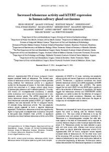
Figure 3. Source of telomerase activity in endometrium. Endometrial epithelial and stromal cells were isolated from endometrial tissues in various menstrual phases (see Materials and Methods), and telomerase activity was examined using 5 mg of protein extract. E, epithelial fractions; S, stromal fractions; N, negative control (lysis buffer only); P, positive control (104 C33A cells).
metrial tissue samples collected at day 3 (during menstruation), day 7 (mid-proliferative phase), day 15 (late proliferative phase), day 20 (early secretory phase), and day 25 (late secretory phase) were fractionated into epithelial glandular cells and stromal cells (see Materials and Methods). TRAP assay revealed high levels of telomerase activity in the fractions of epithelial glandular cells from mid- and late proliferative phases (Figure 3). In contrast, no or only faint activity was observed in these cells in secretory phase and during menstruation. Stromal cell fractions never exhibited telomerase activity at any stage of the menstrual cycle. Thus, telomerase activity was limited to epithelial glandular cells in proliferative phase.
Localization of Human Telomerase RNA Component in Endometrium To further confirm the source of telomerase activity, we next localized human telomerase RNA (hTR) using in situ RNA hybridization. Significant hTR signals were observed in epithelial glandular cells in proliferative phase (Figure 4). However, interestingly, hTR signals were also detected in secretory phase, although less frequently. No or only faint signals were observed in other components, such as stromal cells. These findings further demonstrate the epithelial-specific expression of telomerase activity in human endometrium. Figure 2. Qauntification of telomerase activity in normal endometria and endometrial cancers. Telomerase activity in normal endometria in various menstrual phases and endometrial cancers was determined by stretch PCR assay. Relative telomerase activity was normalized to control activity (100 units) in C33A cells and described in units. EP, early proliferative phase; MP, mid-proliferative phase; LP, late proliferative phase; ES, early secretory phase; LS, late secretory phase; Me, menstruation; At, atrophic endometrium; Ca, endometrial cancer; M, DNA size marker.
Changes in Telomerase Activity during in Vitro Culture of Endometrial Cells To determine the correlation between telomerase activity and proliferation of endometrial cells, the epithelial cells
Telomerase Activity in Human Endometrium 1989 AJP December 1998, Vol. 153, No. 6
Figure 4. Localization of human telomerase RNA in endometrium. Representative results of in situ RNA hybridization for human telomerase RNA are shown. Specific signals were observed in epithelial glandular cells in proliferative-phase endometrium by antisense probes but not by sense probes.
isolated from proliferative-phase endometrium were plated on matrix-coated dishes and cultured. The epithelial cells began to spread over the dishes just after seeding, but soon proliferative activity declined, and significant reduction in proliferative activity was observed after day 4, as confirmed by MTT assay (Figure 5B). Finally, cells became senescent on day 8 or day 9, accompanied by G0/G1 arrest (Figure 5C). Telomerase activity in these cultured cells was monitored. Cells at day 0 exhibited significant telomerase activity, but activity gradually decreased day by day, with no activity detected on day 8
(Figure 5A). Thus, telomerase activity in cultured endometrial cells appeared to be correlated with cellular proliferative activity, and senescence was associated with complete loss of telomerase activity. To clarify whether estrogen affects the growth or telomerase activity of endometrial cells, epithelial cells isolated from proliferative-phase endometrium were cultured in the absence or presence of estrogen. However, neither cell growth nor telomerase activity was affected by estrogen treatment (data not shown). Co-culture with stromal components also had no effect on cell growth or telomerase activity of epithelial cells.
Discussion In this study, we demonstrated epithelial-specific expression of telomerase activity in human endometrium, which was strictly regulated by menstrual phase. Quantitative analysis of telomerase activity revealed that the telomerase activity in late proliferative phase was equivalent to that in endometrial cancer. It is surprising that normal somatic tissues express such strong telomerase activity. Telomerase activation has also been reported in normal somatic cells other than endometrial cells, such as hematopoietic cells, basal stratified skin keratinocytes, and
Figure 5. Telomerase activity, proliferative activity, and cell cycle distribution in cultured endometrial epithelial cells. The epithelial cells isolated from proliferative-phase endometrium were cultured, and cell pellets on days 2, 4, 6, and 8 were subjected to TRAP assay, MTT assay, and cell cycle analysis. A: TRAP assay. N, negative control (lysis buffer only); P, positive control (104 C33A cells). B: MTT assay to evaluate proliferative activity of cells. Relative MTT activity of the cells on days 2, 4, 6, and 8 after isolation was described using a proliferation index (proliferation index of cells on day 0 was defined as 100). C: Flow cytometry for analysis of cell cycle distribution. DNA histograms of cultured epithelial cells on days 2 and day 8 are shown.
1990 Tanaka et al AJP December 1998, Vol. 153, No. 6
intestinal crypt cells, all of which have high regenerative and proliferative activity.11–13 Endometrium proliferates at an extremely high rate in the proliferative phase of the menstrual cycle. These findings suggest that telomerase activation is closely associated with cellular proliferative activity. As proliferative-competent cells progressively lose telomeres due to frequent cell divisions, it is possible that they express high levels of telomerase activity to prevent shortening of telomeres. Our in vitro analysis of endometrial cells revealed that the proliferative activity of epithelial cells decreased in culture and that they become quiescent at day 8 or 9, associated with G0/G1 arrest. This process was accompanied by decrease in telomerase activity, and complete loss of telomerase activity was observed at quiescence. Recent studies have demonstrated that telomerase activity is repressed in cells that exit the cell cycle and enter G0 phase.21,22 Our observations are consistent with the findings of these studies and support the hypothesis that telomerase activity is related to cell proliferation. Belair et al have recently demonstrated that telomerase activity is a biomarker of cell proliferation, rather than malignant transformation.23 Although it remains unclear how telomerase activity is associated with cell proliferation, one possible explanation for this association is that cell proliferation and telomerase activation share signaling pathways. The present findings clearly demonstrate that the source of telomerase activity in endometrium is epithelial glandular cells. Only the epithelial cell fractions from proliferative-phase endometrium exhibited telomerase activity, whereas those from secretory phase and stromal cell fractions did not. This was confirmed by in situ RNA hybridization analysis, demonstrating hTR expression in epithelial cells in proliferative phase. However, in some cases hTR signals were also observed in secretoryphase endometrium without telomerase activity. Recent studies have demonstrated that the expression of the catalytic component of telomerase (human telomerase reverse transcriptase, hTERT) is tightly associated with telomerase activity, whereas that of hTR is more broadly present even in telomerase-negative cells.24 –28 These findings suggest that hTR is commonly expressed in various cell types and that it is required but not sufficient for telomerase activation. The mechanism of this epithelial-specific expression of telomerase activity remains unclear. One possible mechanism is the existence of epithelial-specific transcriptional factors that activate hTERT promoter. Identification and characterization of such factors will contribute to the understanding of molecular mechanisms by which telomerase activity is preferentially expressed in epithelial cells. Another concern of this study is the effect of estrogen on telomerase activity. As estrogen is a potent inducer of endometrial proliferation in vivo, it may play a role in the regulation of telomerase activity. However, estrogen treatment did not affect telomerase activity in cultured epithelial cells. This may be due to the failure of our in vitro culture system to propagate endometrial cells. Successful methods enabling propagation of endometrial cells have been reported for a murine system. Cooke et al
have demonstrated that mitogenic effects of estrogen are mediated by stromal cells with estrogen receptors.29 It has also been reported that the proliferative activity of endometrial epithelial cells was augmented by estrogen when they were co-cultured with stromal cells in mice.30 However, in our assay, co-culture with stromal cells affected neither cell proliferation nor telomerase activity. We do not know whether this is due to differences between human and murine systems. The development of an in vitro assay system permitting endometrial cell proliferation may be needed to elucidate the role of estrogen in telomerase regulation. Further analysis of telomerase regulation in endometrial cells will provide critical insights into the molecular mechanism of telomerase regulation and the role of telomerase activity in human carcinogenesis.
Acknowledgment We thank Drs. C.B. Harley and N.W. Kim (Geron Corp.) for kindly providing hTR plasmid.
References 1. Rhyu MS: Telomeres, telomerase and immortality. J Natl Cancer Inst 1995, 87:884 – 894 2. Watson JD: Origin of concatameric T4 DNA. Nature 1972, 239:197– 201 3. Greider CW, Blackburn EH: A telomeric sequence in the RNA of Tetrahymena telomerase required for telomere repeat synthesis. Nature 1989, 337:331–337 4. Yu GL, Bradley JD, Attardi LD, Blackburn EH: In vivo alteration of telomerase sequences and senescence caused by mutated Tetrahymena telomerase RNAs. Nature 1990, 344:126 –132 5. Counter CM, Avilion AA, LeFeuvre CE, Stewart NG, Greider CW, Harley CB: Telomere shortening associated with chromosome instability is arrested in immortal cells which express telomerase activity. EMBO J 1992, 11:1921–1929 6. Counter CM, Hirte HW, Bacchetti S, Harley CB: Telomerase activity in human ovarian carcinoma. Proc Natl Acad Sci USA 1994, 91:2900 – 2904 7. Kim NW, Piatyszek MA, Prowse KR, Harley CB, West MD, Ho PLC, Coviello GM, Wright WE, Weinrich SL, Shay JW: Specific association of human telomerase activity with immortal cells and cancer. Science 1994, 266:2011–2015 8. Hiyama K, Hiyama E, Ishioka S, Yamakido M, Inai K, Gazdar AF, Piatyszek MA, Shay JW: Telomerase activity in small-cell and non small-cell lung cancers. J Natl Cancer Inst 1995, 87:895–901 9. Hiyama E, Gollahon L, Kataoka, T, Kuroi K, Yokoyama T, Gazdar AF, Hiyama K, Piatyszek MA, Shay JW: Telomerase activity in human breast tumors. J Natl Cancer Inst 1996, 88:116 –122 10. Kyo S, Ueno H, Kanaya T, Inoue M: Telomerase activity in gynecological tumors. Clin Cancer Res 1996, 2:2023–2028 11. Hiyama, K, Hirai, Y, Kyoizumi, S, Akiyama, M, Hiyama, E, Piatyszek, MA, Shay, JW, Ishioka, S, Yamakido M: Activation of telomerase in human lymphocytes and hematopoietic progenitor cells. J Immunol 1995, 155:3711–3715 12. Harle-Bachor C, Boukamp P: Telomerase activity in the regenerative basal layer of the epidermis in human skin and in immortal and carcinoma-derived skin keratinocytes. Proc Natl Acad Sci USA 1996, 93:6476 – 6481 13. Yasumoto S, Kunimura C, Kikuchi K, Tahara H, Ohji H, Yamamoto H, Ide T, Utakoji T: Telomerase activity in normal human epithelial cells. Oncogene 1996, 13:433– 439 14. Kyo S, Takakura M, Kohama T, Inoue M: Telomerase activity in human endometrium. Cancer Res 1997, 57:610 – 614 15. Bradford MM: A rapid and sensitive method for the quantitation of
Telomerase Activity in Human Endometrium 1991 AJP December 1998, Vol. 153, No. 6
16.
17.
18.
19.
20.
21.
22. 23.
microgram quantities of protein utilizing the principle of protein-dye binding. Anal Biochem 1976, 72:248 –254 Tatematsu K, Nakayama J, Danbara M, Shionoya S, Sato H, Omine M, Ishikawa F: A novel quantitative ’stretch PCR assay’ that detects a dramatic increase in telomerase activity during the progression of myeloid leukemias. Oncogene 1996, 13:2265–2274 Kyo, S. Takakura M, Tanaka M, Inoue M: Telomerase activity in cervical cancer is quantitatively distinct from that in its precursor lesions. Int J Cancer 1998, 79:66 –70 Kyo S, Takakura M, Tanaka M, Murakami K, Saito R, Hirano H, Inoue M: Quantitative differences in telomerase activity among malignant, premalignant, and benign ovarian lesions. Clin Cancer Res 1998, 4:399 – 405 Feng J, Funk WD, Wang SS, Weinrich AAA, Chiu CP, Adams RR, Chang E, Allsopp RC, Yu J, Le S, West MD, Harley CB, Andrew WH, Greider CW, Villeponteau B: The RNA component of human telomerase. Science 1995, 269:1236 –1241 Mosmann T: Rapid colorimetric assay for cellular growth and survival: application to proliferation and cytotoxicity assays. J Immunol Methods 1983, 65:55– 63 Holt SE, Aisner DL, Shay JW, Wright WE: Lack of cell cycle regulation of telomerase activity in human cells. Proc Natl Acad Sci USA 1997, 94:10687–10692 Buchkovich KJ, Greider CW: Telomerase regulation during entry into the cell cycle in normal human cells. Mol Biol Cell 1996, 7:1443–1454 Belair CD, Yeager TR, Lopez PM, Reznikoff CA: Telomerase activity: a biomarker of cell proliferation, not malignant transformation. Proc Natl Acad Sci USA 1997, 94:13677–13682
24. Meyerson M, Counter CM, Eaton EN, Ellisen LW, Steiner P, Caddle SD, Ziaugra L, Beijersbergen RL, Davidoff MJ, Liu Q, Bacchetti S, Haber DA, Weinberg RA: HEST2, the putative human telomerase catalytic subunit gene, is up-regulated in tumor cells and during immortalization. Cell 1997, 90:785–795 25. Nakamura TM, Morin GB, Chapman KB, Weinrich SL, Andrews WH, Lingner J, Harley CB, Cech TR: Telomerase catalytic subunit homologs from fission yeast and human. Science 1997, 277:955– 959 26. Nakayama J, Tahara H, Tahara E, Saito M, Ito K, Nakamura H, Nakanishi T, Tahara E, Ide T, Ishikawa F: Telomerase activation by hTRT in human normal fibroblast and hepatocellular carcinoma. Nature Genet 1998, 18:65– 68 27. Takakura M, Kyo S, Kanaya T, Tanaka M, Inoue M: Expression of human telomerase subunits and correlation with telomerase activity in cervical cancer. Cancer Res 1998, 58:1558 –1561 28. Kyo S, Takakura M, Tanaka M, Kanaya T, Sagawa T, Kohama T, Ishikawa H, Nakano T, Shimoya K, Inoue M: Expression of telomerase activity in human chorion. Biochem Biophys Res Commun 1997, 241:498 –503 29. Cooke PS, Buchanan DL, Young P, Seawan T, Brody J, Korach KS, Taylor J, Lubahn DB, Cunha GR: Stromal estrogen receptors mediate mitogenic effects of estradiol on uterine epithelium. Proc Natl Acad Sci USA 1997, 94:6535– 6540 30. Inaba T, Wiest WG, Strickler RC, Mori J: Augmentation of the response of mouse uterine epithelial cells to estradiol by uterine stroma. Endocrinology 1988, 123:1253–1258