with clinical doses of the DNA-damaging drug etoposide. TJ Moriarty1,2 .... lated by analyzing FACS-generated list files using the share- ...... Anticancer. Drugs ...
Leukemia (2002) 16, 1112–1120 2002 Nature Publishing Group All rights reserved 0887-6924/02 $25.00 www.nature.com/leu
Rapid upregulation of telomerase activity in human leukemia HL-60 cells treated with clinical doses of the DNA-damaging drug etoposide TJ Moriarty1,2, S Dupuis2 and C Autexier1,2 1
Department of Anatomy and Cell Biology, McGill University, Montre´al, Que´bec, Canada; and 2The Bloomfield Centre for Research in Aging, Lady Davis Institute for Medical Research, Sir Mortimer B Davis-Jewish General Hospital, Montre´al, Que´bec, Canada
The enzyme telomerase is implicated in cellular resistance to apoptosis, but the mechanism for this resistance remains to be elucidated. The ability of telomerase to synthesize new DNA at telomeres suggests that this enzyme might function in the repair of double-stranded DNA breaks. To distinguish the effects of double-stranded DNA break damage and apoptosis on human telomerase activity, we treated the HL-60 human hematopoietic cancer cell line with clinical doses of the chemotherapeutic drug etoposide (0.5 to 5 M), which allowed us to distinguish between events associated with DNA damageinduced cell cycle arrest, and events associated with apoptosis. Large (three- to seven-fold) upregulation of telomerase activity occurred soon after etoposide treatment (3 h) in S/G2/M-arresting populations; this upregulation was abolished at onset of apoptotic cell death. No upregulation of telomerase activity was observed in cells treated with a larger dose of etoposide (5 M) that caused cells to undergo rapid apoptosis without intervening cell cycle arrests. These observations are consistent with a possible role for telomerase upregulation during the DNA damage response. Leukemia (2002) 16, 1112–1120. DOI: 10.1038/sj/leu/2402522 Keywords: telomerase; DNA damage; apoptosis; leukemia; etoposide
Introduction Telomerase is a unique enzyme that catalyzes the de novo addition of DNA to the 3′ end of telomeres, thereby preventing telomere shortening in the course of successive rounds of DNA replication. It is minimally composed of a template-containing RNA molecule (hTR in human cells), and a protein reverse transcriptase (TERT) which is limiting and necessary for in vivo telomerase activity.1 Telomerase activity is absent in most human somatic cells, but is present in a large majority of immortalized cancer cells.2 Telomere shortening in the absence of telomerase activity can lead to telomeric dysfunction, which is associated with telomeric chromosome fusions, breakages and complex non-reciprocal translocations that are a common feature of human epithelial cancers.3 There is growing evidence that such telomere-mediated events also precede apoptotic cell death,4–6 and combining telomerase inhibition with apoptosis-inducing drug treatments or radiation can cause increased cell death compared to drug treatment alone.7–11 The effects of a number of anti-cancer therapeutic regimens on telomerase activity have been examined. Treatment of human cancer cells and cell lines with certain clinical drugs is associated with decreases in telomerase activity. Telomerase downregulators include the estrogen analogue tamoxifen,12 differentiation inducers such as retinoic acid,13,14 and the Correspondence: C Autexier, Bloomfield Centre for Research in Aging, Lady Davis Institute for Medical Research, Sir Mortimer B Davis-Jewish General Hospital, 3755 chemin Coˆte-Ste-Catherine, Montre´al, Que´bec, H3T 1E2, Canada; Fax: 514-340-8295 Received 30 July 2001; accepted 14 February 2002
DNA-interacting agents daunorubicin, doxorubicin and cisplatin.15–20 However, some of these drugs have complex effects on telomerase activity in treated cells. For example, human testicular cancer cells treated with lethal doses (100 m) of the DNA cross-linker cisplatin exhibit decreased telomerase activity.15 Leukemic cells also downregulate telomerase activity after treatment with cisplatin doses (30 m) larger than the IC50 dose for this drug.16 However, low-dose cisplatin treatment (⬍6.7 m) does not inhibit telomerase activity and is sometimes associated with telomerase upregulation.16,21 Additionally, long-term exposure of colorectal carcinoma cells to cisplatin results in increased telomerase activity, telomere length and drug resistance.22 Cisplatin’s influence on telomerase activity in human cells has been studied more extensively than the effect of other chemotherapeutic drugs. Its apparent dose-dependent effect on telomerase warrants the examination of telomerase activity levels in different cell types subjected to other drug treatments. Other therapeutic regimens reported to elicit telomerase upregulation in treated cells include the DNA-damaging agents bleomycin, 5-fluorouracil and etoposide,16,22–25 and various kinds of radiation.26–34 Telomerase-negative mice with short telomeres are more sensitive to ionizing radiation than their telomerase-positive counterparts, and their cells sustain increased chromosomal damage and apoptosis.11 Thus, telomerase activity and telomere integrity may confer protection against radiation in vivo, and might enhance the resistance of human tumor cells to anti-cancer treatments. The potential role of telomerase upregulation in mediating the resistance of tumor cells to anti-cancer treatments requires further investigation. Our study examined the effect of clinical doses of the chemotherapeutic agent etoposide on telomerase activity in the HL60 human hematopoietic cancer cell line. Etoposide is a topoisomerase II inhibitor that induces double-stranded breaks in DNA. It is commonly used in the treatment of Hodgkin’s disease, non-Hodgkin’s lymphoma, testicular tumors, small cell lung carcinomas, acute lymphoblastic leukemia and childhood neuroblastomas.35,36 Etoposide has been implicated in the development of therapy-related acute myeloid leukemias, specifically acute promyelocytic leukemia, in survivors of primary malignancies.37 However, the events associated with etoposide treatment-related secondary cancers have not been characterized. We treated acute promyelocytic leukemia HL-60 cells with clinically relevant doses of etoposide (0.5 to 5 m)38 and examined telomerase activity at multiple time points up to 48 h. We observed a rapid dose-dependent upregulation of telomerase activity following etoposide treatment, that was not abolished until onset of apoptotic cell death. Apoptosis was preceded by S/G2/M arrests in treated cells that exhibited telomerase upregulation, but not in cells that did not upregulate telomerase. By 24 h after treatment, telomerase activity had increased as much as 20-fold. All etoposide-treated cell
Telomerase upregulation in HL-60 cells treated with etoposide TJ Moriarty et al
populations that upregulated telomerase contained an increased number of cells with hyperdiploid DNA content, whereas treated populations that did not upregulate telomerase contained no cells with a ⬎4N DNA content. However, telomerase hyperactivation preceded the appearance of hyperdiploid cells, suggesting that upstream events leading to the appearance of hyperdiploid DNA content might elicit upregulation of the telomerase enzyme. We concluded that clinically relevant doses of etoposide elicited rapid and sustained telomerase upregulation in a cell cycle arrest-dependent fashion, and that this hyperactivation might constitute part of the regulated DNA damage response in these hematopoietic cancer cells. Materials and methods
Cell culture and drug treatment HL-60 cells were provided by John Th’ng of Cancer Care Ontario. They were grown in RPMI 1640 medium (BioMedia, Montre´al, Canada) supplemented with 20% heat-inactivated FBS (GIBCO-BRL, Burlington, Canada). Seventy-two hours before experiments, cells were diluted to 3 × 10⁵ cells/ml, and were diluted to this density every 24 h before treatment in order to maintain log-phase growth. Cells were strictly maintained at 2–4 × 10⁵ cells/ml during treatment, with DMSOsupplemented medium added to faster-growing controls. Cells were treated with equal volumes of etoposide (Sigma, Oakville, Canada) or DMSO (vehicle), which remained in the medium throughout the experiment. Concentrations of DMSO were used that did not induce differentiation of HL-60 cells (⬍0.03%, vol/vol). At each time point, cells were manually counted after trypan blue staining, and aliquots were collected for cell cycle analysis and telomerase activity based on total cell number (including both viable and non-viable cells).
Cell cycle analysis Two × 105 cells were fixed in 70% ethanol for 45 min, resuspended in phosphate-buffered saline and stored at 4°C. One hour before cell cycle analysis, cells were treated with propidium iodide (50 g/ml) and RNaseA (50 g/ml). The cells in different cell cycle phases were identified by measuring their DNA content using fluorescence-activated cell sorting (FACS) on a Becton Dickinson FACScan machine or on a Beckman Coulter Epics XL machine. Cell cycle distribution was calculated by analyzing FACS-generated list files using the shareware programmeWinMDIv2.8. All flow cytometry data were gated using control samples to eliminate contaminating debris in the subG0 region and pseudo G2/M cell doublets.
Apoptotic DNA fragmentation gels The DNA from 1 × 10⁶ cells was harvested and electrophoresed as previously described.39
Determination of telomerase activity Telomerase extracts were prepared using CHAPS lysis buffer, and telomeric repeat amplification protocol (TRAP) assays
were performed as previously described.40 TRAP primers designed for enhanced quantification (TS, NT and ACX) were included at 20 pmoles per 50 l PCR reaction, as previously described.41 A TSNT internal control substrate41 concentration of 1 × 10⁻²⁰ moles per 50 l TRAP reaction was used to reduce amplification competition with telomerase products. PCR cycling conditions were: 3 min at 94°C, followed by 30 cycles of 30 s at 94°C, 30 s at 60°C and 30 s at 72°C. Five × 105 cells were resuspended in 100 l of lysis buffer, and the protein concentration of extracts was measured using the BioRad Bradford Protein Assay (Mississauga, Canada). TRAP assays were performed on dilutions of selected extracts from every experiment. If subsequent assays on all extracts revealed large amounts of PCR inhibition in individual extracts, these were diluted until inhibition was reduced and quantification of telomerase activity in the linear range with respect to protein input was obtained. TRAP assays were then performed again on all extracts using the new dilution. The dilution usually corresponded to 0.5 to 2 ng protein (50 to 200 cells) per 50 l TRAP reaction. TRAP assays were performed on equal amounts of protein. Telomerase activity was quantified by dividing the signal from the TRAP assay products (generated by amplification of telomerase elongation products using primers TS and ACX) by the internal control signal (generated by amplification of the TSNT internal control substrate using TS and NT primers). Relative telomerase activity was calculated by expressing this ratio for individual extracts as a percentage of the telomerase activity of the 0 h control extract from each treatment (setting the 0 h value at 100% or 1.0). Primer dimers formed between ACX and other primers in the reaction migrate at the lower end of the TRAP ladder. These primer dimers vary in quantity for each set of TRAP assays, independent of primer concentration. To ensure accurate quantification from experiment to experiment, TRAP assay products were measured starting at the first telomerase product above the ACX primer dimer.
1113
Results We investigated the effect of clinical doses of etoposide on telomerase activity, apoptosis, cell cycle progression and necrosis, using the telomerase-positive human promyelocytic leukemia cell line HL-60. Etoposide is used clinically to treat a wide variety of cancers, including acute childhood and adult leukemias.35,36 HL-60 cells, which are highly sensitive to a wide range of DNA-damaging reagents, are an appropriate model system because they are frequently used to examine the regulation of telomerase activity. We wished to examine changes in telomerase activity that are the result of active, regulated cellular processes. Therefore, dose–response experiments were performed to determine the range of etoposide concentrations that would produce substantial but delayed apoptosis and limited, late-occurring necrosis within a 48 h treatment period. The determined concentration range (0.5 to 5 m) closely corresponds to the therapeutic range of serum etoposide concentrations observed in patients treated with standard doses of etoposide (0.5–3 mg/l, or 0.8 to 5 m).38 In addition, critical regulatory events in the DNA damage response often occur in the first few hours after insult, when cells attempt to assess and repair damage before committing to apoptosis.42 Therefore, early time points were collected to capture any potential effects of this early response phase on telomerase activity. Leukemia
Telomerase upregulation in HL-60 cells treated with etoposide TJ Moriarty et al
1114
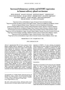
Quantification of telomerase activity PCR-based telomeric repeat amplification protocol (TRAP) assays were first performed using dilutions of cell extracts to determine the protein quantity that would yield optimal quantitative results. The use of nanogram protein quantities (50 to 200 cells) was required for accurate quantification, specifically in extracts containing high levels of telomerase activity (Figure 1a and b). This contrasts with the common use of microgram protein quantities in previous studies using TRAPbased quantification. In the case of the high activity extract shown in Figure 1, increases in extract volume resulted in linear increases in telomerase activity only in reactions containing low protein concentrations of 0.5 to 0.7 ng/l (Figure 1b). We found that the use of microgram quantities of protein was
Figure 1 Analysis and quantification of telomerase activity in HL60 cells containing high levels of telomerase activity. (a) Analysis of telomerase activity by the TRAP assay in dilutions of a cell extract made from HL-60 cells treated with 1 m etoposide for 24 h. TRAP assays were performed with increasing volumes (1, 5, 10 l) of extract diluted to various protein concentrations (0.5, 0.67, 1, 2, 5 ng/l). IC, internal control for PCR. (b) Linearity of TRAP quantification. Telomerase elongation products in (a) were quantified as described in Materials and methods to determine the linear range of telomerase activity with respect to protein concentration. Telomerase activity is plotted as a function of extract volume. Leukemia
saturating, such that no significant changes in activity were detected between extracts that subsequently exhibited a 15fold variation upon dilution to the nanogram level. Representative telomerase activities of etoposide-treated HL-60 cells are shown in Figure 2, and average telomerase activity values from independent experiments are shown graphically in Figure 3. Relative telomerase activity is commonly plotted on a linear scale; however, logarithmic plotting facilitated the visualization of both increases and decreases in activity on one graph, particularly when the range of activity was large (Figure 3: compare linear and logarithmic plots of the same data). All extracts were tested at least twice to confirm the reproducibility of the TRAP results.
Figure 2 Analysis and quantification of telomerase activity in etoposide-treated HL-60 cells. Cell extracts were made from HL-60 cells treated with DMSO (vehicle) and 0.5, 1, 2 and 5 m etoposide, and were analyzed for telomerase activity by TRAP. The telomerase activities shown are representative of telomerase activities from multiple independent etoposide treatments of HL-60 cells. TRAP assays were repeated at least twice for each independent etoposide treatment. Telomerase activity was quantified relative to 0 h controls as described in Materials and methods, and values are shown below each lane.
Telomerase upregulation in HL-60 cells treated with etoposide TJ Moriarty et al
Etoposide-treated HL-60 cells exhibit dose-dependent increases and decreases in telomerase activity
1115
The average relative telomerase activity value for all extracts prepared from DMSO-treated (control) HL-60 cells was 1.03 ± 0.77. DMSO is routinely used as a vehicle for solubilizing etoposide. Unlike recently reported results for ovarian epithelial cell lines,30 we did not observe an increase in telomerase activity following DMSO treatment (Figures 2 and 3: DMSO). Telomerase activity in etoposide-treated cells varied from a 20-fold increase in activity relative to 0 h controls (Figure 2: 2 m, 24 h) to a decrease in activity to less than 10% of controls (Figure 2: 5 m, 36 and 48 h). Telomerase activity was rapidly upregulated 3 to 6 h after etoposide treatment in 0.5 and 1 m-treated cells, with upregulation occurring earlier in cells treated with the higher dose (Figures 2 and 3: 1 m, 3 h; and 0.5 m, 6 h). The magnitude of upregulation also increased with etoposide dose, and was highest in 2 mtreated cells at 24 h (Figures 2 and 3: compare 2, 1 and 0.5 m treatments). The 5 m-treated cells did not exhibit telomerase upregulation at any time point (Figures 2 and 3: 5 m). hTERT protein expression levels did not increase in etoposide-treated cells that upregulated telomerase activity (data not shown). Downregulation of telomerase activity relative to 0 h controls was a late-occurring event at all etoposide doses. Downregulation was detected earliest in 5 m-treated HL-60 cells, where activity began to decline at 24 h after treatment (Figure 3: 5 m). Downregulation of telomerase activity prior to 24 h may be undetectable, since telomerase activity has previously been shown to have a long, 24 h half-life in several cell types.43 Decreases in telomerase activity first appeared at 36 h in 2 m-treated populations, and at 48 h in 1 m-treated cells; however, downregulation was negligible following 0.5 m treatments, even at 48 h (Figure 3: compare 2, 1 and 0.5 m). The magnitude of downregulation was also dosedependent, and decreases in telomerase activity relative to 0 h controls were greatest following treatment with the highest dose of etoposide (Figure 3: compare 5 m to 2, 1 and 0.5 m).
Decreases in telomerase activity in etoposide-treated HL-60 cells follow apoptotic DNA fragmentation, and are associated with late stages of cell death
Figure 3 Etoposide-treated HL-60 cells exhibit dose-dependent increases and decreases in telomerase activity. Cell extracts were made from HL-60 cells treated with DMSO (vehicle; n = 4), 0.5 (n = 3), 1 (n = 2), 2 (n = 1), and 5 m (n = 3) etoposide for up to 48 h. n is the number of independent experiments performed for each treatment. Telomerase activity was analyzed by TRAP, quantified as described in Materials and methods, and expressed relative to the activity of 0 h control cell extracts from each treatment (where 0 h = 1.0). Average telomerase activity values for each time point are represented. Linear plots are provided for reference, since changes in telomerase activity are not usually shown on a logarithmic scale.
Treating cancer cells with higher than the IC50 dose of certain drugs can often result in decreased telomerase activity.16 We measured both apoptosis and necrosis in etoposide-treated HL-60 cells to examine the relationship between changes in telomerase activity and cell death. We distinguished between apoptosis and necrosis to determine if changes in telomerase activity were related to active, regulated cell death (apoptosis) or the passive process of necrosis. The extent of apoptosis in treated cell populations was measured by calculating the percent of total cells with a subG0 DNA content (Figure 4a). Apoptotic DNA fragmentation, indicated by subG0 DNA content, was confirmed by analysis of internucleosomal DNA laddering (Figure 4c) and morphological observations (data not shown). The trypan blue exclusion assay, which detects dead cells with severe membrane damage, was used to measure necrosis. At the etoposide doses used in this study, necrosis did not occur until at least 12 h following DNA fragmentation (Figure 4a and b: compare subG0 curves to time points at which cells are exhibiting ⬎10% necrosis by trypan blue staining). The late onset of necrosis (Figure 4b) and the presLeukemia
Telomerase upregulation in HL-60 cells treated with etoposide TJ Moriarty et al
1116
ence of low molecular weight DNA laddering (Figure 4c) indicated that subG0 DNA fragmentation (Figure 4a) was caused by apoptosis, and was not a by-product of necrosis. Late-onset trypan blue staining likely reflects post-apoptotic necrosis, which is a frequently occurring event in in vitro apoptosis
studies due to the absence of apoptotic cell removal by phagocytosis. The etoposide concentrations chosen for these experiments ranged from doses that elicited an immediate apoptotic DNA fragmentation response as measured by subG0 (Figure 4a: 5 m), to concentrations that did not induce apoptosis (⬎10% of cells in subG0) until 48 h after treatment (Figure 4a: 0.5 m). At all doses except 5 m, cell death was delayed by at least 24 h following etoposide treatment (Figure 4a and b). Declines in telomerase activity below 0 h control values were not coincident with, and did not precede apoptotic DNA fragmentation, but always followed it (compare Figures 3 and 4a). Downregulation of telomerase activity was delayed as much as 21 h after the onset of substantial DNA fragmentation (compare Figures 3 and 4a: 5 m, 3 to 24 h), and no decreases in telomerase activity were detected even when 50% of the cells in a population were in subG0 (compare Figures 3 and 4a: 5 m, 12 h). Therefore, as DNA fragmentation is one of the last events that occurs during apoptosis, we concluded that neither DNA fragmentation nor the preceding apoptotic process is immediately associated with downregulation of telomerase activity. The trypan blue staining results indicated that decreases in telomerase activity relative to 0 h controls were associated with post-apoptotic necrotic membrane permeability. Decreases in activity coincided with increasing membrane permeability (Figures 3 and 4b, compare telomerase activity and trypan blue plots: 1 m, 48 h; 2 m, 36 h; 5 m, 24 h). The magnitude of telomerase downregulation was greatest in populations treated with higher doses of etoposide, which also exhibited the largest increases in trypan blue staining (Figures 3 and 4b: compare 1, 2 and 5 m). This observation is supported by the results of a previous study with leukemic cell lines, that demonstrates a correlation between loss of telomerase activity and plasma membrane damage only at doses significantly exceeding the IC50 of most of the drugs examined.16 In conjunction with the DNA fragmentation data, these results suggest that telomerase activity downregulation in etoposide-treated HL-60 cells is an event associated with late stages of cell death, and not with early events of apoptosis or apoptotic DNA fragmentation.
Figure 4 Apoptotic and necrotic cell death in etoposide-treated HL-60 cells. (a) Analysis of apoptosis in HL-60 cells treated with etoposide over a 48 h time course. DNA fragmentation (apoptosis) was measured by cell cycle analysis and is expressed as % of total cells with subG0 DNA content. Average subG0 values are plotted. n ⭓ 3 for all treatments, where n is the number of independent experiments performed. SubG0 values of ⬎10% indicated the presence of apoptosis. Less than 10% apoptosis occurred in DMSO-treated cells. (b) Analysis of necrosis in HL-60 cells treated with etoposide over a 48 h time course. Necrosis was measured by trypan blue staining and is expressed as % of total cells. Average values are plotted. Trypan blue staining data were collected for all experiments that measured telomerase activity. Trypan blue staining values of ⬎10% indicated the presence of necrotic cell death. Less than 10% necrosis occurred in DMSO-treated cells. (c) Confirmation of apoptosis in etoposidetreated HL-60 cells by analysis of apoptotic DNA fragmentation. Ethidium bromide-stained agarose gel demonstrating the presence of low molecular weight apoptotic DNA laddering in genomic DNA isolated from HL-60 cells treated with 2 m etoposide for 0 to 48 h. The presence of low molecular weight DNA ladders at 24–48 h corresponds to the appearance of large numbers of cells with subG0 DNA content (Figure 4a). 0 m-treated control cells were exposed to vehicle (DMSO). Leukemia
Telomerase upregulation in HL-60 cells treated with etoposide TJ Moriarty et al
Telomerase activity is rapidly upregulated in etoposide-treated HL-60 cells, and upregulation is abolished at onset of cell death Telomerase activity was quickly upregulated in etoposidetreated HL-60 cells, and increased activity appeared earlier with 1 m treatments than 0.5 m treatments (Figure 3: 1 m, 3 h; 0.5 m 6 h). Substantial upregulation of telomerase activity occurred as early as 1 h after treatment with 1 m etoposide (data not shown). Increases in activity first appeared as early as 42 h before the onset of apoptosis (⬎10% cell death) (compare Figures 3 and 4a: 0.5 m 6–48 h). Upregulation of telomerase activity was abolished following an increase in apoptosis (Figures 3 and 4a: 0.5 m 48 h; 1 m 36 h; 2 m 36 h), and was absent in cells that underwent apoptosis soon after treatment (Figure 4a: 5 m). These results reinforce the proposal that telomerase upregulation is an event associated with the early cellular response to etoposide-induced DNA damage, and not with apoptosis.
Telomerase upregulation occurs in etoposide-treated populations that undergo S/G2/M arrests The cell cycle distribution of etoposide-treated HL-60 cells was examined to determine if telomerase upregulation was associated with cell cycle arrests that characteristically occur during the DNA damage response.42 The percent of total cells in different cell cycle phases was determined by calculating the number of cells with 2N, 2 to 4N and 4N DNA content (G0/G1, S and G2/M, respectively), as measured by FACS analysis. The normal cell cycle distribution of DMSO-treated control cells was used to define cell cycle arrests in etoposidetreated cells. The proportion of cells with an abnormal ⬎4N DNA content was also measured, since the presence of hyperdiploid cells can be indicative of genomic damage, including chromosome fusions. Etoposide treatment elicited two types of cell cycle responses, including: (1) S/G2/M arrests followed by the appearance of cells with hyperdiploid DNA content and apoptotic DNA fragmentation (Figure 5: 0.5, 1 and 2 m); and (2) immediate apoptosis without preceding cell cycle arrest (Figure 5: 5 m). HL-60 cells are p53-negative,44 and G1 arrest did not occur at any etoposide dose. Etoposide doses that elicited telomerase upregulation did not cause apoptosis until at least 24 h after treatment (compare Figures 3 and 5). In these cases delayed cell death was preceded by S/G2/M arrests (Figure 5: 0.5, 1 and 2 m). Etoposide doses that induced rapid apoptosis without intervening S/G2/M arrests did not upregulate telomerase activity (compare Figures 3 and 5: 5 m), even at time points as early as 1 h after treatment (data not shown). We concluded that events associated with or leading to cell cycle arrest may be important for telomerase upregulation. However, telomerase activity is not normally cell cycle-regulated in human cells,43 and upregulation of telomerase activity in etoposide-treated HL-60 cells sometimes preceded S/G2/M arrests (compare Figures 3 and 5: 1 m, 3 h). Therefore, we concluded that rapid telomerase upregulation in etoposide-treated HL-60 cells was likely to be associated with elements of the cellular DNA damage response preceding apoptosis, and that telomerase upregulation might be associated with events occurring upstream of S/G2/M arrests.
Telomerase upregulation occurs in etoposide-treated populations which contain cells with hyperdiploid DNA content
1117
Another event which occurred in etoposide-treated HL-60 cells that upregulated telomerase activity was the appearance of cells with hyperdiploid DNA content (Figure 5: 0.5, 1 and 2 m). Populations treated with 0.5, 1 and 2 m concentrations of etoposide also contained large numbers of giant cells, many of which were polyploid (data not shown). Telomerase was hyperactivated only in response to etoposide doses that elicited S/G2/M arrests, hyperdiploid DNA content and subsequent apoptosis in treated cells (Figure 5: 0.5, 1 and 2 m). Cells treated with a 5 m dose of etoposide did not become hyperdiploid (Figure 5), and did not upregulate telomerase activity. However, telomerase upregulation preceded the appearance of hyperdiploid cells (compare Figure 3: 0.5 m 3–6 h; 1 m 3 h with Figure 5: 0.5 m 12–24 h; 1 m 6–12 h), suggesting that telomerase hyperactivation was not a response to abnormal DNA content. To determine if telomerase hyperactivation constituted a response to altered telomere length, we analyzed telomere length by terminal restriction fragment (TRF) analysis. Telomere length was not grossly altered in cells treated with 1 and 2 m doses of etoposide (data not shown), although small changes in telomere length or integrity would not be easily detected by this method. Discussion We treated the HL-60 human leukemia cell line with clinically relevant doses of the commonly used anti-cancer drug etoposide to characterize its effects on telomerase activity, apoptosis, cell cycle progression and necrosis. Telomerase activity did not decline in treated cells until late stages of cell death, after substantial membrane damage. Moreover, telomerase activity was quickly upregulated in cells that underwent cell-cycle arrests in response to etoposide treatment. Telomerase activity declined to pretreatment levels with the onset of cell death, and telomerase activity was not upregulated at an etoposide dose that caused rapid cell death without intervening cell cycle arrest. However, telomerase hyperactivation was not dependent on cell cycle-specific regulation, since upregulation of telomerase activity sometimes preceded cell cycle arrests. This is the first report of early (3 h) upregulation of the human telomerase enzyme in response to the clinical drug etoposide. Early upregulation has previously been reported in irradiated mouse and human hematopoietic cell lines.26,31 In agreement with our results, several radiation studies also noted large increases in telomerase activity 24 h after treatment, as well as dose-dependent hyperactivation26–28,31,32 that plateaus at higher doses.26,28 The magnitude of telomerase upregulation observed in our study is similar to the increases in activity reported for X-irradiated human colorectal carcinoma cell lines.32 In contrast to the increased telomerase activity previously reported for etoposide-treated pancreatic cancer cells,25 we observed a more complex, biphasic pattern of telomerase regulation in etoposide-treated HL-60 cells. This pattern was characterized by an early upregulation of telomerase activity followed by decreases in activity to control levels or below following cell death. In addition, hTERT protein expression levels did not increase in etoposide-treated HL-60 cells that rapidly upregulated telomerase activity, although telomerase upregulation coincides with increased hTERT mRNA Leukemia
Telomerase upregulation in HL-60 cells treated with etoposide TJ Moriarty et al
1118
Figure 5 Telomerase upregulation in etoposide-treated HL-60 cells is dependent on S/G2/M cell cycle arrests and is related to the presence of hyperdiploid DNA content. Telomerase upregulation occurs in etoposide-treated populations undergoing S/G2/M cell cycle arrests, but not in populations that bypass these arrests. Representative cell cycle profiles of HL-60 cells treated with DMSO (vehicle control) and 0.5, 1 and 5 m etoposide for a total of 48 h. Cell cycle phases are identified in the top left panel. Polyploidy (hyperdiploid DNA content) is indicated by arrows in the 0.5, 1 and 2 m plots. Left panels (DMSO, 0.5 and 5 m) show cell cycle data that were collected using a different FACScan machine than data shown in right panels (DMSO, 1 and 2 m). All samples were analyzed with a gate set using 0 h controls. n ⭓ 4 for all treatments, where n is the number of independent experiments.
expression in etoposide-treated pancreatic cancer cells.25 These observations suggest that rapid etoposide-induced telomerase upregulation in HL-60 cells is mediated differently to telomerase hyperactivation in etoposide-treated pancreatic cancer cells. The absence of increased hTERT expression in HL-60 cells and the rapidity of telomerase hyperactivation suggest that telomerase upregulation might be mediated by post-translational mechanisms such as phosphorylation and/or nuclear localization. Rapid transcription- and translationindependent regulation is a hallmark of the DNA damage response.42 Such post-translational mechanisms are also responsible for telomerase activation in human T lymphocytes, where transcriptional regulation is not the major mechanism controlling telomerase activity.45 In fact, rapid radiation- and genotoxic drug-induced telomerase upregulation has been reported only in hematopoietic cells (Refs 26, 31 and this study). Unlike most somatic cells in the human body, hematopoietic cells can express active telomerase, and telomerase may be regulated differently in this tissue type than in other somatic and cancer cells.45 Thus, it will be essential to determine if the early telomerase upregulation that we observe Leukemia
is unique to hematopoietic lineages, and to characterize the molecular mechanisms mediating rapid telomerase hyperactivation. The cellular event(s) which stimulate telomerase hyperactivation are unknown. One hypothesis is that telomerase upregulation may constitute part of the cellular response to DNA damage, as telomerase upregulation has been reported only in cells exposed to DNA-damaging treatments such as radiation.26–28,31–33 This hypothesis is supported by our data, which showed that telomerase upregulation occurred only in cells that undergo an extensive period of cell cycle arrest prior to cell death, a type of response that is characteristic of the DNA damage repair process.42 Double-stranded DNA breakinducing treatments result in broken chromosomes that may mimic exposed or dysfunctional telomeres. Both telomere damage and chromosome breakage result in chromosome fusions.3,5 Interestingly, our data indicated that telomerase upregulation occurred only in response to etoposide doses that also caused the appearance of cells with hyperdiploid DNA content and polyploid morphology, implying that etoposide treatment resulted in extensive genomic damage. How-
Telomerase upregulation in HL-60 cells treated with etoposide TJ Moriarty et al
ever, using terminal restriction fragment (TRF) analysis we did not observe changes in the telomere length of etoposidetreated cells. Late generation telomerase-negative mice have previously been observed to develop substantial numbers of telomeric chromosome fusions following doxorubicin or radiation treatments,10,11 although no significant changes in telomere length are observed when telomeres are measured shortly after radiation treatment.11 Previous studies of radiation- and genotoxic drug-induced telomerase upregulation did not detect telomere shortening in treated cells.11,22,31,32 Similarly, in vivo etoposide treatment does not appear to alter the telomere length of HeLa cells, although etoposide treatment mediates in vitro cleavage of telomeric DNA.46 It remains possible that telomerase upregulation could be mediated by small changes in telomere length undetectable by TRF analysis, or by other forms of DNA or telomeric damage. However, the events precipitating telomerase upregulation have yet to be identified. Etoposide-mediated telomerase upregulation is of potential clinical importance, as the etoposide doses used in this study correspond closely to the therapeutic range of serum etoposide concentrations. One recent study reports that hTERT expression in telomerase-negative cells decreases their sensitivity to topoisomerase inhibitors such as etoposide,9 and long-term cultivation of colorectal carcinoma cells with the genotoxic drugs cisplatin and 5-fluorouracil results in gradual lengthening of telomeres and enhanced drug resistance.22 A clinical study of chemotherapy and epithelial ovarian cancer reports no increase in telomerase activity in cancer cells from treatment responders, whereas cancer cells from 58.3% of treatment nonresponders show an increase in telomerase activity after treatment.47 In addition, repeated exposure of the epidermis of hairless mice to DNA-damaging UV irradiation elicits a progressive increase in telomerase activity that culminates in a 45-fold enhancement of activity in carcinomas.29 Chromosome healing, the de novo addition of telomeric repeats to broken chromosomes, occurs in vertebrates and other organisms; in ciliates and plants, this process is telomerase-mediated.33,48–51 Thus, upregulation of telomerase activity in reponse to DNA-damaging stimuli suggests a potential functional role for this enzyme in response to DNA and/or telomeric damage. We have reported a rapid, dose-dependent increase in telomerase activity in a human hematopoietic cancer cell line treated with clinical doses of etoposide, and this upregulation was not abolished until cell death. Telomerase upregulation appeared to occur in the context of the cellular DNA damage response, a hypothesis which is supported by previous reports of telomerase hyperactivation in response to DNA-damaging treatments. Thus, the potential role of telomerase in the development of cancer cell resistance to clinical treatments such as etoposide warrants future experimental and clinical examination. Acknowledgements We thank E Petroulakis, R Marcotte, J Th’ng and O Tounekti for suggestions related to tissue culture time courses and cell cycle analysis; A LeBlanc for helpful discussions related to apoptosis; and K McDonnell and C Lacelle for training and technical assistance with cell cycle analysis. We also thank R Marcotte, O Tounekti, A LeBlanc and members of the Autexier laboratory for critical reading of the manuscript. This work was supported by a grant from the Cancer Research Society to C Autexier.
References
1119
1 Liu Y, Snow B, Hande M, Yeung D, Erdmann N, Wakeham A, Itie A, Siderovski D, Lansdorp P, Robinson M, Harrington L. The telomerase reverse transcriptase is limiting and necessary for telomerase function in vivo. Curr Biol 2000; 10: 1459–1462. 2 Shay JW, Bacchetti S. A survey of telomerase activity in human cancer. Eur J Cancer 1997; 33: 787–791. 3 Artandi S, Chang S, Lee S, Alson S, Gottlieb G, Chin L, DePinho R. Telomere dysfunction promotes non-reciprocal translocations and epithelial cancers in mice. Nature 2000; 406: 641–645. 4 Zhang X, Mar V, Harrington L, Robinson MO. Telomere shortening and apoptosis in telomerase-inhibited human tumor cells. Genes Dev 1999; 13: 2388–2399. 5 Karlseder J, Broccoli D, Dai Y, Hardy S, de Lange T. p53- and ATM-dependent apoptosis induced by telomeres lacking TRF2. Science 1999; 283: 1321–1325. 6 Pathak S, Risin S, Brown N, Berry K. Telomeric association of chromosomes is an early manifestation of programmed cell death. Int J Oncol 1994; 4: 323–328. 7 Kondo Y, Kondo S, Tanaka Y, Haqqi T, Barna BP, Cowell JK. Inhibition of telomerase increases the susceptibility of human malignant glioblastoma cells to cisplatin-induced apoptosis. Oncogene 1998; 16: 2243–2248. 8 Fu W, Begley JG, Killen MW, Mattson MP. Anti-apoptotic role of telomerase in pheochromocytoma cells. J Biol Chem 1999; 274: 7264–7271. 9 Ludwig A, Saretzki G, Holm P, Tiemann F, Lorenz M, Emrich T, Harley C, Zglinicki Tv. Ribozyme cleavage of telomerase mRNA sensitizes breast epithelial cells to inhibitors of topoisomerase. Cancer Res 2001; 61: 3053–3061. 10 Lee K-H, Rudolph K, Ju Y-J, Greenberg R, Dannizzaro L, Chin L, Weiler S, DePinho R. Telomere dysfunction alters the chemotherapeutic profile of transformed cells. Proc Natl Acad Sci USA 2001; 98: 3381–3386. 11 Goytisolo F, Samper E, Martin-Caballero J, Finnon P, Herrera E, Flores J, Bouffler S, Blasco M. Short telomeres result in organismal hypersensitivity to ionizing radiation in mammals. J Exp Med 2000; 192: 1625–1636. 12 Aldous WK, Marean AJ, DeHart MJ, Matej LA, Moore KH. Effects of tamoxifen on telomerase activity in breast carcinoma cell lines. Cancer 1999; 85: 1523–1529. 13 Reichman T, Albanell J, Wang X, Moore M, Studzinski G. Downregulation of telomerase activity in HL-60 cells by differentiating agents is accompanied by increased expression of telomeraseassociated protein. J Cell Biochem 1997; 67: 13–23. 14 Bestilny L, Brown C, Miura Y, Robertson L, Riabowol K. Selective inhibition of telomerase activity during terminal differentiation of immortal cell lines. Cancer Res 1996; 56: 3796–3802. 15 Burger AM, Double JA, Newell DR. Inhibition of telomerase activity by cisplatin in human testicular cancer cells. Eur J Cancer 1997; 33: 638–644. 16 Akiyama M, Horiguchi-Yamada J, Saito S, Hoshi Y, Yamada O, Mizoguchi H, Yamada H. Cytostatic concentrations of anticancer agents do not affect telomerase activity of leukaemic cells in vitro. Eur J Cancer 1999; 35: 309–315. 17 Ogretmen B, Schady D, Usta J, Wood R, Kraveka J, Luberto C, Birbes H, Hannun Y, Obeid L. Role of ceramide in mediating the inhibition of telomerase activity in A549 human lung adenocarcinoma cells. J Biol Chem 2001; 276: 24901–24910. 18 Asai A, Kiyozuka Y, Yoshida R, Fujik T, Hioki K, Tsubura A. Telomere length, telomerase activity and telomerase RNA expression in human esophageal cancer cells: correlation with cell proliferation, differentiation and chemosensitivity to anticancer drugs. Anticancer Res 1998; 18: 1465–1472. 19 Faraoni I, Turriziani M, Masci G, Vecchis LD, Shay J, Bonmassar E, Graziani G. Decline in telomerase activity as a measure of tumor cell killing by antineoplastic agents in vitro. Clin Cancer Res 1997; 3: 579–585. 20 Faraoni I, Graziani G, Turriziani M, Masci G, Mezzetti M, Testori A, Veronesi U, Bonmassar E. Suppression of telomerase activity as an indicator of drug-induced cytotoxicity against cancer cells: in vitro studies with fresh human tumor samples. Lab Invest 1999; 79: 993–1005. 21 Wang X, Wong S, Pan J, Tsao S, Fung K, Kwong D, Sham J, Nicholls J. Evidence of cisplatin-induced senescent-like growth arrest Leukemia
Telomerase upregulation in HL-60 cells treated with etoposide TJ Moriarty et al
1120 22
23 24
25
26
27 28 29
30 31 32 33 34
35
36
Leukemia
in nasopharyngeal carcinoma cells. Cancer Res 1998; 58: 5019–5022. Kuranaga N, Shinomiya N, Mochizuki H. Long-term cultivation of colorectal carcinoma cells with anti-cancer drugs induces drug resistance and telomere elongation: an in vitro study. BMC Cancer 2001; 1. Nozaki Y, Liu T, Hatano K, Gharaee-Kermani M, Phan S. Induction of telomerase activity in fibroblasts from bleomycin-injured lungs. Am J Respir Cell Mol Biol 2000; 23: 460–465. Sato N, Mizumoto K, Kusumoto M, Niiyama H, Maehara N, Ogawa T, Tanaka M. 9-Hydroxyellipticine inhibits telomerase activity in human pancreatic cancer cells. FEBS Lett 1998; 441: 318–321. Sato N, Mizumoto K, Kusumoto M, Hishio S, Maehara N, Urashima T, Ogawa T, Tanaka M. Up-regulation of telomerase activity in human pancreatic cancer cells after exposure to etoposide. Br J Cancer 2000; 82: 1819–1826. Finnon P, Silver A, Bouffler S. Upregulation of telomerase activity by X-irradiation in mouse leukaemia cells is independent of Tert, Terc, Tnks and Myc transcription. Carcinogenesis 2000; 21: 573–578. Hande M, Balajee A, Natarajan A. Induction of telomerase activity by UV-irradiation in Chinese hamster cells. Oncogene 1997; 15: 1747–1752. Leteurtre F, Li X, Gluckman E, Carosella E. Telomerase activity during the cell cycle and in gamma-irradiated hematopoietic cells. Leukemia 1997; 11: 1681–1689. Balasubramanian S, Kim K-H, Ahmad N, Mukhtar H. Activation of telomerase and its association with G1-phase of the cell cycle during UVB-induced skin tumorigenesis in SKH-1 hairless mouse. Oncogene 1999; 18: 1297–1302. Matte MA-D, Cheng J, Kruk P. Ultraviolet irradiation- and dimethyl sulfoxide-induced telomerase activity in ovarian epithelial cell lines. Exp Cell Res 2001; 267: 13–27. Neuhof D, Ruess A, Wenz F, Weber K. Induction of telomerase activity by irradiation in human lymphoblasts. Radiation Res 2001; 155: 693–697. Joo O, Hande M, Lansdorp P, Natarajan A. Induction of telomerase activity and chromosome aberrations in human tumour cell lines following X-irradiation. Mutat Res 1998; 401: 121–131. Hande M, Lansdorp P, Natarajan A. Induction of telomerase activity by in vivo X-irradiation of mouse splenocytes and its possible role in chromosome healing. Mutat Res 1998; 404: 205–214. Terashima M, Ogawa Y, Toda K, Nishioka A, Inomata T, Kubonishi I, Taguchi H, Yoshida S, Shizuta Y. Effects of irradiation on telomerase activity in human lymphoma and myeloma cell lines. Intl J Mol Med 1998; 2: 567–571. Bishop J, Lowenthal R, Joshua D, Matthews J, Todd D, Cobcroft R, Whiteside M, Kronenberg H, Ma D, Dodds A. Etoposide in acute nonlymphocytic leukemia. Australian Leukemia Study Group. Blood 1990; 75: 27–32. Lodge A, Hall A, Reid M, McIntosh G, Steward M, Anderson J,
37 38
39
40
41 42 43 44 45
46 47
48 49 50 51
Horne C, Angus B. Topoisomerase IIalpha and IIbeta expression in childhood acute lymphoblastic leukaemia: relation to prognostic factors and clinical outcome. J Clin Pathol 2001; 54: 31–36. Kudo K, Yoshida H, Kiyoi H, Numata S, Horibe K, Naoe T. Etoposide-related acute promyelocytic leukemia. Leukemia 1998; 12: 1171–1175. Wurthwein G, Krumpelmann S, Tillmann B, Real E, Schulze-Westhoff P, Jurgens H, Boos J. Population pharmacokinetic approach to compare oral and i.v. administration of etoposide. Anticancer Drugs 1999; 10: 807–814. Tounekti O, Belehradek JJ, Mir L. Relationships between DNA fragmentation, chromatin condensation, and changes in flow cytometry profiles detected during apoptosis. Exp Cell Res 1995; 217: 506–516. Kim NW, Piatyszek MA, Prowse KR, Harley CB, West MD, Ho PLC, Coviello GM, Wright WE, Weinrich SL, Shay JW. Specific association of human telomerase activity with immortal cells and cancer. Science 1994; 266: 2011–2015. Kim NW, Wu F. Advances in quantification and characterization of telomerase activity by the telomeric repeat amplification protocol (TRAP). Nucleic Acids Res 1997; 25: 2595–2597. Rich T, Allen R, Wyllie A. Defying death after DNA damage. Nature 2000; 407: 777–783. Holt S, Aisner D, Shay J, Wright W. Lack of cell cycle regulation of telomerase activity in human cells. Proc Natl Acad Sci USA 1997; 94: 10687–10692. Collins S. The HL-60 promyelocytic leukemia cell line: proliferation, differentiation, and cellular oncogene expression. Blood 1987; 70: 1233–1244. Liu K, Hodes R, Weng N. Cutting edge: telomerase activation in human T lymphocytes does not require increase in telomerase reverse transcriptase (hTERT) protein but is associated with hTERT phosphorylation and nuclear translocation. J Immunol 2001; 166: 4826–4830. Yoon H, Choi I, Kang M, Kim S, Muller M, Spitzner J, Chung I. DNA topoisomerase II cleavage of telomeres in vitro and in vivo. Biochim Biophys Acta 1998; 1395: 110–120. Takahashi M, Kigawa J, Oishi T, Itamochi H, Shimada M, Sato S, Kamazawa S, Akeshima R, Terakawa N. Alteration of telomerase activity in ovarian cancer cells after chemotherapy. Gynecol Obstet Invest 2000; 49: 204–208. Sprung C, Reynolds G, Jasin M, Murnane J. Chromosome healing in mouse embryonic stem cells. Proc Natl Acad Sci USA 1999; 96: 6781–6786. Hanish JP, Yanowitz JL, de Lange T. Stringent sequence requirements for the formation of human telomeres. Proc Natl Acad Sci USA 1995; 91: 8861–8865. Wang H, Blackburn EH. De novo telomere addition by Tetrahymena telomerase in vitro. EMBO J 1997; 16: 866–879. Bednenko J, Melek M, Greene E, Shippen D. Developmentally regulated initiation of DNA synthesis by telomerase: evidence for factor-assisted de novo telomere formation. EMBO J 1997; 16: 2507–2518.