Jun 21, 2004 - muscle and the GDP-forming enzyme from liver (John- ..... Przybyla-Zawislak, B., Dennis, R. A., Zakharkin, S. O., and McCammon, M. T..
THE JOURNAL OF BIOLOGICAL CHEMISTRY © 2004 by The American Society for Biochemistry and Molecular Biology, Inc.
Vol. 279, No. 35, Issue of August 27, pp. 36621–36624, 2004 Printed in U.S.A.
Expression of Two Succinyl-CoA Synthetases with Different Nucleotide Specificities in Mammalian Tissues* Received for publication, June 21, 2004, and in revised form, July 1, 2004 Published, JBC Papers in Press, July 2, 2004, DOI 10.1074/jbc.M406884200
David O. Lambeth‡, Kristin N. Tews, Steven Adkins, Dean Frohlich, and Barry I. Milavetz From the Department of Biochemistry and Molecular Biology, University of North Dakota School of Medicine and Health Sciences, Grand Forks, North Dakota 58202
For nearly 50 years, succinyl-CoA synthetase in animals was thought to be specific for guanine nucleotides. Recently, we purified and characterized both an ADPforming succinyl-CoA synthetase from pigeon breast muscle and the GDP-forming enzyme from liver (Johnson, J. D., Muhonen, W. W., and Lambeth, D. O. (1998) J. Biol. Chem. 273, 27573–27579). Using the sequences of the pigeon enzymes as queries in BLAST searches, we obtained genetic evidence that both enzymes are expressed in a wide range of animal species (Johnson, J. D., Mehus, J. G., Tews, K., Milavetz, B. I., and Lambeth, D. O. (1998) J. Biol. Chem. 273, 27580 –27586). Here we extend those observations by presenting data from Western and Northern blots and enzymatic assays showing that both proteins are widely expressed in mammals with the relative amounts varying from tissue to tissue. We suggest that both succinyl-CoA synthetases catalyze the reverse reaction in the citric acid cycle in which the ADP-forming enzyme augments ATP production, whereas the GDP-forming enzyme supports GTPdependent anabolic processes. Widely accepted shuttle mechanisms are invoked to explain how transport of P-enolpyruvate across mitochondrial membranes can transfer high energy phosphate between the cytosol and mitochondrial matrix.
In earlier reports (1, 2), we reviewed data prior to 1990 suggesting that animals have an ADP-forming succinyl-CoA synthetase (A-SCS1; EC 6.2.1.5) in addition to the GDP-forming succinyl-CoA synthetase (G-SCS; EC 6.2.1.4) discovered in 1954 (3), almost 20 years after formulation of the citric acid cycle by Krebs and Johnson (4). We definitively showed by enzyme purification and characterization that GDP-forming and ADP-forming enzymes occur in pigeon liver and breast muscle, respectively (1). The two enzymes were found to contain the same ␣ subunit but different  subunits that share 54% sequence identity (1, 2). This indicated, contrary to previous reports (5), that the  subunit of the heterodimer determines nucleotide specificity. * This work was supported by the National Science Foundation Grant 9817172. The costs of publication of this article were defrayed in part by the payment of page charges. This article must therefore be hereby marked “advertisement” in accordance with 18 U.S.C. Section 1734 solely to indicate this fact. ‡ To whom correspondence should be addressed: Dept. of Biochemistry and Molecular Biology, University of North Dakota School of Medicine and Health Sciences, P. O. Box 9037, Grand Forks, ND 58202. Tel.: 701-777-2759; Fax: 701-777-3894; E-mail: dlambeth@medicine. nodak.edu. 1 The abbreviations used are: A-SCS, ADP-forming succinyl-CoA synthetase; G-SCS, GDP-forming succinyl-CoA synthetase; A-,  subunit of A-SCS; G-,  subunit of G-SCS; CHAPS, 3-[(3-cholamidopropyl)dimethylammonio]-1-propanesulfonate; DTNB, 5,5⬘-dithiobis(2-nitrobenzoic acid.; RT, reverse transcriptase. This paper is available on line at http://www.jbc.org
Using each  sequence in pigeon as a query in BLAST searches, we found that aligning expressed sequence tags in mouse and human fell into groups of closely and distantly related sequences. The group that aligned closely with the G- subunit aligned more distantly with A- and vice versa. This strongly indicated that the two  subunits arose by gene duplication and also provided evidence that both types of  subunits are expressed in mammals. Early determination of the coding regions of the A- and G- mRNAs in mouse and human has permitted the appropriate assignment of nucleotide specificity to hundreds of expressed sequence tag and mRNA sequences that have been deposited in GenBankTM and other public databases. We also reported data obtained by semiquantitative RT-PCR that confirmed expression of both types of  subunits in several mouse and human tissues (2). The relative amounts of messages varied with tissue type, from being mostly G- in liver to being mostly A- in brain and testis (2). We also reported that the apparent enzyme activity of A-SCS was unaccountably low in tissue homogenates according to the methods of assay used (2). Here results obtained by using Western blotting and a different enzyme assay show that A-SCS is prominently expressed at the protein level in a wide range of mammalian tissues. Knowledge that both ATP- and GDP-forming succinyl-CoA synthetases occur in mammalian tissues has prompted us to further consider the role of the GDP-forming enzyme. The long held assumption that GTP produced by the citric acid cycle is transphosphorylated with ADP in the nucleoside diphosphate kinase reaction is no longer attractive since we now know that A-SCS can synthesize ATP directly. Under “Discussion,” we propose that currently accepted shuttles between mitochondrial matrix and cytosol would allow the phosphorylation potential of P-enolpyruvate to be transferred from mitochondrial matrix to cytosol and vice versa. EXPERIMENTAL PROCEDURES
MTN Multiple Tissue Northern Blot—An MTN multiple tissue Northern blot membrane kit from Clontech that contained eight different adult mouse tissues was probed for messages for succinyl-CoA synthetase subunits. 32P-labeled probes of about 400 bases were prepared by using the MAXIscript in vitro transcription kit from Ambion. Primers used for preparing the probes are listed in Table I. The transcripts were cloned into the pBluescript II KS (⫹/⫺) vector from Stratagene. A NorthernMax kit from Ambion was used to hybridize probes to the membrane. Hybridization was carried out for at least 2 h at 70 °C for the A- and G- probes and 68 °C for the ␣ probe. Membranes were washed twice with 2⫻ SSC (0.3 M NaCl, 0.03 M sodium citrate, pH 7.0), 0.1% SDS for 5 min at room temperature with agitation and then twice with 0.1⫻ SSC containing 0.1% SDS for 15 min at 42 °C. The blots were wrapped in plastic and exposed to film. Observed bands were quantified by densitometry. Western Blots—Antipeptide antibodies to SCS subunits were prepared by immunizing chicken (␣ subunit, Gallus Immunotech, Ontario, Canada) or rabbit (both  subunits, Bioworld, Dublin, OH) using the following sequences: KEAHVDVKFQLPI, A- C-terminal;
36621
36622
Expression of Succinyl-CoA Synthetases in Mammals TABLE I Primers used to prepare probes for Northern blots
Primera
Sequence
Accession no.
Position
G- F G- R A- F A- R ␣F ␣R PAF PAR
5⬘-TGACGGATCCCCCTAAAGTAGTGGGAGAGTTGGCTCA-3⬘ 5⬘-TGACGAATTCGATGGTACAGCTTCGTAATCTGATCTG-3⬘ 5⬘-TGACGGATCCTGAAGATGTGGCTGCCGAGAACCCTGA-3⬘ 5⬘-TGACGAATTCTACTAGACATCCTATGCTCCCATCGAG-3⬘ 5⬘-TGACGGATCCAGAAAACAGGAGCAACGGCTTCTGTCA-3⬘ 5⬘-TGACGAATTCGGGTCACCTCCAATGCCAATACACAAG-3⬘ 5⬘-TGACGGATCCGCTTCCTCTACAGAATGTCACTTGCAG-3⬘ 5⬘-TGACGAATTCACCACCACAGAATCATGACGACTTGCA-3⬘
AF058956 AF058956 AF058955 AF058955 AF144101 AF144101 AF058955 AF058955
243–269 581–607 465–491 829–855 311–337 648–674 1518–1544 1859–1885
a F and R refer to the forward and reverse primers for the subunits indicated to the left. PAF and PAR are primers for an A- probe that was used to test for polyadenylation sites (Fig. 2). The placements of the probes are shown relative to a sequence in GenBankTM.
DAAKKAVASVAKK, G- C-terminal, and GSYTASRKHIYID, N-terminal of expected processed protein for ␣ (6). These peptide sequences are conserved in human, rat, and mouse. The antibodies generated were affinity-purified from serum by the vendor. Mitochondria were isolated from tissues of rat and mouse by using standard procedures for homogenization and centrifugation. Pelleted mitochondria were stored frozen at ⫺20 °C until analyzed by enzymatic assay or SDS-PAGE. Human tissues analyzed by Western blotting were obtained from Clontech as Protein Medley samples prepared by using SDS to solubilize proteins in whole tissue homogenates. For Western blots, 10 g of mitochondrial protein (rat and mouse) or 20 g of Protein Medley (human) were separated by SDS-PAGE using 11% polyacrylamide gels. Following transfer of proteins to polyvinylidene difluoride membranes, the membranes were probed using a 1:2000 dilution of primary antibody and then developed using a 1:70,000 dilution of secondary antibodies. The SCS  proteins were detected by using goat anti-rabbit antibodies conjugated to horseradish peroxidase that were purchased from Bio-Rad. Rabbit anti-chicken antibody from Jackson ImmunoResearch Laboratories was used to detect the ␣ subunit. Bands were visualized using the SuperSignal West Dura extended duration substrate from Pierce and analyzed by a Lumi-Imager from Roche Applied Science that used Lumi-Analyst 3.0 software. Assays of A-SCS and G-SCS activities were performed at 30 °C using a modification of the procedure of Alarcon et al. (7). Frozen mitochondrial pellets were extracted in 20 mM potassium phosphate buffer, pH 7.2, containing 0.4% CHAPS. The complete assay mixture in a volume of 1 ml contained 50 mM potassium phosphate, pH 7.2, 10 mM MgCl2, 0.2 mM succinyl-CoA, 2 mM ADP (for A-SCS) or 1 mM GDP (for G-SCS), and 0.2 mM DTNB). The reactions were initiated by adding succinylCoA and DTNB) in quick succession. Rates were corrected by subtracting the rate observed when ADP or GDP was deleted. The formation of thionitrobenzoate was followed at 412 nm. Controls demonstrated that the observed succinyl-CoA synthetase activities were dependent on magnesium, succinyl-CoA, and nucleoside diphosphate. RESULTS
Following evidence obtained by RT-PCR and analysis of expressed sequence tags in databases that the gene for A-SCS is widely expressed in animal tissues (2), we have further studied the relative levels of A-SCS and G-SCS in selected tissues by using enzymatic assays and Western and Northern blots. The levels of mRNA for the ␣ and two  SCS subunits in eight mouse tissues were determined by Northern blots as shown in Fig. 1. Two transcripts of ⬃2.5 and 1.7 kb were noted for A- with the relative densitometric units shown in Fig. 1 being based on the smaller transcript only. The 1.7-kb transcript accounted for nearly the entire ␣ message in testis and liver. The amount of the 2.5-kb transcript exceeded that of the smaller one in kidney and was also prominent in heart. Expression of the 1.7-kb transcript was strongest in testis and heart followed by kidney and liver. BLAST of the available expressed sequence tags against accession number AF058955 for A- in mouse suggested that the two different length transcripts for A- arose from alternative polyadenylation signals. Examination of the sequence in accession number AF058955 (which lacks the 5⬘-untranslated region) shows alternative polyadenylation signals TATAAA beginning at 1459 and 1475. This was confirmed by Northern blots that compared results obtained by using a probe recog-
FIG. 1. Northern blots of SCS subunits in mouse tissues. The numbers below each lane represent the relative densitometric units adjusted for differences in loading, as determined by the -actin signal, and normalized to a maximum value of 1.0 for each tissue. The values for A- were calculated for the 1.7-kb transcript only. Sk. Mus., skeletal muscle.
nizing the middle of both transcripts with one that was distal to the 3⬘end of the shorter transcripts. The latter probe detected the 2.5-kb transcript only (Fig. 2). A single transcript of ⬃2.5 kb was found for G-. The level of message was highest in kidney followed by liver and heart. It was detected at a very low level in testis, where the message for A- was the highest of the eight tissues examined. The same ␣ subunit is found in both A-SCS and G-SCS enzymes (1). Northern blots revealed a single transcript of 1.5 kb with the strongest expression of ␣ observed in heart followed by kidney, liver, and testis. Western blots were used to study the relative amounts of the subunits of A-SCS and G-SCS in mouse, rat, and human tissues. Antipeptide antibodies were used that recognize the Cterminal sequences of the  subunits and the N-terminal sequence of the processed ␣ subunit in human, rat, and mouse. Fig. 3 shows the Western blots for each subunit in these three animal species. The results indicate that the A- subunit is expressed most highly in testis, brain, heart, and kidney. The G- subunit is highly expressed in liver, kidney, and heart but barely detected in brain and testis. As reported in a prior study (2), we experienced difficulties in reliably measuring the enzymatic activity of A-SCS using either a high pressure liquid chromatography-based method (8) or the spectrophotometric method of Cha and Parks (9). Although the origin of the difficulties is not understood, we were able to get consistent data by using a different assay as described below. Mitochondria were isolated by differential centrifugation in isotonic medium and stored as frozen pellets following decantation of the supernatant after the final wash. The pellets were extracted by resuspension in hypotonic phosphate buffer containing 0.4% CHAPS. After centrifugation, the
Expression of Succinyl-CoA Synthetases in Mammals
36623
TABLE II Specific activities of A-SCS and G-SCS in extracts of rat mitochondria Specific activities are expressed as units/mg of mitochondrial protein where one unit of activity converts one mol of succinyl-CoA to CoA/ minute. Shown are the nucleotide-stimulated rates of conversion of succinyl-CoA to CoASH as followed by the reduction of DTNB. Observed rates were corrected for the nonenzymatic rate of succinyl-CoA hydrolysis by running a control assay in parallel.
FIG. 2. Northern blots of mouse heart and testis using two different probes. The experiment was designed to show that the different size transcripts seen in Fig. 1 are due to different length 3⬘-untranslated regions arising from alternative polyadenylation sites. A shows the result using a probe placed within the coding region. The probe used in B recognized a region 3⬘ to the suspected early polyadenylation signals (Table I).
Tissue
A-SCS
G-SCS
Heart Testis Brain Kidney Liver
0.229 0.095 0.037 0.124 0.053
0.123 0.014 0.001 0.227 0.139
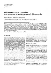
FIG. 4. Scheme showing possible P-enolpyruvate-pyruvate and P-enolpyruvate-malate cycles. FIG. 3. Western blots of SCS subunits in rat, mouse, and human. Mitochondria were isolated from tissues of rat and mouse, and 10 g of mitochondrial protein were applied in each lane. For human, each lane contained 15 g of SDS-solubilized protein derived from a whole tissue homogenate (see “Experimental Procedures”). H, heart; T, testis; B, brain; K, kidney; L, liver; SM, skeletal muscle.
extract was immediately assayed by using a modification of the procedure of Alaracon et al. (7) in which DTNB) is used to follow the formation of CoA from succinyl-CoA (see “Experimental Procedures”). Due to the high absorbance of thionitrobenzoate at 412 nm, the assay is highly sensitive. The results obtained are shown in Table II. The relative amounts of A-SCS and G-SCS vary, from mostly G-SCS in liver to mostly A-SCS in brain and testis. Kidney mitochondria show high activities of both enzymes with the activity of G-SCS exceeding that of A-SCS. The activities are nearly equal in rat heart mitochondria. The results obtained are consistent with the relative intensities of the bands seen in Western blots. Western blots and enzymatic assays of subcellular and submitochondrial fractions showed that A-SCS and G-SCS are both localized in the matrix fraction of mitochondria (data not shown). The possibility that A-SCS might be tetrameric was investigated by chromatography of a kidney extract on Superose 12. Fractions were analyzed by Western blotting, which showed that A-SCS and G-SCS eluted together with an apparent molecular weight consistent with heterodimers. DISCUSSION
In earlier work, we established by RT-PCR that messages encoding the  chains of A-SCS and G-SCS are well represented in a variety of tissues in mouse and human (2). Here we present data for the enzymatic activities of A-SCS and G-SCS
in several tissues of rat. These data together with the results of RT-PCR and Western blots obtained for rat, mouse, and human demonstrate that the relative protein amounts of A-SCS and G-SCS varies with tissue source. The G- subunit is much more highly expressed in anabolic tissues such as liver and kidney, whereas A- predominates in testis and brain. Both enzymes are located in the matrix of mitochondria, where they could participate in the citric acid cycle. We previously found that G-SCS is by far the predominant enzyme detected by enzyme assay in pigeon liver, whereas A-SCS predominates in pigeon breast muscle (1). Since the discovery of G-SCS 50 years ago, most textbooks and reviews have shown that GTP produced by substrate-level phosphorylation in the citric acid cycle undergoes a transphosphorylation with ADP catalyzed by NDP kinase. This presumption is now unattractive because A-SCS is available to form ATP directly. The significance of GTP production by substratelevel phosphorylation has been considered previously (10, 11), and the following discussion is intended to provide a framework for further work. A review of sequenced genomes reveals that genes for two  subunits of SCS are found in some species of eubacteria (for example, Nitrosomonas europaea and Geobacter metallireducens) and archaebacteria (Archaeoglobus fulgidus). In each species, the two  sequences align with 50 – 60% identity, which is similar to that found for vertebrates. Thus the origin of the two  genes in metazoan species may have originated from the endosymbiont that gave rise to mitochondria. Limited data on the nucleotide specificity of succinyl-CoA synthetase in bacterial species suggest that most bacterial enzymes will use both adenine and 6-oxopurine nucleotides
36624
Expression of Succinyl-CoA Synthetases in Mammals
with adenine usually, but not always, being the best (12, 13). Fungal species sequenced thus far have only one gene for the  subunit, and the enzyme in Saccharomyces cerevisiae is specific for ATP (14). The presence of highly specific succinyl-CoA synthetases in metazoan species may be related to the development of specialized tissues and the need for the functional separation of the adenine and guanine nucleotide pools. In yeast, both succinyl-CoA synthetase and P-enolpyruvate carboxykinase are specific for ATP (14). Given that both cytosolic and mitochondrial P-enolpyruvate carboxykinases in animals are GTP-forming, it is attractive to speculate that a functional linkage between P-enolpyruvate carboxykinase and G-SCS is mediated through GTP. Following the discovery of the catabolic role of the citric acid cycle in oxidative tissues, the involvement of cycle enzymes in various anabolic and amphibolic processes has become increasingly appreciated (15). Although ATP supplies the free energy for many biological processes, anabolic reactions are generally supported by GTP, UTP, or CTP (16). G-SCS could directly support anabolic functions of the citric acid cycle within mitochondria by providing GTP for the GTP-requiring steps of protein synthesis as well as the synthesis of P-enolpyruvate by mitochondrial Penolpyruvate carboxykinase. There is no known mechanism for the export of GTP by mitochondria. However, intramitochondrial GTP could support the synthesis of P-enolpyruvate by mitochondrial P-enolpyruvate carboxykinase. Such P-enolpyruvate could then be transported to cytosol by the tricarboxylate or adenine nucleotide transporters (17). In cytosol, as illustrated in Fig. 4, P-enolpyruvate could be used for glucogenesis, for the phosphorylation of GDP by cytosolic P-enolpyruvate carboxykinase, or to synthesize ATP by going through the pyruvate kinase reaction. The latter reaction would increase the adenylate charge and could be a part of “pyruvate cycling,” the rate of which can exceed by severalfold the rate of flux through the citric acid cycle and the synthesis of glucose (18). Rather than being an energy-wasting, futile cycle, such cycling of P-enolpyruvate and pyruvate may be a mechanism for transferring phosphorylation potential from mitochondria to cytosol (19). GTP synthesized from P-enolpyruvate by the reversal of P-enolpyruvate carboxykinase in cytosol could directly support cytosolic protein synthesis and various GTP-binding regulatory proteins. Transport of P-enolpyruvate to the cytosol might occur primarily when the cell is sufficiently energetic to carry out anabolic reactions. The functional role of cytosolic P-enolpyruvate carboxykinase has been thought to be glucogenesis, and the high level of the cytosolic enzyme found in the major gluconeogenic tissues, liver and kidney, is consistent with that role. However, there has long been doubt about whether the primary role of mitochondrial P-enolpyruvate carboxykinase is to support gluconeogenesis (20). The more sensitive techniques of molecular biology have shown that G-SCS and both forms of P-enolpyruvate carboxykinase are expressed at lower levels than those found in kidney and liver in a wide array of immune, nervous, muscle, and secretory tissues.2 Consistent with the reversibility of the P-enolpyruvate carboxykinase reaction in isolated mitochondria (22), an anaplerotic function for the mi2 See bioinfo.weizmann.ac.il/cards-bin/carddisp?_symbol⫽SUCLA2 and bioinfo.weizmann.ac.il/cards-bin/carddisp?_symbol⫽SUCLG2.
tochondrial enzyme has been suggested. Thus the potential involvement of the P-enolpyruvate carboxykinases in roles other than glucose synthesis, including ones in which the reversal of the enzyme is required, should receive attention. The importance of A-SCS presumably lies with the catabolic functions of the citric acid cycle in which the oxidation of acetyl groups provides reducing equivalents for oxidative phosphorylation. The prevalence of A-SCS in the mitochondria of highly catabolic tissues is consistent with this role. The synthesis of an ATP by substrate-level phosphorylation would augment ATP produced by oxidative phosphorylation by about 10%. In the switch from the bloodstream to the procyclic form of Trypanosome brucei, the procyclic form becomes heavily dependent on oxidative metabolism for energy. The switch is accompanied by the development of mitochondria, and the amount of A-SCS strongly increases along with other citric acid cycle enzymes (23). However, the finding in a yeast two-hybrid screen that A-SCS is an interacting protein with aminolevulinate synthase suggests that A-SCS can also support anabolic reactions (21). In summary, the presence of A-SCS in animals is now well established. Ironically, its known presence should refocus interest in the metabolic and regulatory roles of G-SCS. Discovery of how these two synthetases function in the same mitochondria would lead to a better understanding of how the cell balances and regulates anabolism and catabolism. We expect that the mechanisms underlying regulation involve proteinprotein interactions with the two SCS enzymes being associated with different metabolic enzymes. Current techniques for gene manipulation should be used in the design of experiments that will address the relevant questions. REFERENCES 1. Johnson, J. D., Muhonen, W. W., and Lambeth, D. O. (1998) J. Biol. Chem. 273, 27573–27579 2. Johnson, J. D., Mehus, J. G., Tews, K., Milavetz, B. I., and Lambeth, D. O. (1998) J. Biol. Chem. 273, 27580 –27586 3. Sanadi, D. R., Gibson, D. M., and Ayengar, P. (1954) Biochim. Biophys. Acta 14, 434 – 436 4. Krebs, H. A., and Johnson, W. A. (1937) Enzymologia 4, 148 –156 5. Pearson, P. H., and Bridger, W. A. (1975) J. Biol. Chem. 250, 8524 – 8529 6. Majumdar, R., and Bridger, W. A (1990) Biochem. Cell Biol. 68, 292–299 7. Alarcon, C., Wicksteed, B., Prentki, M., Corkey, B. E., and Rhodes, C. J. (2002) Diabetes 51, 2496 –2504 8. Lambeth, D. O., and Muhonen, W. W. (1993) Anal. Biochem. 209, 192–198 9. Cha, S., and Parks, R. E., Jr., (1964) J. Biol. Chem. 239, 1961–1967 10. Ottaway, J. H., McClellan, J. A., and Saunderson, C. L. (1981) Int. J. Biochem. 13, 401– 410 11. Lambeth, D. O. (2002) IUBMB Life 54, 143–144 12. Weitzman, P. D. J., and Jaskowska-Hodges, H. (1982) FEBS Lett. 143, 237–240 13. Kelly, C. J., and Sungman, C. (1977) Arch. Biochem. Biophys. 178, 208 –217 14. Przybyla-Zawislak, B., Dennis, R. A., Zakharkin, S. O., and McCammon, M. T. (1998) Eur. J. Biochem. 258, 736 –743 15. Owen, O. E., Kalhan, S. C., and Hanson, R. W. (2002) J. Biol. Chem. 277, 30409 –30412 16. Atkinson, D. E. (1977) Cellular Energy Metabolism and Its Regulation, pp. 32–35, Academic Press, New York 17. Kleineke, J., Sauer, H., and Soling, H. D. (1973) FEBS Lett. 29, 82– 86 18. Jones, J. G., Solomon, M. A., Cole, S. M., Sherry, A. D., and Malloy, C. R. (2001) Am. J. Physiol. 281, E848 –E856 19. Drahota, Z., Rauchova, H., Mikova, M., and Bass, A. (1981) FEBS Lett. 157, 347–349 20. Modaressi, S., Brechtel, K., Christ, B., and Jungermann, K. (1998) Biochem. J. 333, 359 –366 21. Furuyama, K., and Sassa, S. (2000)) J. Clin. Invest. 105, 757–764 22. Wilson, D. F., Erecinska, M., and Schramm, V. L. (1983) J. Biol. Chem. 258, 10464 –10473 23. Jenkins, T. M., Eisenthal, R., and Weitzman, P. D. (1988) Biochem. Biophys. Res. Commun. 151, 257–261