BIOLOGY OF REPRODUCTION 65, 1548–1557 (2001)
Tissue-Specific Expression of Two Structurally Different Estrogen Receptor Alpha Isoforms along the Female Reproductive Axis of an Oviparous Species, the Rainbow Trout1 Arnaud Menuet, Isabelle Anglade, Gilles Flouriot, Farzad Pakdel, and Olivier Kah2 Endocrinologie Mole´culaire de la Reproduction, UMR CNRS 6026, Campus de Beaulieu, 35042 Rennes Cedex, France
ABSTRACT In oviparous species, in addition to a full-length estrogen receptor alpha (ERa), another ERa isoform lacking the A domain and exhibiting a ligand-independent transactivation function has been consistently reported. Although both isoforms are expressed in the liver, their respective sites of expression in other potential target tissues are unknown. In contrast to the situation in Xenopus and chicken, the two isoforms of rainbow trout (Oncorhynchus mykiss) are generated from two classes of transcripts with different 59 untranslated sequences issued from the same gene by alternative splicing and promoter usage. The aim of this study was to take advantage of the unique organization of the rainbow trout ERa gene to investigate the tissue distribution of these two messenger species along the reproductive axis of female trout. The S1 nuclease assay and in situ hybridization were used, with probes specific for each of the transcripts. Reverse transcription polymerase chain reaction (RT-PCR) with primers specific for each of the isoforms also was performed. The data indicated that the full-length ERa is expressed in liver, brain, pituitary, and ovary, whereas expression of the isoform with the truncated A domain is restricted to the liver, demonstrating a tissue-specific expression of these two ERa isoforms. The presence of a short liver-specific isoform in oviparous species suggests its role in the development and/or maintenance of the unique function of the liver in the vitellogenesis process.
estradiol receptor, hypothalamus, oocyte development, ovary, pituitary
INTRODUCTION
In vertebrates, estradiol (E 2) exerts a wide range of effects on many physiologic processes such as growth, development, or cell differentiation, but it is best known for being a key actor at all levels of the brain-pituitary-gonad axis in both males and females. These effects are mediated by specific nuclear receptors, the estrogen receptors (ER) [1]. These proteins, which belong to the nuclear receptor superfamily, are able to regulate the transcription of target genes and possess a modular organization in six domains from A to F. In the C-terminal region, the E domain is clearly involved in hormone binding and plays a key role in the ligand-dependent transactivation function (AF2). The C domain permits specific interactions with DNA by binding to specific cis-elements called estrogen-responsive eleThis work was supported by the CNRS, the INRA, the French Ministry for Education, Research and Technology, and the Foundation Langlois. 2 Correspondence. FAX: 33 2 99 28 67 94; e-mail:
[email protected] 1
Received: 8 January 2001. First decision: 25 January 2001. Accepted: 27 June 2001. Q 2001 by the Society for the Study of Reproduction, Inc. ISSN: 0006-3363. http://www.biolreprod.org
ments (ERE) and possesses a dimerization capacity. In the N-terminal region, the A/B domain has a ligand-independent transactivation function (AF1) [2, 3]. In mammals, two subtypes of ER have been characterized by molecular cloning, ERa and ERb, which are generated by two different genes [4–6], and for each subtype, different isoforms have been reported [7, 8]. These isoforms are generated from either an alternative exon splicing or usage of different promoters of a single gene [9, 10], and their tissue-specific expression [1] could explain the pleiotropic roles of estrogens on different target tissues. In addition to these functions, in oviparous species E 2 plays a crucial role in the liver by stimulating the synthesis of vitellogenin, the main precursor of the oocyte reserves [11]. In teleosts, an ERa subtype was first characterized in rainbow trout (Oncorhynchus mykiss) [12] and then in other species such as the tilapia [13], the channel catfish [14], the Japanese eel [15], the red sea bream [16], and the gilthead sea bream [17]. More recently, the presence of an ERb has been reported in the gilthead sea bream [18], the channel catfish [19], the rainbow trout (GenBank AJ289883), the goldfish [20], and the zebrafish (GenBank AF349414). In several species (e.g., the Atlantic croaker [21], goldfish [22], and zebrafish (GenBank AF349413), a third form, either ERg or ERb2, has been described. A characteristic shared by all fish ERa cloned until now is the lack of an A domain, but very recently the existence of two ERa isoforms having different estrogen dependencies and issuing from the same gene was reported in rainbow trout [23]. In the rainbow trout, the short isoform (rtERaS, 65 kDa) corresponds to the cDNA previously cloned from the liver [24], whereas the long isoform (rtERaL, 71 kDa) obtained from rainbow trout ovary possesses an extra 45 residues in the N-terminal region [23]. Very recent data in channel catfish also indicate the presence of several N-terminal ERa variants [25]. Similarly, two isoforms of ERa, including one lacking an A domain, have been reported in amphibians [26] and chicken [27], suggesting that the existence of the short ERa isoform could be a common feature of oviparous species. Functional analysis of both ERa isoforms in chicken and rainbow trout showed that the short form exhibits basal estrogen-independent transactivation activity that can be further increased in the presence of E 2, whereas the long form is characterized by strictly estrogen-dependent transcriptional activity [23, 27]. In a recent comparative study of the human and rainbow trout ERa, a conserved a-helicoidal structure that plays a key role in AF1 activity was identified at the beginning of the B domain [28]. The same authors showed that in the absence of ligand the presence of the A domain could repress this AF1 hormone-independent transactivation function [28]. The identification of this new isoform of rtER raises the
1548
ESTROGEN RECEPTOR ALPHA ISOFORMS IN TROUT
1549
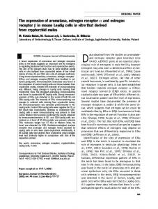
FIG. 1. Schematic representation of the rtERa gene, transcripts, and resulting proteins (adapted from [23]). The positions of the transcription start points of rtERaS (11) and rtERaL (119) transcripts are noted. S1 probes A and B were used for the S1 nuclease mapping. Riboprobes C, L, and S were used for in situ hybridization. Riboprobes S and L are specific for exon 1 and exon 2a, respectively, whereas riboprobe C hybridizes with all rtERa mRNAs.
question of the respective sites of expression of the two forms. All studies aiming at localizing ER mRNAs or proteins were based on riboprobes or antibodies corresponding to the hormone-binding domain, which is common to both rtERaS and rtERaL [29–33], and thus did not allow discrimination between the two mRNAs or proteins. Considering the difference in the structural characteristics of the two rtERa proteins, it is of importance to investigate the tissue specificity, if any, in the expression of these two forms. The truncated form and the full-length form are encoded by two classes of transcripts derived from a single ERa gene. In chicken, the truncated receptor is encoded by a class of mRNA whose initiation site is in the open reading frame of the second class [27], whereas in trout an alternative usage of two promoters generates two classes of transcripts, rtERaL mRNA and rtERaS mRNA1,2, which differ significantly in their 59 untranslated region (Fig. 1) [23]. This unique organization of the rainbow trout ERa gene thus permits generation of specific probes of each class of mRNA encoding two structurally different proteins. Here, we describe in an oviparous species the tissue distribution of the mRNAs corresponding to two different isoforms in different E 2 target tissues. We used nuclease S1 mapping to identify which transcripts are expressed in the different reproductive organs, and these results were confirmed by in situ hybridization and reverse transcription polymerase chain reaction (RT-PCR) techniques, demonstrating the tissue-specific expression of the two mRNA species. MATERIALS AND METHODS
Animals Rainbow trout were supplied by the INRA fish farm (Le Drennec, Finiste`re, France) and kept in the laboratory in a recirculating water system at 12–158C under an artificial light regime mimicking the natural photoperiod (468N). The fish were fed a trout diet ad libitum and were treated in agreement with the European Union regulation concerning the protection of experimental animals. For RNA isolation, fish were killed by de-
capitation, and for in situ hybridization they were perfused intracardially under anesthesia. Immature (100 g), previtellogenic (650 g; gonadosomatic index [GSI]: 0.2%), early vitellogenic (800 g; GSI: 0.3%–0.4%), vitellogenic (1200 g; GSI: 5%), and mature (1500 g; GSI: 12%) females were used. An i.p. injection of E 2 (1.5 mg/kg in ethanol/saline, v/v) was given to three immature, three previtellogenic, and early vitellogenic females 48 h before death. Control fish were given the vehicle alone.
RNA Isolation Total RNA from liver, brain, pituitary, and ovary was extracted with TRIzol (Gibco-BRL, Eggenstein, Germany) as described by the manufacturer. Forebrain, pituitary, liver, and ovary total RNA corresponded to a pool of two to nine fish. The quality of total RNA was checked by agarose gel electrophoresis, and quantification was performed by spectrophotometry at 260 nm.
S1 Nuclease Assay A modified S1 nuclease protection assay was performed as previously described [34]. The method involves the use of biotinylated single-stranded DNA template to prepare highly labeled single-stranded DNA probes by extension from a specific primer with the T7 DNA polymerase in the presence of [a-32P]dCTP (3000 Ci/mmol). These probes were hybridized with an appropriate RNA sample and digested by S1 nuclease (Boehringer Mannheim, Mannheim, Germany). The vectors containing complete rtERaS or rtERaL cDNAs were used to obtain PCR products corresponding to the region from 180 base pairs (bp) in exon 1 to 1510 bp in exon 2 or from 1120 bp in exon 2a to 1595 bp in exon 2, respectively (Fig. 1) [23]. These DNA fragments were subcloned in the PCRTM2.1 vector (Invitrogen, San Diego, CA) downstream of T7 and upstream of the M13 reverse primer. To generate the probes, a PCR was performed with biotinylated T7 primer and the M13 reverse primer. The biotinylated PCR products were purified with the High Pure PCR product purification kit (Boehringer Manheim). Products were then bound to streptavidin-coated magnetic beads (Dynal, Great Neck, NY), and nonbiotinylated DNA strands were eluted in 0.1 M NaOH. The labeled probes were obtained in the presence of [a-32P]dCTP (3000 Ci/mmol) by extending primer 5 (59TCTCCAGGTAGTATGACTGGCTGG-39), complementary to the 39 end of exon 2 and annealed to the biotinylated single-stranded template. The probes were then eluted by an alkaline solution and purified on a 4% denaturing polyacrylamide/urea gel. An amount corresponding to 2 3 105 cpm was coprecipitated with different quantities of RNA (30 mg for liver mRNA samples; 100 mg for ovaries, forebrain, and pituitary mRNA and yeast tRNA samples) and resuspended in 20 ml of hybridization buffer. The templates were then denaturated at 808C for 10 min and incubated at
1550
MENUET ET AL.
538C overnight. Following S1 digestion, the samples were electrophoresed through a denaturing polyacrylamide/urea gel and exposed to a hyperfilmbMax (Amersham International, Uppsala, Sweden) plate for 24 h (liver) or 15 days (brain, pituitary, ovary, and yeast).
Qualitative RT-PCR Complementary DNA was obtained from 1 mg of total RNA following reverse transcription with 50 U of Expand reverse transcriptase (Boehringer Mannheim) and random hexamers. To amplify rtERaL cDNA, PCR was performed with forward primer 6 located in exon 2a (59-GTGAGCCAGTCTAAACCAAGCTG-39) and reverse primer 7 in exon 3 (59GTACCTCGTCTCGTTGGCCAT-39). These primers generated a product 628 bp in length. A forward primer 8 located in exon 1 (59-TCAACACAACAATGCTCATGAT-39) and reverse primer 7 were used to obtain a PCR product of 578 bp corresponding to rtERaS cDNA. Thirty-five cycles of amplification were carried out under the following conditions: denaturation at 948C for 30 sec, annealing at 598C for 30 sec, and extension at 728C for 1 min. The amplification products were analyzed on a 1.5% agarose gel stained with ethidium bromide.
Riboprobe Synthesis For in situ hybridization, three riboprobes were used (Fig. 1) [23]. To generate riboprobe C, a fragment of 776 bp corresponding to the E/F domain of the rtERa was inserted in the site ApaI of the plasmid pBS (Bluescribe; Stratagene, La Jolla, CA). The plasmid was linearized with EcoRI to give the antisense riboprobe using RNA polymerase T3. For the sense riboprobe synthesis by RNA polymerase T7, after linearization by KpnI the plasmid was pretreated with Klenow for blunt-end formation of recessed ends. For riboprobe L, a genomic PCR product corresponding to the region of 256 bp in intron 1 to 1172 bp in exon 2a was obtained with primer 1 (59-ACGAGAAAAGAGAGAGAGAGA-39) and primer 2 (59-CTGTCTGACCAGCATCACATT-39) and was subcloned into PCR 2.1. Likewise, for riboprobe S, a PCR product corresponding to the region of 258 bp in proximal promoter rtERaS to 1140 bp in exon 1 was obtained with primer 3 (59-AGATACACTACTATCAATATCGAT-39) and primer 4 (59-TTAATGACGTTATGGATCAAT-39) and inserted into PCR 2.1. After determination by PCR of the orientation of each inserted fragment, plasmids were linearized by BamHI. Sense and antisense riboprobes L and S were synthesized by RNA polymerase T7, using the RNA transcription kit (Boehringer Mannheim). The linearized DNA template (1 mg) was incubated for 1 h at 378C in a solution containing transcription buffer (13), rATP (0.4 mM), rCTP (0.4 mM), rGTP (0.4 mM), 35S-UTP (5 mCi/ ml), RNase inhibitor (1.6 U/ml), and the adapted RNA polymerase and adjusted to 20 ml with sterile water. The DNA template was digested with 1 ml RQ-1 DNase (1 U/ml) for 15 min at 378C. After incubation, 10 mg yeast tRNA dissolved in 8% formamide was added. Fragments were separated on a Sephadex G50 column as previously described [35]. For in situ hybridization, anesthetized rainbow trout (phenoxy ethanol, 4 ml/10 L fresh water) were perfused intracardially with 0.65% NaCl and 4% paraformaldehyde in PBS (pH 7.4). Brain, pituitary, ovary, and liver were removed and maintained in the same fixative overnight at 48C. After dehydration in ethanol, tissues were embedded in Paraplast and transversally sectioned at 6 mm. Adjacent sections were mounted on parallel TESPA-treated slides (Sigma) and rehydrated prior to hybridization.
In Situ Hybridization Radioactive in situ hybridization was performed as previously described [35]. Tissues were prepared for hybridization by rehydration, treatment with proteinase K (2 mg/ml for brain and pituitary, 20 mg/ml for liver and ovary), acetylation, and dehydration. After being air dried, tissue sections were hybridized under coverslips with the radioactive probe diluted in hybridization buffer (2 3 104 cpm riboprobe/ml; 70 ml on each slide) and incubated for 12–14 h at 558C in a humid chamber. After hybridization, coverslips were removed by bathing in 53 standard saline citrate (SSC; 13: 15 mM sodium citrate, 150 mM NaCl, pH 7) with 10 mM dithiothreitol (DTT) at 558C for 30 min. The slides were washed twice in 23 SSC, 50% formamide, and 10 mM DTT at 658C. After a rinse in NTE buffer (10 mM Tris-HCl, 0.5 M NaCl, 85 mM EDTA, pH 8), sections were treated with RNase A (20 mg/ml NTE; 30 min at 378C) and then rinsed again with NTE buffer before another wash in 23 SSC, 50% formamide, and 10 mM DTT at 658C for 30 min. Before autoradiography, the tissue was rinsed in 23 SSC (15 min) and 0.13 SSC (15 min) at room temperature and dehydrated in an ethanol series containing 0.3 M ammonium acetate. Autoradiography was performed by apposing the slides
to hyperfilm-bMax to produce film autoradiographs for analysis or by dipping the slides into Ilford K5 nuclear track emulsion. After exposure in the dark for 5–18 days at 48C, slides were developed, counterstained with toluidine blue (0.01%), and coversliped in Depex (Gurr). The slides were observed and photographed with a Provis photomicroscope (Olympus France, Rungis, France) using Ilford 400 ASA or Agfapan 25 ASA for bright-field and dark-field photographs, respectively. The specificity of the hybridization signal was systematically checked by hybridizing sense probes on parallel sections.
RESULTS
S1 Nuclease Assay
In the liver, the rtERa gene generates two classes of transcripts, rtERaS mRNA1,2 and rtERaL mRNA, by alternative splicing and by promoter usage (Fig. 1) [23]. The mRNA of rtERaL encodes a protein of 622 amino acids, whereas mRNA1,2 rtERaS codes for an N-terminus truncated protein of 577 amino acids that is 100% identical to the C-terminal portion of the rtERaL protein. To determine the pattern of distribution of these two classes of transcripts in a panel of E 2 target tissues (forebrain, pituitary, liver, and ovary) and to investigate whether any other splicing events occur, S1 nuclease assays were performed. Two single-stranded DNA S1 probes, probe A and probe B specific for rtERaL and rtERaS, respectively, were used. Because rtERa mRNA expression levels strongly differ between tissues, films were exposed for either 24 h (liver) or 15 days (brain, pituitary, and ovary). Yeast tRNA was used as a negative control. After 24 h of exposure, hybridization of probe A with total RNA from liver of E 2-treated and untreated previtellogenic female trout and S1 digestion showed three protected fragments, whereas no signal was detected with yeast tRNA (Fig. 2A). The signal obtained at 475 bp corresponds in size to a total protection of rtERaspecific sequences of probe A, confirming the existence of rtERaL mRNA in the liver. In addition to this band, two specific products of 408 and 366 bp were also detected, resulting from a partial protection of probe A by, respectively, rtERaS mRNA2 and rtERaS mRNA1, which remains homologous to probe A as far as the two acceptor splice sites of exon 2a. Hybridization with probe B followed by S1 digestion and a 24-h exposure, resulted in two protected fragments in liver mRNA samples (treated or not treated with E 2; Fig. 2B). The fragment of 430 bp indicates full protection of probe B by mRNA1 rtERaS, corresponding to exons 1 and 2, and the shortest band of 366 bp indicates partial protection by mRNA rtERaL. The same pattern was obtained with RNA from mature females and E 2-injected males (data not shown). After 15 days of autoradiography, only one protected mRNA fragment was detectable with both probes in the brain and pituitary of mature females or in the ovary of immature females. With probe A, this fragment results from the protection of the full-length probe (exons 2a and 2; Fig. 2A), whereas with probe B, only the fragment corresponding to exon 2 is protected (Fig. 2B). These results indicate that only rtERaL mRNAs are expressed in the brain, pituitary, and ovary. Clear protected fragments were obtained in the ovary and pituitary, but in the forebrain the band intensity was lower, suggesting that rtERaL mRNA expression is low (Fig. 2, A and B). The absence of other protected fragments with probe A suggests that no other 59 splicing mechanisms are at work in these tissues. Whereas E 2 treatment caused a clear increase in the amount of both mRNAs in the liver, the difference was not obvious in the
ESTROGEN RECEPTOR ALPHA ISOFORMS IN TROUT
1551
FIG. 2. Trout tissue specificity of each class of transcripts, rtERaS and rtERaL, as shown by S1 nuclease assay. Total RNA from pituitary, forebrain, ovary (100 mg), and liver (30 mg) were incubated with S1 probe A (A) or probe B (B), submitted to S1 digestion, and separated on a 4% polyacrylamide gel. All samples are from females: brain and pituitary correspond to mature fish, ovary (1E 2, 2E 2) to immature fish, and liver (1E 2, 2E 2) to previtellogenic fish. Films were exposed for 15 days for pituitary, forebrain, and ovary and for 24 h for liver. Yeast tRNA was used as a negative control. The origin of the different protected fragments is schematically represented on the left of each panel. A) Partially protected fragments of 408 (*) and 366 (**) bp corresponding to mRNA2 and mRNA1, respectively, of rtERaS detected only in the liver. A fully protected fragment of 475 bp, corresponding to mRNA of rtERaL, is observed in all tissues. . indicates the undigested probe. B) Partially protected fragment of 366 bp, corresponding to mRNA of rtERaL, in all tissues. A fully protected fragment of 430 bp and corresponding to mRNA of rtERaS was detected only in the liver. . indicates the undigested probe.
ovary. No signal was observed in the ovary of previtellogenic females (GSI: 0.2%; oocyte diameter , 1 mm; data not shown). In Situ Hybridization
The results obtained by S1 nuclease assay were in part confirmed by in situ hybridization on paraffin-embedded sections using three riboprobes. Riboprobe C (776 bp) corresponds to the C-terminal domain of rtERa and hybridizes with both short and long mRNA species. Riboprobe S (198 bp) corresponds to exon 1 and hybridizes only with rtERaS mRNAs, and riboprobe L (238 bp) corresponds to exon 2a and thus is specific of rtERaL mRNAs (Fig. 1) [23]. The liver of E 2-treated and untreated previtellogenic females was used as a positive control because both mRNA populations are expressed in this organ [23]. With riboprobe C, a strong signal was detected in both E 2-treated and untreated samples (Fig. 3A). Riboprobes L and S also resulted in a positive hybridization signal as compared with the respective controls (Fig. 3, B and C). A lower signal was obtained with riboprobes L and S probably because these probes are three times shorter than riboprobe C or because
riboprobe C recognizes both short and long transcripts. In all cases, no signal was detected following hybridization of adjacent sections with the corresponding sense riboprobes (Fig. 3). These results established that the three probes were suitable for the detection of the corresponding mRNAs by in situ hybridization. As expected, the distribution of total rtERa mRNA as revealed by riboprobe C in the brain and the pituitary of mature females was similar to that previously reported in rainbow trout [29]. A clear signal was detected in the preoptic area at the level of the anterior parvocellular preoptic nucleus (Fig. 4) and in the mediobasal hypothalamus at the level of the posterior nucleus lateralis tuberis (Fig. 5). Hybridization of adjacent sections with riboprobe L resulted in exactly the same pattern of distribution of the long rtERaL mRNA, although the signal was lower, probably because of the shorter probe size. However, absolutely no signal was obtained with riboprobe S, indicating the absence or very low expression of rtERaS mRNA in these regions (compare Fig. 4, A–C; Fig. 4, D–F; and Fig. 5, A– C). Adjacent sections treated with the corresponding sense probes did not exhibit any signal (data not shown). FIG. 3. X-ray film images of in situ hybridization in trout liver using riboprobes C (A), L (B), and S (C). The upper panels (2E 2) correspond to control previtellogenic females (650 g; GSI: 0.2%), and the lower panels correspond to E 2-treated previtellogenic females. AS, Antisense probe; S, sense probe. Bar 5 2 mm.
1552
MENUET ET AL.
FIG. 4. In situ hybridization of parallel sections in the preoptic region of mature female trout hybridized with riboprobe C (A and D), riboprobe L (B and E), and riboprobe S (C and F). Panels A, B, and C correspond to the anterior preoptic recess (rpo), and D, E, and F correspond to the level of maximal extension of the preoptic recess. A specific signal was obtained in the anterior nucleus preopticus parvicellularis (Ppa) with riboprobes C and L, but no signal was detected with riboprobe S. Bar 5 100 mm.
Similar data were obtained in the pituitary gland, where a hybridization signal was obtained in the proximal pars distalis with riboprobes C (Fig. 5D) and L (Fig. 5E), but no signal was detected on adjacent sections with riboprobe S (Fig. 5F). No hybridization was detected when using the corresponding sense probes (data not shown). The presence of rtERa mRNAs was also investigated in the ovary of E 2-treated and untreated previtellogenic and
early vitellogenic females. Using riboprobe C, a weak but consistent signal was detected in the follicular layer surrounding the oocytes (Fig. 6A) only in previtellogenic animals. This signal seemed to be increased by E 2 treatment (Fig. 6B). At the histologic level, this labeling clearly corresponds primarily to the granulosa cells (Fig. 6D); the thecal and interstitial cells have fewer silver grains. Hybridization with the sense probe on adjacent sections showed no signal
ESTROGEN RECEPTOR ALPHA ISOFORMS IN TROUT
1553
FIG. 5. In situ hybridization of parallel sections in the trout mediobasal hypothalamus (A–C) and pituitary (D–F) of mature females hybridized with riboprobe C (A and D), riboprobe L (B and E), and riboprobe S (C and F). A specific signal was obtained in the nucleus lateralis tuberis (nlt) and in cells of the proximal pars distalis (PPD) with riboprobes C and L, but no signal was detected with riboprobe S. III, Third ventricle. Bars 5 200 mm (A–C) and 15 mm (D–F).
(Fig. 6, C and E). No labeling could be detected with riboprobe L or riboprobe S, again probably because of their lower specific activities (data not shown). As already observed with the S1 nuclease assay, no in situ hybridization signal was detected in the ovary of previtellogenic females. Qualitative RT-PCR
To confirm expression of rtERaL mRNA in the ovary during the reproductive cycle, qualitative RT-PCR was
used. Single-stranded cDNAs were synthesized by random priming of total RNA from ovaries from immature, previtellogenic, vitellogenic, and mature fish. The liver from previtellogenic E 2-treated and untreated females was used as a positive control. As expected, short and long rtERa amplification products of 578 bp and 628 bp, respectively, were evident in the liver (Fig. 7). Whatever the stage, only amplification products corresponding to rtERaL were visible in the ovary.
1554
MENUET ET AL.
FIG. 6. In situ hybridization of ovaries from a control (A) and E 2-treated (B and C) early vitellogenic female trout using riboprobe C. A signal was detected in the follicular layers with the antisense probe (A, B, and E) but not with the sense probe (C and F). Gr, Granulosa cells; oo, oocyte; th, thecal cells. Bars 5 200 mm (A–C) and 35 mm (E and F).
DISCUSSION
The existence of two rtERa mRNA isoforms differing in their N-terminus has been reported in Xenopus [26], chicken [27, 36], rainbow trout [23], and channel catfish [25] and thus seems to be a common feature of oviparous species. However, although in these species the two isoforms are expressed in the liver, information concerning their tissue expression is limited because in Xenopus and chicken the structure of the gene does not allow manufacture of probes specific for each of the two isoforms. However, the unique structure of the rtERa gene, in which the 59 untranslated regions of the two mRNAs are transcribed from two different exons [23], permits study of the tissue specificity of expression of the two mRNA populations us-
ing probes without overlapping sequences. Using a combination of S1 nuclease assay, in situ hybridization, and RTPCR, tissue-specific expression of two rtERa mRNA isoforms was demonstrated along the female reproductive axis: the rtERaL mRNA species was detected in the liver, brain, pituitary, and ovary, and the rtERaS transcripts were restricted to the liver. With the exception of the liver, expression of rtERa in other tissues was low, as shown by in situ hybridization, but despite the dilution of the rtERa transcripts in a complex mRNA mixture, S1 nuclease assays still allowed detection of a specific signal, highlighting the usefulness of this method. The results indicate that in the different tissues investigated no other splicing mechanisms between exon 1 and exon 2 occur upstream of the
FIG. 7. Qualitative RT-PCR analysis of the rtERa long (lanes 1–7) and rtERa short (lanes 8–14) isoforms in the trout ovary at different stages of maturation. M, markers, bp number is indicated on the left. Lanes 1 and 8: liver, positive control (E 2 treated); lanes 2 and 9: liver, positive control (untreated); lanes 3 and 10: immature; lanes 4 and 11: previtellogenic; lanes 5 and 12: vitellogenic; lanes 6 and 13: mature; lanes 7 and 14: negative control (water).
ESTROGEN RECEPTOR ALPHA ISOFORMS IN TROUT
gene, but we cannot exclude the possible downstream existence of truncated mRNAs, as suggested by the fact that a 1.4-kilobase band was detected in the rainbow trout pituitary by Northern blot [24]. Very short N-terminal-deleted transcripts have been reported in both fishes [25] and mammals [37], but neither these isoforms nor the rtERb would be recognized by our probes. In the brain and pituitary, the present results confirm data obtained previously by in situ hybridization and showing that rtERa mRNAs are localized in portions of the ventral telencephalon, the anterior ventral preoptic region, the mediobasal hypothalamus, and the proximal pars distalis of the pituitary [29]. However, both nuclease S1 and in situ hybridization, using probes specific of the two mRNAs species, indicate that only the long isoform, rtERaL mRNA corresponding to the full-length receptor, is active in these regions. The long mRNAs are likely to be translated into a functional full-length receptor because immunohistochemical studies using antibodies against the hormone-binding domain resulted in the same brain and pituitary distribution as that of the rtERaL mRNAs in the present study [30– 33]. Therefore, all these data indicate that a full-length rtERa is expressed in the brain and pituitary of the rainbow trout. At present, there is little information on the expression of ERb in the brain and the pituitary of fishes, but central expression of ERb and ERg has been reported in the Atlantic croaker [21]. In this species, preliminary in situ hybridization data indicated that ERa, ERb, and ERg mRNAs have a similar but slightly different distribution in the preoptic region and mediobasal hypothalamus [21]. Further studies will be necessary to firmly establish the expression patterns of the different ERs in the brain of teleosts. In mammals, ERb mRNAs have also been detected in the brain and pituitary [38]. The presence of ERa in fish ovary has been revealed by RT-PCR or Northern blot in several species, notably the rainbow trout [39, 40], the gilthead sea bream [18], and the channel catfish [41], but the precise localization and the potential functions of ovarian ERs remain unknown. Fulllength rtERa has been cloned from an ovarian cDNA library [23], and our nuclease S1 assays showed a clear band, corresponding to rtERaL, in the ovary of immature (oocyte diameter , 100 mm) but not in previtellogenic females (oocyte diameter , 1 mm). However, by in situ hybridization, a weak positive signal could be obtained only with riboprobe C in early vitellogenic ovaries (mean oocyte diameter: 1.3 mm) but not in previtellogenic females. These data agree with those of a recent study in rainbow trout, based on quantitative RT-PCR, showing that ovarian ER a mRNAs are found during two distinct periods of ovarian development: in previtellogenic fish with ovarian follicle diameter of #100 mm and in midvitellogenic females with ovarian follicle diameter .1000 mm [39]. However, the primers used in that study did not discriminate between short and long rtERa mRNA species. Our qualitative RTPCR data indicate that at all stages only rtERaL mRNA is expressed in the ovary. These data together suggest that only the full-length rtERa is present in the ovary, but the low expression level probably explains why riboprobe L did not reveal an in situ hybridization signal. Nevertheless, our in situ hybridization results indicate that these transcripts are localized within the follicular layers surrounding the oocytes, more specifically in the granulosa cells. This finding is the first evidence of ERa mRNA in the ovary of a fish. A role of rtERa in sex determination is possible because E 2 treatment of male fish causes intersex [42] or
1555
sex reversal [40]. The pattern of expression of rtERa in the ovary is opposite that of the vitellogenin receptor, whose maximum expression decreases when oocytes reach a diameter of 1000 mm [43]. Thus, ERa in the fish ovary may be involved in the regulation of vitellogenin receptor expression. The question remains whether, as shown in the liver [12, 44, 45], rtERa in the ovary is upregulated by E 2 itself. In addition to rtERa, there is recent evidence in fishes for ovarian expression of rtERb, as reported in tilapia [46], channel catfish [14, 41], gilthead sea bream [18], and goldfish [19, 20]. At present, there is no detailed information regarding the precise ovarian distribution of ERb in teleosts. In mammals, there appears to be a higher level of ovarian expression of ERb as compared with ERa, but there are discrepancies in the literature concerning the cellular distribution of ERa and ERb [38, 47, 48]. The two rtERa isoforms differ strikingly with respect to their transcriptional activity. Similar to ERa from other vertebrates, the full-length receptor exhibits a strictly estrogendependent transcriptional activity, whereas the short isoform has a hormone-independent transactivation capacity, representing 15%–25% of the total receptor activity depending on the cell and promoter context [49]. Steroid receptors possess two transactivation functions, AF1 and AF2, located in the B and E domains, respectively [50]. Recent data have demonstrated that AF1 relies on the presence in the B domain of an alpha helicoidal structure, which is conserved from fishes to mammals, and that AF1 is repressed in the presence of the A domain [28], which explains the hormone-independent activity of the A-truncated rtERa. Until now, there has been no precise information regarding the respective physiologic functions of those two isoforms, whose existence seems to be a characteristic of oviparous species. However, given that the short ERa isoform appears to be mainly expressed in the liver, such receptors could be somehow involved in driving the liver toward its vitellogenic function in the adult and/or during development in oviparous species, according to estrogenindependent mechanisms [23]. Experiments are in progress to study the expression and function of these isoforms during development. In mammals, the existence of several ERa truncated isoforms also has been reported, but these isoforms have been mainly studied in cancer tissues and cell lines. The human ERa gene generates several mRNA variants (A–F mRNA), which differ in their 59 untranslated region but code for the same ERa protein of 66 kDa [51]. Recently, a novel isoform of 46 kDa encoded by the same gene has been described. This isoform is characterized by the lack of an A/ B domain and could play a key role in cell proliferation [7]. In the rat, ERa isoforms lacking the second zinc finger of domain C (isoform S3), part of the hormone-binding domain (S4), or both (S3-4) are transiently expressed in the pituitary during development 4 days earlier than are the full-length ERa isoforms. These S3 and S4 isoforms could block estrogen responsiveness and/or modulate gene transcription in an estrogen-independent fashion [52]. In the adult female rat, TERP-1 (a truncated ERa lacking the A– C domains) is specifically expressed in the pituitary on the day of estrus [37]. The ability of TERP-1 to both enhance and inhibit ER-dependent promoter activity suggests that TERP-1 may play a physiologic role in estrogen feedback in the rat pituitary. The existence of various tissue-specific ER isoforms could partly explain the pleiotropic effects of E 2 in a variety of target organs.
1556
MENUET ET AL.
Two structurally different rtERa isoforms with different E 2 dependencies are differentially expressed along the reproductive axis of the rainbow trout. The liver specifically expresses an A domain-truncated rtERa, which might be involved in the establishment and/or maintenance of the liver vitellogenic function in oviparous species. Because most ERa cDNA sequences from fishes have been obtained from the liver, a full-length ERa may exist in other teleost species, and the differential distribution of the two ERa isoforms reported in rainbow trout may be a common characteristic of all fishes and other oviparous vertebrates. REFERENCES 1. Couse JF, Korach KS. Estrogen receptor null mice: what have we learned and where will they lead us? Endocrinol Rev 1999; 20:358– 417. 2. Klinge CM. Estrogen receptor interaction with co-activators and corepressors. Steroids 2000; 65:227–251. 3. Evans RM. The steroid and thyroid hormone receptor superfamily. Science 1988; 240:889–895 4. Green S, Walter P, Kumar V, Krust A, Bornert JM, Argos P, Chambon P. Human estrogen receptor cDNA: sequence, expression and homology to v-erb-A. Nature 1986; 320:134–139. 5. Kuiper GG, Enmark E, Pelto-Huikko M, Nilsson S, Gustafsson JA. Cloning of a novel receptor expressed in rat prostate and ovary. Proc Natl Acad Sci U S A 1996; 93:5925–5930. 6. Katzenellenbogen BS, Korach KS. A new actor in the estrogen receptor drama: enter ER-beta. Endocrinology 1997; 138:861–862. 7. Flouriot G, Brand H, Denger S, Metivier R, Kos M, Reid G, SonntagBuck V, Gannon F. Identification of a new isoform of the human estrogen receptor-alpha (hER-alpha) that is encoded by distinct transcripts and that is able to repress hER-alpha activation function 1. EMBO J 2000; 19:4688–4700. 8. Moore JT, McKee DD, Slentz-Kesler K, Moore LB, Jones SA, Horne EL, Su JL, Kliewer SA, Lehmann JM, Willson TM. Cloning and characterization of human estrogen receptor beta isoforms. Biochem Biophys Res Commun 1998; 247:75–78. 9. Friend KE, Ang LW, Shupnik MA. Estrogen regulates the expression of several different estrogen receptor mRNA isoforms in rat pituitary. Proc Natl Acad Sci U S A 1995; 92:4367–4371. 10. Chu S, Fuller PJ. Identification of a splice variant of the rat estrogen receptor beta gene. Mol Cell Endocrinol 1997; 132:195–199. 11. Flouriot G, Pakdel F, Ducouret B, Ledrean Y, Valotaire Y. Differential regulation of two genes implicated in fish reproduction: vitellogenin and estrogen receptor genes. Mol Reprod Dev 1997; 48:317–323. 12. Pakdel F, Le Guellec C, Vaillant C, Le Roux MG, Valotaire Y. Identification and estrogen induction of two estrogen receptors (ER) mRNA ribonucleic acids in the rainbow trout liver: sequence homology with other ERs. Mol Endocrinol 1989; 3:44–51. 13. Tan NS, Lam TJ, Ding JL. Molecular cloning and sequencing of the hormone-binding domain of Oreochromis aureus estrogen receptor gene. DNA Seq 1995; 5:359–370. 14. Xia Z, Patino R, Gale WL, Maule AG, Densmore LD. Cloning, in vitro expression, and novel phylogenetic classification of a channel catfish estrogen receptor. Gen Comp Endocrinol 1999; 113:360–368. 15. Todo T, Adachi S, Yamauchi K. Molecular cloning and characterization of Japanese eel estrogen receptor cDNA. Mol Cell Endocrinol 1996; 119:37–45. 16. Touhata K, Kinoshita M, Toyohara H, Sakaguchi M. Sequence and expression of a cDNA encoding the red seabream estrogen receptor. Fish Sci 1998; 64:131–135. 17. Munoz-Cueto JA, Burzawa-Gerard E, Kah O, Valotaire Y, Pakdel F. Cloning and sequencing of the gilthead sea bream estrogen receptor cDNA. DNA Seq 1999; 10:75–84. 18. Socorro S, Power DM, Olsson PE, Canario AV. Two estrogen receptors expressed in the teleost fish, Sparus aurata: cDNA cloning, characterization and tissue distribution. J Endocrinol 2000; 166:293–306. 19. Xia Z, Gale WL, Chang X, Langenau D, Patino R, Maule AG, Densmore LD. Phylogenetic sequence analysis, recombinant expression, and tissue distribution of a channel catfish estrogen receptor beta. Gen Comp Endocrinol 2000; 118:139–149. 20. Tchoudakova A, Pathak S, Callard GV. Molecular cloning of an estrogen receptor beta subtype from the goldfish, Carassius auratus. Gen Comp Endocrinol 1999; 113:388–400.
21. Hawkins MB, Thornton JW, Crews D, Skipper JK, Dotte A, Thomas P. Identification of a third distinct estrogen receptor and reclassification of estrogen receptors in teleosts. Proc Natl Acad Sci U S A 2000; 97:10751–10756. 22. Ma CH, Dong KW, Yu KL. cDNA cloning and expression of a novel estrogen receptor beta-subtype in goldfish (Carassius auratus). Biochim Biophys Acta 2000; 1490:145–152. 23. Pakdel F, Metivier R, Flouriot G, Valotaire Y. Two estrogen receptor (ER) isoforms with different estrogen dependencies are generated from the trout ER gene. Endocrinology 2000; 141:571–580. 24. Pakdel F, Le Gac F, Le Goff P, Valotaire Y. Full-length sequence and in vitro expression of rainbow trout estrogen receptor cDNA. Mol Cell Endocrinol 1990; 71:195–204. 25. Patino R, Xia Z, Gale W, Wu C, Maule AG, Chang X. Novel transcripts of the estrogen receptor a gene in channel catfish. Gen Comp Endocrinol 2000; 120:314–325. 26. Claret FX, Chapel S, Garces J, Tsai-Pflugfelder M, Bertholet C, Shapiro DJ, Wittek R, Wahli W. Two functional forms of the Xenopus laevis estrogen receptor translated from a single mRNA species. J Biol Chem 1994; 269:14047–14055. 27. Griffin C, Flouriot G, Sonntag-Buck V, Gannon F. Two functionally different protein isoforms are produced from the chicken estrogen receptor-alpha gene. Mol Endocrinol 1999; 13:1571–1587. 28. Me´tivier R, Petit FG, Valotaire Y, Pakdel F. Function of N-terminal transactivation domain of the estrogen receptor requires a potential ahelical structure and is negatively regulated by the A domain. Mol Endocrinol 2000; 14:1849–1871. 29. Salbert G, Bonnec G, Le Goff P, Boujard D, Valotaire Y, Jego P. Localization of the estradiol receptor mRNA in the forebrain of the rainbow trout. Mol Cell Endocrinol 1991; 76:173–180. 30. Pakdel F, Petit F, Anglade I, Kah O, Delaunay F, Bailhache T, Valotaire Y. Overexpression of rainbow trout estrogen receptor domains in Escherichia coli: characterization and utilization in the production of antibodies for immunoblotting and immunocytochemistry. Mol Cell Endocrinol 1994; 104:81–93. 31. Anglade I, Pakdel F, Bailhache T, Petit F, Salbert G, Jego P, Valotaire Y, Kah O. Distribution of estrogen receptor-immunoreactive cells in the brain of the rainbow trout (Oncorhynchus mykiss). J Neuroendocrinol 1994; 6:573–583. 32. Navas JM, Anglade I, Bailhache T, Pakdel F, Breton B, Jego P, Kah O. Do gonadotrophin-releasing hormone neurons express estrogen receptors in the rainbow trout? A double immunohistochemical study. J Comp Neurol 1995; 363:461–474. 33. Linard B, Anglade I, Corio M, Navas JM, Pakdel F, Saligaut C, Kah O. Estrogen receptors are expressed in a subset of tyrosine hydroxylase-positive neurons of the anterior preoptic region in the rainbow trout. Neuroendocrinology 1996; 63:156–165. 34. Flouriot G, Nestor P, Kenealy M, Pope C, Gannon F. An S1 nuclease mapping method for detection of low abundance transcripts. Ann Biochem 1996; 237:159–161. 35. Mazurais D, Brierley I, Anglade I, Drew J, Randall C, Bromage N, Michel D, Kah O, Williams LM. Central melatonin receptors in the rainbow trout: comparative distribution of ligand binding and gene expression. J Comp Neurol 1999; 409:313–324. 36. Griffin C, Flouriot G, Sonntag-Buck V, Nestor P, Gannon F. Identification of novel chicken estrogen receptor-alpha mRNA ribonucleic acid isoforms generated by alternative splicing and promoter usage. Endocrinology 1998; 139:4614–4125. 37. Schreihofer DA, Resnick EM, Soh AY, Shupnik MA. Transcriptional regulation by a naturally occurring truncated rat estrogen receptor (ER), truncated ER product-1 (TERP-1). Mol Endocrinol 1999; 13: 320–329. 38. Shughrue PJ, Lane MV, Scrimo PJ, Merchenthaler I. Comparative distribution of estrogen receptor-a (ERa) and b (ERb) mRNA in the rat pituitary, gonad and reproductive tract. Steroids 1998; 63:498–504. 39. Nagler JJ, Krisfalusi M, Cyr DG. Quantification of rainbow trout (Oncorhynchus mykiss) estrogen receptor-alpha mRNA RNA and its expression in the ovary during the reproductive cycle. J Mol Endocrinol 2000; 25:243–251. 40. Guiguen Y, Baroiller JF, Ricordel MJ, Iseki K, McMeel OM, Martin SA, Fostier A. Involvement of estrogens in the process of sex differentiation in two fish species: the rainbow trout (Oncorhynchus mykiss) and a tilapia (Oreochromis niloticus). Mol Reprod Dev 1999; 54:154– 162. 41. Xia Z, Gale WL, Chang X, Langenau D, Patino R, Maule AG, Densmore LD. Phylogenetic sequence analysis, recombinant expression,
ESTROGEN RECEPTOR ALPHA ISOFORMS IN TROUT
42.
43.
44.
45.
46.
and tissue distribution of a channel catfish estrogen receptor beta. Gen Comp Endocrinol 2000; 118:139–149. Krisfalusi M, Nagler JJ. Induction of gonadal intersex in genotypic male rainbow trout (Oncorhynchus mykiss) embryos following immersion in estradiol-17beta. Mol Reprod Dev 2000; 56:495–501. Perazzolo LM, Coward K, Davail B, Normand E, Tyler CR, Pakdel F, Schneider WJ, Le Menn F. Expression and localization of mRNA ribonucleic acid for the vitellogenin receptor in ovarian follicles throughout oogenesis in the rainbow trout, Oncorhynchus mykiss. Biol Reprod 1999; 60:1057–1068. Le Drean Y, Lazennec G, Kern L, Saligaut D, Pakdel F, Valotaire Y. Characterization of an estrogen-responsive element implicated in regulation of the rainbow trout estrogen receptor gene. J Mol Endocrinol 1995; 15:37–47. Lazennec G, Kern L, Salbert G, Saligaut D, Valotaire Y. Cooperatioon between the human estrogen receptor and MCF-7 cell-specific transcription factors elicits high activity of an estrogen inducible enhancer from the trout ER gene promoter. Mol Endocrinol 1996; 10:1116– 1126. Chang XT, Kobayashi T, Todo T, Ikeuchi T, Yoshiura Y, Kajiura-Kobayashi H, Morrey C, Nagahama Y. Molecular cloning of estrogen
47. 48.
49. 50. 51.
52.
1557
receptors alpha and beta in the ovary of a teleost fish, the tilapia (Oreochromis niloticus). Zool Sci 1999; 16:653–658. Wang H, Eriksson H, Sahlin L. Estrogen receptors alpha and beta in the female reproductive tract of the rat during the estrous cycle. Biol Reprod 2000; 63:1331–1340. Bao B, Kumar N, Karp RM, Garverick HA, Sundaram K. Estrogen receptor-beta expression in relation to the expression of luteinizing hormone receptor and cytochrome P450 enzymes in rat ovarian follicles. Biol Reprod 2000; 63:1747–1755. Petit F, Valotaire Y, Pakdel F. Differential functional activities of rainbow trout and human estrogen receptors expressed in the yeast Saccharomyces cerevisiae. Eur J Biochem 1995; 233:584–592. Gronemeyer H, Laudet V. Transcription factors 3: nuclear receptors. CCQ 1995; 2:1173–1308. Flouriot G, Griffin C, Kenealy M, Sonntag-Buck V, Gannon F. Differentially expressed mRNA RNA isoforms of the human estrogen receptor-alpha gene are generated by alternative splicing and promoter usage. Mol Endocrinol 1998; 12:1939–1954. Pasqualini C, Guivarc’h D, Boxberg YV, Nothias F, Vincent JD, Vernier P. Stage- and region-specific expression of estrogen receptor alpha isoforms during ontogeny of the pituitary gland. Endocrinology 1999; 140:2781–2789.