Journal of General Virology (2007), 88, 758–769
DOI 10.1099/vir.0.82623-0
Extensive sequence variation exists among isolates of murine cytomegalovirus within members of the m02 family of genes Alexandra J. Corbett,1,2 Catherine A. Forbes,1,2 Dorian Moro33 and Anthony A. Scalzo1,2 Correspondence Anthony A. Scalzo
[email protected]
1
Immunology and Virology Program, Centre for Ophthalmology and Visual Science, University of Western Australia, Nedlands, WA 6009, Australia
2
Centre for Experimental Immunology, Lions Eye Institute, 2 Verdun Street, Nedlands, WA 6009, Australia
3
School of Natural Sciences, Edith Cowan University, Joondalup, WA 6027, Australia
Received 5 October 2006 Accepted 16 November 2006
Murine cytomegalovirus (MCMV) is a widely used model for human cytomegalovirus (HCMV) and has facilitated many important discoveries about the biology of CMVs. Most of these studies are conducted using the laboratory MCMV strains Smith and K181. However, wild-derived isolates of MCMV, like HCMV clinical isolates, exhibit genetic variation from laboratory strains, particularly at the ends of their genomes in areas containing known or putative immune-evasion and tropism genes. This study analysed the nucleotide sequence of the m02–m05 region, within the m02 gene family, of a number of laboratory and wild-derived MCMV isolates, and found a large degree of variation in both the sequence and arrangement of genes. A new open reading frame (ORF), designated m03.5, was found to be present in a number of wild isolates of MCMV in place of m03. Two distinct isolates, W8 and W8211, were found to possess both m03 and m03.5. Both m03 and m03.5 had early transcription kinetics and the encoded proteins could be detected on the cell surface, consistent with a possible role in immune evasion through binding to host-cell proteins. These data show that gene duplication and sequence variation occur within different isolates of MCMV found in the wild. As this variation among strains may alter the function of genes, these findings should be considered when analysing gene function or host–virus interactions in laboratory models.
INTRODUCTION Murine cytomegalovirus (MCMV) has been used extensively as a model for human cytomegalovirus (HCMV) infection, as strict species specificity makes in vivo HCMV studies difficult. CMVs are betaherpesviruses with slow replication cycles that are capable of persisting for the lifetime of their respective hosts (Ho, 1991). These viruses have large double-stranded DNA genomes (~230 kb) and encode many immune-modulating proteins. The two viruses share many biological characteristics and considerable genetic homology, and the MCMV model has facilitated many important discoveries about the pathology and the immune control of CMV infections. 3Present address: Chevron Australia Pty Ltd, QV1 Building, 250 St George’s Terrace, Perth, WA 6000, Australia. The GenBank/EMBL/DDBJ accession numbers for the sequences determined in this study are EF031243–EF031268. A supplementary table showing primers used for RT-PCR analysis of m03, m03.5 and m04 is available in JGV Online.
758
Most MCMV studies are conducted using the laboratory strains Smith or K181, both of which have been passaged extensively in vitro since their isolation. Variations in the genetic and biological properties of these strains have been reported (Boname & Chantler, 1992; Hudson et al., 1988; Misra & Hudson, 1980). In addition, CMVs have evolved alongside their hosts over millions of years, resulting in significant genetic and biological variation among MCMV wild-derived isolates (Booth et al., 1993; Lyons et al., 1996; Smith et al., 2006; Voigt et al., 2003; Xu et al., 1996), as well as extensive genetic variation among HCMV clinical isolates, which has implications for the immune control and pathogenesis of different HCMV strains (reviewed by Pignatelli et al., 2004). The MCMV Smith strain is predicted to encode at least 170 open reading frames (ORFs) and, although the sequence of the Smith MCMV genome has been determined (Brocchieri et al., 2005; Rawlinson et al., 1996), many of these have not been characterized. Homologues of approximately 80 HCMV ORFs are encoded in the central region of the MCMV genome, 0008-2623 G 2007 SGM
Printed in Great Britain
m02 family sequence variation in MCMV wild isolates
whereas the left and right ends are not homologous to HCMV. These non-homologous regions of both viruses contain genes that are ‘non-essential’ for growth in vitro and include a number of clearly defined immune-evasion functions (reviewed by Alcami & Koszinowski, 2000; Hengel et al., 1998). Genetic variation in these regions therefore has implications for immune control of these viruses in their respective hosts. The functions of a number of these immuneevasion genes are also likely to be host-genotype specific. This is perhaps not widely appreciated, as most MCMV studies analyse the laboratory strains Smith and K181 in inbred strains of mice. Such model systems may not accurately represent the spectrum of interactions in the natural hosts. The MCMV genome contains two families of glycoproteins (the m02 and m145 gene families) at opposite ends of the genome. The m02 gene family includes m02–m16 and forms three subgroups (m02–m06, m07–m10 and m11–m16) based on conserved cysteine residues (Rawlinson et al., 1996). This gene family is ‘non-essential’ for MCMV growth in vitro, and members play a role in natural killer (NK) cell-mediated immune escape in vivo (Oliveira et al., 2002) and in subverting T-cell responses (reviewed by Hengel et al., 1999). The m04 gene product (gp34) binds to major histocompatibility complex (MHC) class I molecules in the endoplasmic reticulum (ER) and on the cell surface (Kavanagh et al., 2001; Kleijnen et al., 1997). The m06 gene encodes a protein that downregulates the surface expression of MHC class I proteins by redirecting them to lysosomal compartments (Reusch et al., 1999). The functions of m02, m03 and m05 have not been determined. When m02 alone is knocked out, there is no substantial phenotype in vivo, suggesting that this gene has no significant immune-evasion role in the C57BL/6 mouse strain (Oliveira et al., 2002). Thus, some m02 family members have functions that have been defined by studies in inbred mice, whereas the roles of others in host–virus interactions are not evident based on the limited analyses performed so far. In the current study, we compared the sequence of the m02– m05 gene region of seven MCMV strains, comprising the laboratory strains Smith and K181 and five wild-derived isolates. These viruses were found to have three distinct gene arrangements within this region. We have described a new ORF, designated m03.5, which is present in a number of distinct MCMV isolates either in place of, or in addition to, m03. Individual ORFs were sequenced for additional wildderived isolates and showed a high degree of variation within some genes. The transcriptional kinetics and surface expression of m03 and m03.5 were determined. Our observations raise the possibility that the sequence variation in these genes has been selected for by functional binding to heterogeneous host-cell surface molecules.
METHODS Viruses and viral DNA preparation. The MCMV laboratory
strains used were Smith (obtained from E. S. Mocarski, Stanford University, USA) and K181-Perth (abbreviated to K181). A number http://vir.sgmjournals.org
of wild-derived isolates (G1F, G2, G3B, G4, G5, G6, K4, K6, K17B, N1, N5, W2, W8 and W8211) have been described previously (Booth et al., 1993). Isolates MI2A, MI2C, MI3A, MI4A, MI6A, MI4B, MI4C, MI6C, TR8A and TR8B were derived by sequential limiting dilution purification from salivary glands from feral house mice trapped on Macquarie Island, Australia (54u 309 S 158u 579 E). Tissue culture virus stocks were produced by propagation in mouse embryonic fibroblasts (MEFs) and titres determined by standard plaque assay, as described previously (Farrell & Shellam, 1989). Viral DNA was produced from infected MEFs, as described previously (Xu et al., 1996). Cell culture. COS-7 cells were maintained in Dulbecco’s modified Eagle’s medium (Invitrogen) containing 10 % fetal calf serum (Invitrogen), 2 mM glutamine, 1 mM sodium pyruvate, 100 U penicillin ml21 and 40 mg gentamicin ml21. Primary MEFs were produced by trypsin dispersion of 15–17-day-old embryos from outbred ARC/S mice (Animal Resources Centre, Murdoch, Western Australia), as described previously (Chalmer et al., 1977), and were maintained in minimal essential medium (MEM; Invitrogen) supplemented with 10 % normal calf serum (NCS; Invitrogen). Immediately prior to infection, the culture medium was replaced with MEM supplemented with 2 % NCS. Sequencing analysis. In order to sequence the complete m02–
m05 region, overlapping ~1–3 kb amplicons were produced using primers based on the published Smith sequence and subsequently on sequences obtained from the wild-derived isolates. PCR amplicons from duplicate reactions were sequenced using the Applied Biosystems Big-Dye Terminator version 3.1 sequencing kit, and at least two exact matches from separate reactions were obtained at each nucleotide position. Sequences obtained from K181 or wildderived isolates were compared with the published Smith sequence (GenBank accession no. U68299; Rawlinson et al., 1996). Sequence alignments (CLUSTAL W), translations, sequence identity and similarity matrices (BLOSUM62) and phylogenetic analyses (neighbour-joining method) were performed using the BioEdit Sequence Alignment Editor suite of programs (Isis Pharmaceuticals; Hall, 1999). Signal peptide, transmembrane (TM) region and N-linked glycosylation site predictions were performed using SignalP version 3.1 (Bendtsen et al., 2004; Nielsen & Krogh, 1998; Nielsen et al., 1997), HMMTOP version 2.0 (Tusnady & Simon, 1998, 2001) and CBS NetNGlyc 1.0 servers, respectively. Annotation of ORFs was based on the published annotation for the Smith MCMV strain (Rawlinson et al., 1996). The initiation ATG codon used for analysis was predicted based on conservation among strains in each case. In all cases, predicted poly(A) motifs, which closely matched the consensus AATAAA, were found downstream of the predicted ORFs. Analysis of transcription kinetics by RT-PCR. For analysis of
the transcription kinetics of m03, m03.5 and m04, MEFs were infected with Smith, MI6A or W8211 tissue culture virus stock with centrifugal enhancement, resulting in an m.o.i. of 3. Total RNA was extracted using RNA-Bee (Tel-Test) from MEFs harvested at 2, 4, 8 and 24 h post-infection (p.i.) or uninfected MEFs. Cells harvested at 8 h were infected in the presence of phosphonoacetic acid (PAA; 250 mg ml21). Cells harvested at 4 and 24 h p.i. were infected in both the presence and absence of cycloheximide (50 mg ml21) and PAA, respectively. Total RNA was treated with DNA-free (Ambion) to remove contaminating DNA. First-strand cDNA synthesis was performed using an oligo-d(T)17 adaptor primer [59-GACTCGACGTCGACATCGA(T)17-39; Frohman et al., 1988] and avian myeloblastosis virus reverse transcriptase (RT; Promega). RT-PCR was performed on RT+ and RT2 samples for each gene using specific primers. ie1-specific PCR was included as a control for immediate-early (IE) transcription. b-Actin RT-PCR was included as a loading control. The primers used are listed in Supplementary Table S1, available in JGV Online. 759
A. J. Corbett and others Construction of FLAG-tagged proteins. In order to construct N-terminal FLAG-tagged proteins, the MI6A m04 ORF (minus the predicted signal peptide) was amplified from viral DNA using primers containing BamHI and NruI sites and cloned into the pEF5CMVLFlag vector (Voigt et al., 2003), which contains a human CMV leader peptide. The MI6A CMV leader FLAG–m04 cassette was then reamplified from this construct with flanking primers containing XhoI and EcoRI sites to facilitate cloning into the MigR1 vector (Pear et al., 1998). The MI6A m04 ORF in the MigR1 construct was then replaced with the m03 (Smith or W8211), m03.5 (MI6A or W8211) or m04 (Smith, K181, G4, G1F or W8211) ORF amplified using forward primers containing a BamHI or BclI site and reverse primers containing an EcoRI or EcoRV site. Analysis of expression of FLAG-tagged proteins by flow cytometry. COS-7 cells (26105) were transiently transfected with 1.5 mg plasmid DNA using FuGene 6 transfection reagent (Roche
Diagnostics). At 2 days post-transfection, cells were detached with 50 mM EDTA and blocked with 10 % normal goat serum before being stained with anti-FLAG monoclonal antibody (mAb) (mouse M2 clone; Immunex Corporation), followed by Cy5-conjugated anti-mouse IgG (#115-176-062; Jackson Laboratories) and analysed by flow cytometry using a BD FACSCanto (BD Biosciences). Live cells were gated based on exclusion of 7-aminoactinomycin D (Sigma) and green fluorescent protein (GFP) expression was used to identify transfected cells. Analysis of protein expression by immunofluorescence. For
immunofluorescence analyses using FLAG-tagged proteins, COS-7 cells (56104) seeded on glass coverslips were transiently transfected with 0.5 mg plasmid DNA using FuGene 6 transfection reagent. At 2 days post-transfection, cells were washed with PBS, fixed with 4 % paraformaldehyde and permeabilized with 0.1 % Triton X-100 before being stained with anti-FLAG mAb M2, followed by biotinylated anti-mouse IgG (#115-066-071; Jackson Laboratories) and streptavidin–Alexa Fluor 546 (#S-11225; Invitrogen). Cells were analysed by standard epifluorescence and images were captured digitally using a Zeiss Axioskop microscope and Northern Eclipse image analysis software (Empix Imaging).
RESULTS Sequence variation and gene duplication in the m02–m05 region In order to determine the extent of genetic variation among MCMV strains within the m02–m05 region, overlapping PCR amplicons were produced and sequenced from duplicate PCRs. This region was found to display a high degree of variation in both the DNA and protein sequences. In particular, the m03–m04 gene region of MCMV shows marked sequence variation. In addition, the gene arrangement in this region was found to differ among strains. In the laboratory strains Smith and K181, which have previously been completely or partially sequenced, the m02, m03, m04 and m05 genes lie in a head-to-tail fashion. In contrast, there was significant variation in some of the wild-derived MCMV isolates (Fig. 1a). Of the five wild-derived isolates for which the complete m02–m05 region was sequenced, only G4 had the same gene arrangement as the laboratory strains. Two of the isolates analysed, W8 and W8211, possessed m03 and also contained another ORF in tandem, which we designated m03.5 because it is most closely related to m03 based on comparison with the Smith sequence. In contrast, 760
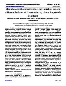
Fig. 1. (a) Schematic of the m02–m05 region ORF arrangement for the Smith (GenBank accession no. U68299), K181, G4, G1F, MI6A, W8211 and W8 strains of MCMV. The upper part of the figure shows the Smith HindIII digestion map (Ebeling et al., 1983). Homologous ORFs are indicated by pattern. Arrows show the direction of translation. (b) Phylogenetic tree for the m02–m05 region nucleotide sequence for seven MCMV strains.
MI6A and G1F did not possess m03 and only contained m03.5. These gene arrangement groupings were reflected in a phylogenetic analysis based on the complete DNA sequence across this region (Fig. 1b). Conservation of the m02 ORF All seven MCMV strains sequenced in this study were found to contain an m02 ORF. Amino acid sequence identity with the published Smith m02 sequence (Rawlinson et al., 1996) ranged from 92.9 % for the G1F isolate to 97.9 % for the K181 strain (data not shown). The sequences of the predicted signal peptides, TM domains and cysteine motifs were mostly conserved among strains. Sequence comparison of the m03 and m03.5 ORFs Of the five wild-derived isolates analysed, only G4 had the same m02, m03, m04 and m05 gene arrangement as the two laboratory strains (Fig. 1a). Two isolates, W8 and W8211, had both m03 and an additional ORF, m03.5, which lay between m03 and m04. All other strains analysed had only m03.5 and the m03 ORF was absent. Sequence comparisons of m03 showed that amino acid sequence identity with Smith m03 ranged from 86.4 to 93.0 % (Fig. 2a). For all Journal of General Virology 88
m02 family sequence variation in MCMV wild isolates
Fig. 2. Alignment of predicted protein sequences for m03 (a) and m03.5 (b). Dots indicate conserved residues. Dashes indicate gaps introduced into alignments. The percentage identity and similarity with the G1F m03.5 and Smith m03 sequences are shown. Predicted N-linked glycosylation sites are underlined. Predicted signal peptides, TM regions and conserved motifs are boxed. (c) Phylogenetic tree for the m03 and m03.5 protein sequences.
http://vir.sgmjournals.org
761
A. J. Corbett and others
strains containing this ORF, the encoded protein was predicted to be a type I TM glycoprotein, with an Nterminal signal peptide of 33 aa. The NAXWXXE/HWo and CXLXXCL/PW/Ro motifs described previously (Rawlinson et al., 1996), as well as the seven predicted N-linked glycosylation sites in m03, were conserved in all strains analysed. Isolates W8 and W8211 were found to contain two genes between m02 and m04. We considered this second ORF to be a new gene and not simply a highly variant form of m03, as it was present in addition to m03 in at least two independent MCMV isolates previously shown to have distinct RFLP profiles (Booth et al., 1993), and have given it the designation m03.5, as it has closest homology to m03 based on nucleotide and protein BLAST searches of the GenBank database among all MCMV ORFs. A complete m03.5 ORF sequence was obtained for seven isolates (Fig. 2b). In addition to those strains for which the complete m02–m05 region was analysed, 11 more MCMV wild-derived isolates have so far been found to encode an m03.5 ORF (data not shown). All strains analysed so far encoded at least one of the m03 or m03.5 ORFs. The sequence similarity suggests that these have arisen via a gene duplication event and that most strains have subsequently lost one ORF. Although m03.5 is most closely related to m03 based on BLAST analysis, these genes are quite distinct (Fig. 2c). The m03.5 predicted protein contains the NAXWXXE/HWo and CXLXXCL/PW/Ro motifs typical of the m02 gene family (Rawlinson et al., 1996). Amino acid sequence identity (with G1F m03.5) ranged from 73.7 to 76.9 % (Fig. 2b). Sequence identity with Smith m03 ranged from 42.3 to 45.8 % (and similarity ranged from 55.1 to 59.7 %), again suggesting that m03.5 is a distinct gene. Two possible initiation ATG codons were present, neither of which matched the Kozak consensus sequence ACCATGG (Kozak, 1986). The second had homology with m03 and therefore this was used for the alignment. Six N-linked glycosylation motifs were conserved among all of the strains, with five more being variably present among these strains. The predicted signal peptide and TM regions were conserved, except for G1F m03.5, which had a shorter predicted TM region. The cytoplasmic tail was truncated in the W8, W8211 (by seven residues) and G1F (by three residues) isolates. m04/gp34 sequence comparison The m04/gp34 sequence was found to be highly variable among the MCMV strains analysed, with amino acid sequence identity with Smith m04/gp34 ranging from 39.4 to 93.6 % (Fig. 3a). Indeed, in order to sequence m04, primer sets based on the m03 and m05 Smith sequences were used to amplify larger products, as the region directly flanking m04 was highly variable and Smith-based m04 primers did not work for many strains. In a recent report, Smith et al. (2006) also failed to amplify the m04 gene in wild-derived MCMV isolates with strain Smith-based primers directly flanking the gene, suggesting that these isolates also contained m04 762
sequences that were highly divergent from the Smith strain. The m04/gp34 sequences fell into six distinct groups (Fig. 3b). The type I TM structure was conserved with the exception of the MI6A and N1 isolates, which each had two predicted TM regions. However, based on the hydrophobicity plots and comparison with the very similar isolates G3B and MI2A, we believe this prediction of additional TM domains using the HMMTOP version 2.0 server (Tusnady & Simon, 1998, 2001) to be incorrect. Most of the m04/gp34 sequence variation occurred in the extracellular region, with the predicted TM region and short cytoplasmic tail being relatively conserved. The position and number of predicted N-linked glycosylation sites varied greatly, with different strains containing between one and five sites. Experimental evidence has shown Smith m04/gp34 to contain three N-linked glycans (Kleijnen et al., 1997). The NAXWXXE/HWo and CXLXXCL/PW/Ro m02 family motifs described previously (Rawlinson et al., 1996) were conserved in all strains. m04/gp34 contained an antigenic peptide, 243YGPSLYRRF251, recognized by cytotoxic T cells (CTLs) (Holtappels et al., 2000b). This was conserved in seven strains, including Smith, G1F, G4 and K181. Five strains, including W8211 and W8, contained the sequence FGPSLYRRF, and five strains, including MI6A, had the sequence FGPSLCRRF. Both of these sequences fit the H-2Dd motif XGPXXXXXL/I/F described previously (Holtappels et al., 2000a, b) and are thus likely to be recognized by m04/gp34-specific CTLs. m05 sequence comparison The m05 sequences analysed formed two distinct groups (Fig. 4a). The first group, containing ten strains, showed a high level of sequence identity with Smith m05 (83.9.1–93.9 %). The second group contained two strains, W8 and W8211, having sequence identity with Smith m05 of 41.6 and 41.0 %, respectively. Interestingly, these were the same strains found to have both m03 and m03.5 and which fell into a distinct m04/gp34 sequence group. The m05 protein, like other members encoded by the m02 gene family, is a type I TM glycoprotein with an N-terminal signal peptide, a long extracellular region, a TM region and a short cytoplasmic tail. This structure, as well as the NAXWXXE/ HWo and CXLXXCL/PW/Ro motifs described previously (Rawlinson et al., 1996), were conserved in all strains, including W8 and W8211. The m05 predicted protein consisted of 332 aa (including the signal peptide) except for W8 and W8211, where shorter proteins of 282 and 289 aa, respectively, were predicted. The W8 and W8211 m05 sequences contained two predicted N-linked glycosylation motifs. These sites were conserved in some of the other strains, which also contained three to five additional sites. Sequence variation in intragenic regions: clues to genetic events In addition to the marked variation among MCMV strains within the predicted coding regions, the intragenic regions showed a high level of variation in both length and Journal of General Virology 88
m02 family sequence variation in MCMV wild isolates
Fig. 3. (a) Alignment of predicted protein sequences for m04. Dots indicate conserved residues. Dashes indicate gaps introduced into alignments. The percentage identity and similarity with the Smith sequence are shown. Predicted N-linked glycosylation sites are underlined. Predicted signal peptides, TM regions and conserved motifs are boxed. For strains with identical sequences, only one is shown. (b) Phylogenetic tree for the m04 protein sequences.
http://vir.sgmjournals.org
763
A. J. Corbett and others
nucleotide sequence (data not shown). Homology between the m02–m03 and m03–m03.5 regions in both W8 and W8211, along with homology within the genes themselves, suggests that the m03 and m03.5 ORFs have arisen from a gene duplication event and subsequently diverged. Similar gene duplication is thought to be a common mechanism for evolution in herpesviruses and other organisms (Arav-Boger et al., 2005; Chee et al., 1990; Lesniewski et al., 2006; Rawlinson et al., 1996; Sahagun-Ruiz et al., 2004). These regions may then have allowed homologous recombination resulting in the loss of one gene in the majority of strains. A 21 nt triple repeat (CGCCTGAGCCTACCTCGAGAG) was found in the m02–m03 region (nt 995–1015) of the G4 strain. This may be an indication of the capacity of MCMV to evolve through DNA duplication events. Direct repeats are present at the ends of the MCMV genome and are also clustered in the m02 gene family within the m08, m11 and m12 ORFs (Rawlinson et al., 1996). Short (1–3 nt) repeats are common in HCMV (Davis et al., 1999) and longer direct repeats are present in other herpesviruses such as Epstein– Barr virus (Dambaugh & Kieff, 1982). Assessment of the transcriptional kinetics of m03, m03.5 and m04 After infection, MCMV (and HCMV) genes are expressed in a temporally controlled manner in the IE, early and late phases of replication. In order to analyse the kinetics of m03 and m03.5 expression, RT-PCR was performed on total RNA prepared from infected MEFs at various time points p.i. (Fig. 5). Both m03 (Smith or W8211 strains) and m03.5 (MI6A or W8211 strains) were found to be produced as early transcripts, but delayed when compared with m04 (m03 and m03.5 were present at 8 but not 4 h p.i., as for m04). Interestingly, in W8211-infected cells, both m03 and m03.5 transcripts were produced and these had identical kinetics. This was not due to the amplification of nonspecific PCR products, as the W8211 m03 and m03.5 product sizes were 840 and 897 bp, respectively. The m04 gene is transcribed at early times (Holtappels et al., 2000a). As this gene exhibited marked sequence variation among strains, we performed RT-PCR for m04 from MI6A and W8211 wild isolates, as well as Smith MCMV. In all three viruses, m04 transcription was evident at early times, with some transcripts present at late times. MI6A m04 transcripts appeared with slower kinetics than Smith or W8211. Kinetic differences may be due to differences in the promoter regions. These have not been mapped, but intragenic regions likely to contain these sequences differ among these strains (data not shown). However, as these viruses grow differently in vitro, a closer analysis of the kinetics is needed using a quantitative approach to determine whether there is a real difference in the transcriptional control of m04. The m02 family glycoproteins m03, m03.5 and m04 all reach the cell surface In order to analyse the cellular expression of these molecules, Smith m03, W8211 m03, MI6A m03.5 and 764
W8211 m03.5 ORFs were cloned in frame with an Nterminal leader peptide and FLAG tag in the MigR1 expression vector. Both m03–FLAG and m03.5–FLAG proteins could be detected in fixed COS-7 cells by immunofluorescence after transient transfection (Fig. 6b) and surface expression could be detected by flow cytometry (Fig. 6a) The immunofluorescence staining pattern was identical for m03 and m03.5 from variant strains. The m04encoded protein gp34 binds to MHC class I molecules in the ER and on the cell surface (Kavanagh et al., 2001; Kleijnen et al., 1997). m04/gp34 has been expressed previously and detected intracellularly by immunofluorescence using a haemagglutinin tag (Oliveira et al., 2002). As m04/gp34 showed a high degree of amino acid sequence variation, we analysed its expression by flow cytometry using FLAGtagged constructs. The m04–FLAG proteins from strains in each sequence group, A–F (Fig. 3b), could also be detected on the cell surface by anti-FLAG staining after transient transfection in COS-7 cells (Fig. 6a), indicating that m04/ gp34 from each strain is expressed in a similar fashion, despite sequence variation. In addition, immunofluorescence staining patterns of fixed, transfected COS cells for FLAG-tagged m04 constructs from all strains tested (Fig. 6b) were similar to those previously reported (Oliveira et al., 2002).
DISCUSSION In this study, sequence variation was analysed over the m02– m05 gene region for seven MCMV strains, including two commonly used laboratory strains and five wild-derived isolates, as well as individual ORFs for additional wildderived isolates. This region was found to exhibit significant genetic variation among MCMV laboratory strains and wild-derived isolates, with most of the variation occurring in the m03–m04 gene region. The gene arrangement differed among strains, such that three different genotypes were present. We identified the m03.5 ORF, present in a number of isolates instead of, or in addition to, m03. Conserved cysteine motifs, N-linked glycosylation sites and protein structure, as well as sequence homology to m03, identified m03.5 as a member of the m02 gene family. Homology to m03 in both the gene and intragenic regions suggests that these ORFs have arisen via a gene duplication event and that one ORF has subsequently been lost in some isolates. The conservation of at least one of these ORFs, along with the loss of one in most strains, suggests that if the m03.5encoded protein is expressed in the host after infection, it could share a (presently undefined) redundant function with m03. The W8 and W8211 strains may not have yet had significant evolutionary pressure to lose one ORF or, alternatively, if these proteins do have divergent functions, they may confer survival advantages upon these viruses in certain outbred mouse populations. Transcripts for both m03 and m03.5 were expressed early, but with delayed kinetics when compared with m04, and this did not vary significantly among the strains. Interestingly, in W8211, which has both m03 and m03.5, both genes were transcribed Journal of General Virology 88
m02 family sequence variation in MCMV wild isolates
Fig. 4. (a) Alignment of predicted protein sequences for m05. Dots indicate conserved residues. Dashes indicate gaps introduced into alignments. The percentage identity and similarity with the Smith sequence are shown. Predicted N-linked glycosylation sites are underlined. Predicted signal peptides, TM regions and conserved motifs are boxed. (b) Phylogenetic tree for m05 protein sequences.
http://vir.sgmjournals.org
765
A. J. Corbett and others
Fig. 5. RT-PCR analysis of MEFs infected with the Smith, MI6A or W8211 strain of MCMV for the m03 (a), m03.5 (b), m04 (c) and ie1 (d, infection with Smith strain only) genes. RT-PCR of b-actin (e) was used as a loading control. No product was present for any of the RT” or H2O controls (not shown). Numbers indicate h p.i. C, Cycloheximide; P, PAA; ND, not done.
with identical kinetics. This suggests that the two encoded proteins both have the potential to be expressed and supports the argument that m03 and m03.5 are separate ORFs and not simply highly variable forms of the same ORF. At this stage however, the potential function of these proteins remains undefined. ORFs within the m02 gene family of MCMV lie in a head-totail fashion and do not overlap. Multiple alternative initiation ATGs were found to be present for the m02 (Smith, K181), m03.5 (G1F, MI6A, W8, W8211), m04 (G1F, MI6A, W8211) and m05 (all strains) ORFs. We used those previously annotated for Smith (Rawlinson et al., 1996) for our alignments, with the exception of m02 where we used the second ATG (in Smith), due to its conservation among the isolates. This second ATG is not within a Kozak consensus sequence. Our analysis of other genes including m03, m04 and m05 revealed that Kozak sequences are not common in the MCMV genome, at least within this subset of the m02 gene family. Experimental confirmation is still required for the majority of MCMV ORFs, and annotation of the MCMV genome will continue to be revised as sequence data become available. The shorter m05 ORFs predicted for the W8 and W8211 strains appear to be true m05 ORFs based on homology and gene arrangement. We sequenced the m06 gene for a selection of strains (data not shown) and found this to be highly conserved, consistent with a previous study (Smith et al., 2006). The percentage amino acid identity with Smith m06 ranged from 96.2 (G1F) to 98.2 % (K181). W8211 m06 shared 97.6 % amino acid sequence identity with Smith m06, whilst W8211 m05 shared only 17.6 % sequence identity with Smith m06, compared with 39.5 % identity with Smith m05. A number of m02 family members have been found previously to be expressed intracellularly in an overexpression system (Oliveira et al., 2002). The expression of m03 has not been reported previously. FLAG-tagged m03 and m03.5 proteins from all strains tested could be detected 766
on the cell surface of transfected cells with a FLAG-specific mAb, suggesting that, if the native proteins are similarly expressed in infected cells, then these proteins may bind to ligands in trans on other cells in a similar fashion to the m145 family member m157 (Arase et al., 2002; Smith et al., 2002) or to soluble ligands. Alternatively, they may bind host molecules in cis, as is the case for m04/gp34 (Kavanagh et al., 2001; Kleijnen et al., 1997). If m03 and m03.5 proteins are expressed and interact with host molecules, then the sequence variation observed is likely to be reflected in the binding of these host proteins. In addition to intracellular expression, m04/gp34 could be detected on the cell surface by flow cytometry and this did not vary among strains, despite a high degree of sequence variation. This is also the case for m157, which exhibits a similar degree of variation (C. A. Forbes, A. J. Corbett & A. A. Scalzo, unpublished data; Voigt et al., 2003). The m04/gp34 protein is expressed in the ER of mouse cells and traffics to the surface only in complex with MHC class I molecules (Kavanagh et al., 2001; Kleijnen et al., 1997). The extensive amino acid sequence variation observed would be consistent with m04/gp34 from different MCMV strains being capable of binding different MHC class I molecules and may indicate adaptation to the diverse array of MHC class I molecules in a heterogeneous host population. Alternatively, these variant m04/gp34 proteins may bind to host proteins other than MHC class I and therefore may be under different selective pressures. The m04/gp34 protein sequences analysed fell into six distinct groups (Fig. 3b), suggesting possible selection by different host H2 haplotypes. Similarly, the significant variability in the m03 and m03.5 ORFs and the probable gene duplication event leading to their evolution may reflect an adaptation of the virus to genetically diverse wild mouse host populations and may give a clue to the function of these genes as potentially interacting with variant host molecules. Our data are consistent with previous reports that the regions at the extremities of CMV genomes, which in MCMV include the m02 and m145 gene families, are Journal of General Virology 88
m02 family sequence variation in MCMV wild isolates
Fig. 6. Expression of m03, m03.5 and m04 proteins. COS-7 cells were transiently transfected with the FLAG-tagged m03, m03.5 or m04 construct and analysed for expression of FLAG by flow cytometry (a) or immunofluorescence (b) after 48 h. GFP was expressed independently as a marker of transfection and plots were gated on GFP+ live cells. Solid lines, transfected cells; filled histograms, empty vector controls. In (b), GFP fluorescence is green and anti-FLAG staining is red.
highly divergent. Phylogenetic analyses produced different groupings of strains for each of the individual m02 gene family members (Figs 1–4), suggesting that there is limited linkage between these genes. Within genes, regions responsible for maintaining protein structure and function (e.g. the TM domain) were conserved and the extracellular domains were most divergent. ORFs with http://vir.sgmjournals.org
immune-modulatory function may have developed polymorphisms as a means of evading the host immune system in genetically diverse populations. Polymorphisms in antigenic determinants recognized by CTLs may be useful to the virus in terms of evading this response. However, in the case of m04/gp34, the antigenic determinant is conserved. In contrast, the ie1 epitope was not conserved 767
A. J. Corbett and others
in wild MCMV isolates (Lyons et al., 1996). As the m04/gp34 peptide is derived from the inner part of the TM region and cytoplasmic domain, it may be essential for function and not readily altered by the virus. A recent report shows that the TM domain of m04/gp34 is essential for association with Kb heavy chains (Lu et al., 2006). MCMV immune evasion genes are likely to be host-strain specific (e.g. m04/gp34, which interacts with host MHC class I molecules), and genes from different isolates may have evolved to function in individual host strains. The m157 protein was originally reported to be responsible for a dominant resistant phenotype in C57BL/6 mice by binding to the Ly49H NK cell-activation receptor (Arase et al., 2002; Brown et al., 2001; Scalzo et al., 1990, 1992; Smith et al., 2002), but the binding of m157 to the Ly49I inhibitory receptor in 129/J mice may have a different effect (Bubic et al., 2004). The localized genetic variation in m03–m05 among relatively conserved m02 and m06 genes suggests that amino acid changes have been selected by functional constraints such as interactions with variant host proteins. Recent reports have shown that MCMV can mutate rapidly in vivo under functional selective pressure from m157– Ly49H interactions (French et al., 2004; Voigt et al., 2003). However, undertaking studies to test this hypothesis for these m02 family genes would depend on first characterizing the putative host receptors. Most MCMV studies are conducted using MCMV laboratory strains and inbred mice, which may not reflect the high degree of natural variability in viruses or hosts. The analysis of variation in the immune-evasion genes highlights the need for caution in assigning functions in host–virus combinations that do not fully reflect the spectrum of viral or host variability in nature. Genetic variation in CMVs also has implications for the development of antiviral therapies or vaccines that will be effective against the complete range of HCMV strains, and for PCR-based diagnosis of HCMV infection.
Bendtsen, J. D., Nielsen, H., von Heijne, G. & Brunak, S. (2004).
Improved prediction of signal peptides: SignalP 3.0. J Mol Biol 340, 783–795. Boname, J. M. & Chantler, J. K. (1992). Characterization of a strain
of murine cytomegalovirus which fails to grow in the salivary glands of mice. J Gen Virol 73, 2021–2029. Booth, T. W., Scalzo, A. A., Carrello, C., Lyons, P. A., Farrell, H. E., Singleton, G. R. & Shellam, G. R. (1993). Molecular and biological
characterization of new strains of murine cytomegalovirus isolated from wild mice. Arch Virol 132, 209–220. Brocchieri, L., Kledal, T. N., Karlin, S. & Mocarski, E. S. (2005).
Predicting coding potential from genome sequence: application to betaherpesviruses infecting rats and mice. J Virol 79, 7570–7596. Brown, M. G., Dokun, A. O., Heusel, J. W., Smith, H. R., Beckman, D. L., Blattenberger, E. A., Dubbelde, C. E., Stone, L. R., Scalzo, A. A. & Yokoyama, W. M. (2001). Vital involvement of a natural killer
cell activation receptor in resistance to viral infection. Science 292, 934–937. Bubic, I., Wagner, M., Krmpotic, A., Saulig, T., Kim, S., Yokoyama, W. M., Jonjic, S. & Koszinowski, U. H. (2004). Gain of virulence
caused by loss of a gene in murine cytomegalovirus. J Virol 78, 7536–7544. Chalmer, J. E., Mackenzie, J. S. & Stanley, N. F. (1977). Resistance to
murine cytomegalovirus linked to the major histocompatibility complex of the mouse. J Gen Virol 37, 107–114. Chee, M. S., Bankier, A. T., Beck, S., Bohni, R., Brown, C. M., Cerny, R., Horsnell, T., Hutchison, C. A., III, Kouzarides, T. & other authors (1990). Analysis of the protein-coding content of the sequence of
human cytomegalovirus strain AD169. Curr Top Microbiol Immunol 154, 125–169. Dambaugh, T. R. & Kieff, E. (1982). Identification and nucleotide
sequences of two similar tandem direct repeats in Epstein–Barr virus DNA. J Virol 44, 823–833. Davis, C. L., Field, D., Metzgar, D., Saiz, R., Morin, P. A., Smith, I. L., Spector, S. A. & Wills, C. (1999). Numerous length polymorphisms
at short tandem repeats in human cytomegalovirus. J Virol 73, 6265– 6270. Ebeling, A., Keil, G. M., Knust, E. & Koszinowski, U. H. (1983).
Molecular cloning and physical mapping of murine cytomegalovirus DNA. J Virol 47, 421–433. Farrell, H. E. & Shellam, G. R. (1989). Immunoblot analysis of the
ACKNOWLEDGEMENTS This work was supported by grants from the NH & MRC and the Australian Antarctic Division. A. J. C. was the recipient of a WA & MG Saw Medical Research Fellowship from the University of Western Australia. A. A. S. was supported by an NH & MRC Senior Research Fellowship. The authors would like to thank Nicole Harvey (University of Western Australia) for technical assistance and Ed Mocarski (Stanford University) for critical reading of the manuscript.
antibody response to murine cytomegalovirus in genetically resistant and susceptible mice. J Gen Virol 70, 2573–2586. French, A. R., Pingel, J. T., Wagner, M., Bubic, I., Yang, L., Kim, S., Koszinowski, U., Jonjic, S. & Yokoyama, W. M. (2004). Escape of
mutant double-stranded DNA virus from innate immune control. Immunity 20, 747–756. Frohman, M. A., Dush, M. K. & Martin, G. R. (1988). Rapid
production of full-length cDNAs from rare transcripts: amplification using a single gene-specific oligonucleotide primer. Proc Natl Acad Sci U S A 85, 8998–9002. Hall, T. A. (1999). BioEdit: a user-friendly biological sequence
REFERENCES
alignment editor and analysis program for Windows 95/98/NT. Nucleic Acids Symp Ser 41, 95–98.
Alcami, A. & Koszinowski, U. H. (2000). Viral mechanisms of
Hengel, H., Brune, W. & Koszinowski, U. H. (1998). Immune evasion
immune evasion. Immunol Today 21, 447–455.
by cytomegalovirus – survival strategies of a highly adapted opportunist. Trends Microbiol 6, 190–197.
Arase, H., Mocarski, E. S., Campbell, A. E., Hill, A. B. & Lanier, L. L. (2002). Direct recognition of cytomegalovirus by activating and
inhibitory NK cell receptors. Science 296, 1323–1326.
Hengel, H., Reusch, U., Gutermann, A., Ziegler, H., Jonjic, S., Lucin, P. & Koszinowski, U. H. (1999). Cytomegaloviral control of
Arav-Boger, R., Zong, J. C. & Foster, C. B. (2005). Loss of linkage
MHC class I function in the mouse. Immunol Rev 168, 167–176.
disequilibrium and accelerated protein divergence in duplicated cytomegalovirus chemokine genes. Virus Genes 31, 65–72.
Ho, M. (1991). Cytomegalovirus Biology and Infection, 2nd edn. New
768
York: Plenum Medical Book Company. Journal of General Virology 88
m02 family sequence variation in MCMV wild isolates
Holtappels, R., Thomas, D., Podlech, J., Geginat, G., Steffens, H.-P. & Reddehase, M. J. (2000a). The putative natural killer decoy early gene
m04 (gp34) of murine cytomegalovirus encodes an antigenic peptide recognized by protective antiviral CD8 T cells. J Virol 74, 1871–1884. Holtappels, R., Thomas, D. & Reddehase, M. J. (2000b).
Identification of a Kd-restricted antigenic peptide encoded by murine cytomegalovirus early gene M84. J Gen Virol 81, 3037–3042. Hudson, J. B., Walker, D. G. & Altamirano, M. (1988). Analysis in
vitro of two biologically distinct strains of murine cytomegalovirus. Arch Virol 102, 289–295. Kavanagh, D. G., Koszinowski, U. H. & Hill, A. B. (2001). The murine
cytomegalovirus immune evasion protein m4/gp34 forms biochemically distinct complexes with class I MHC at the cell surface and in a pre-Golgi compartment. J Immunol 167, 3894–3902. Kleijnen, M. F., Huppa, J. B., Lucin, P., Mukherjee, S., Farrell, H., Campbell, A. E., Koszinowski, U. H., Hill, A. B. & Ploegh, H. L. (1997). A mouse cytomegalovirus glycoprotein, gp34, forms a
complex with folded class I MHC molecules in the ER which is not retained but is transported to the cell surface. EMBO J 16, 685–694. Kozak, M. (1986). Point mutations define a sequence flanking the
AUG initiator codon that modulates translation by eukaryotic ribosomes. Cell 44, 283–292. Lesniewski, M., Das, S., Skomorovska-Prokvolit, Y., Wang, F.-Z. & Pellett, P. E. (2006). Primate cytomegalovirus US12 gene family: a
distinct and diverse clade of seven-transmembrane proteins. Virology 354, 286–298.
Pear, W. S., Miller, J. P., Xu, L., Pui, J. C., Soffer, B., Quackenbush, R. C., Pendergast, A. M., Bronson, R., Aster, J. C. & other authors (1998). Efficient and rapid induction of a chronic myelogenous
leukemia-like myeloproliferative disease in mice receiving P210 bcr/ abl-transduced bone marrow. Blood 92, 3780–3792. Pignatelli, S., Dal Monte, P., Rossini, G. & Landini, M. P. (2004).
Genetic polymorphisms among human cytomegalovirus (HCMV) wild-type strains. Rev Med Virol 14, 383–410. Rawlinson, W. D., Farrell, H. E. & Barrell, B. G. (1996). Analysis of
the complete DNA sequence of murine cytomegalovirus. J Virol 70, 8833–8849. Reusch, U., Muranyi, W., Lucin, P., Burgert, H. G., Hengel, H. & Koszinowski, U. H. (1999). A cytomegalovirus glycoprotein re-routes
MHC class I complexes to lysosomes for degradation. EMBO J 18, 1081–1091. Sahagun-Ruiz, A., Sierra-Honigmann, A. M., Krause, P. & Murphy, P. M. (2004). Simian cytomegalovirus encodes five rapidly evolving
chemokine receptor homologues. Virus Genes 28, 71–83. Scalzo, A. A., Fitzgerald, N. A., Simmons, A., La Vista, A. B. & Shellam, G. R. (1990). Cmv-1, a genetic locus that controls murine
cytomegalovirus replication in the spleen. J Exp Med 171, 1469–1483. Scalzo, A. A., Fitzgerald, N. A., Wallace, C. R., Gibbons, A. E., Smart, Y. C., Burton, R. C. & Shellam, G. R. (1992). The effect of the Cmv-1
resistance gene, which is linked to the natural killer cell gene complex, is mediated by natural killer cells. J Immunol 149, 581–589.
Lu, X., Kavanagh, D. G. & Hill, A. B. (2006). Cellular and molecular requirements for association of the murine cytomegalovirus protein m4/gp34 with major histocompatibility complex class I molecules. J Virol 80, 6048–6055.
Smith, H. R., Heusel, J. W., Mehta, I. K., Kim, S., Dorner, B. G., Naidenko, O. V., Iizuka, K., Furukawa, H., Beckman, D. L. & other authors (2002).
Lyons, P. A., Allan, J. E., Carrello, C., Shellam, G. R. & Scalzo, A. A. (1996). Effect of natural sequence variation at the H-2Ld-restricted
Smith, L. M., Shellam, G. R. & Redwood, A. J. (2006). Genes of
CD8+ T cell epitope of the murine cytomegalovirus ie1-encoded pp89 on T cell recognition. J Gen Virol 77, 2615–2623. Misra, V. & Hudson, J. B. (1980). Minor base sequence differences
between the genomes of two strains of murine cytomegalovirus differing in virulence. Arch Virol 64, 1–8.
Recognition of a virus-encoded ligand by a natural killer cell activation receptor. Proc Natl Acad Sci U S A 99, 8826–8831. murine cytomegalovirus exist as a number of distinct genotypes. Virology 352, 450–465. Tusnady, G. E. & Simon, I. (1998). Principles governing amino acid
composition of integral membrane proteins: application to topology prediction. J Mol Biol 283, 489–506. Tusnady, G. E. & Simon, I. (2001). The HMMTOP transmembrane
Nielsen, H. & Krogh, A. (1998). Prediction of signal peptides and
topology prediction server. Bioinformatics 17, 849–850.
signal anchors by a hidden Markov model. Proc Int Conf Intell Syst Mol Biol 6, 122–130.
Voigt, V., Forbes, C. A., Tonkin, J. N., Degli-Esposti, M. A., Smith, H. R., Yokoyama, W. M. & Scalzo, A. A. (2003). Murine cytomegalovirus
Nielsen, H., Engelbrecht, J., Brunak, S. & von Heijne, G. (1997).
m157 mutation and variation leads to immune evasion of natural killer cells. Proc Natl Acad Sci U S A 100, 13483–13488.
Identification of prokaryotic and eukaryotic signal peptides and prediction of their cleavage sites. Protein Eng 10, 1–6. Oliveira, S. A., Park, S.-H., Lee, P., Bendelac, A. & Shenk, T. E. (2002). Murine cytomegalovirus m02 gene family protects against
natural killer cell-mediated immune surveillance. J Virol 76, 885–894.
http://vir.sgmjournals.org
Xu, J., Lyons, P. A., Carter, M. D., Booth, T. W., Davis-Poynter, N. J., Shellam, G. R. & Scalzo, A. A. (1996). Assessment of antigenicity and
genetic variation of glycoprotein B of murine cytomegalovirus. J Gen Virol 77, 49–59.
769