Abstract. Random Decision Forest-based approaches have previously shown promising performance in the domain of brain tumor segmenta- tion. We extend ...
Extremely randomized trees based brain tumor segmentation Michael Goetz1 , Christian Weber1,2 , Josiah Bloecher1 , Bram Stieltjes2 , Hans-Peter Meinzer1 , and Klaus Maier-Hein1 1
Medical and Biological Informatics, German Cancer Research Center (DKFZ), Heidelberg, Germany 2 Quantitative Image-based Disease Characterization, DKFZ, Heidelberg, Germany
Abstract. Random Decision Forest-based approaches have previously shown promising performance in the domain of brain tumor segmentation. We extend this idea by using an ExtraTree-classifier. Several features are calculated based on normalized T1, T2, T1 with contrast agent and T2 Flair MR-images. With these features an ExtraTree-classifier is trained and used to predict different tissue classes on voxel level. The results are compared to other state-of-the-art approaches by participating at the BraTS 2013 challenge.
1
Introduction
The segmentation of brain tumors is an important prerequisite in different scenarios related to treatment controlling, radiotherapy planning and longitudinal studies. Manual segmentation is not only time-consuming and prone to errors, but additionally complicated by the fact that the necessary information is distributed over different MR-contrasts. Therefore a lot of research has been done to improve the segmentation process and create automatic segmentation methods based on multimodal MR images. A promising approach is the use of Random Decision Forests like as done in the works of Reza et al. [1], Tustison et al. [1] and Zikic et al. [2], which learn the appearance of tumorous and healthy tissue using this method. While the proposed solution is similar to those mentioned before it differs mainly in the used classifier. Instead of Random Decision Forests [4] we use Extremely randomized Trees (ExtraTrees) [3] which are similar to Random Decision Forests but introduce more randomness during the training phase. It has previously been shown that this often improves the variance / bias trade-off and gives slightly better results than Random Decision Forests do [3].
2 2.1
Method Preprocessing
The preprocessing pipeline for our experiments consisted of two steps. First the N4 bias field correction algorithm [5] was used to correct nonuniformity within
each MR-file. In a second step the histogram was normalized. This is especially challenging in the case of brain tumor MR images. In addition to the usual MRartefacts which cause bright areas in parts of the image, the large variability of brain tumors has a massive influence on the histogram. Figure 1 shows some exemplary non-normalized histograms. It can be clearly seen that they differ not only in range of values but also in shape. Normalizing these histograms to match a template histogram as it is done by the pice-wise linear normalization [6] can lead to a wrong result if the shapes are too different.
Fig. 1. Exemplary histograms of 3 non-normalized MR-Flair-images out of the BraTSdataset. The histogram is over the complete non-zero image.
To overcome these problems a simple normalization to the image mode, e.g. the gray-value of the highest histogram bin, was used. This was done by subtracting the mode from each gray-value and then normalizing the standard derivation to 1.
2.2
Features
54 features were calculated for each voxel and each modality. The features of all modalities were then combined into the final feature vector.
Gray Value: The gray value of each voxel was used as a feature. The images were also filtered with gaussian filters with a sigma of 3 and 7 voxel-lengths and the corresponding gray values were used as features.
Local Histogram: A local histogram was calculated within a radius of 5 voxels; each of the 11 bins were used as features.
First order statistics: Within a radius of 3 voxels the mean, variance, skewness, kurtosis, minimum and maximum of all gray values were added as features.
Second order statistics: A co-occurrence matrix [7] filled with all values within a radius of 3 was used to calculate the second order statistics for the three main directions. The features extracted from the co-occurrence matrix were energy, entropy, correlation, inertia, clustershade, clusterprominence, harralick feature, and the difference of moments. Histogram based segmentations: The output segmentation of some widely used parameter-less automatic threshold methods implemented in ITK [8] were used as features, namely Huang, Intermode, Isodata, Kittler, Li, Entropy, Moments and Otsu. For all except the Otsu-threshold a two-class problem is assumed. For the Otsu, a two-, a three- and a four-class problem were assumed. 2.3
Classifier
An Extremely Randomized Trees (ExtraTrees) [3] classifier was used. This classifier is similar to Random Decision Forests but differs in how the randomness is introduced during the training. To train an ExtraTrees-classifier multiple trees are trained, each tree is trained on all training data. Similar to the Random Decision Forest the best split at a node is found by analyzing a subset of all available features. Instead of searching for the best threshold for each feature a single threshold for each feature is selected at random. From these random splits the one that leads to the highest increase in the used score is then selected. The higher grade of randomness during the training yields more independent trees and thus further decreases the variance [3]. Due to that ExtraTrees tend to give slightly better results than Random Decision Forests. For the training of the classifier 5% of the training data were randomly sampled to reduce the training time. The classifier is then trained combining the features described above to a 208-dimensional feature vector. 2.4
Experiments
The results were evaluated by participating in the BraTS 2013 challenge. A classifier is trained on the 20 training datasets using all available modalities, namely T2 Flair, T1, T1 with contrast agent and T2. With the so-trained classifier the 10 high-grade glioma evaluation datasets are labeled and the results are evaluated by the provided online tool. For the evaluation the overlap with 3 labels is measured using the DICEscore. The first label, the complete tumor, includes necrosis, edema and both enhancing and non-enhancing tumor. The second label, tumor core, is the same as the complete tumor but without edema. Finally, the label enhancing tumor is evaluated.
3
Results
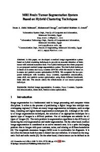
Table 1 provides the DICE-scores for the test cases. Figure 2 and Figure 3 depict exemplary slices of the original images and the retrieved segmentations.
Normalized Flair
Segmentation
HG306
HG305
HG304
HG303
HG302
HG301
T2 Flair
Fig. 2. Example slices from patients HG0301 to HG0306. The first column shows the original Flair image, the second the Flair image normalized with N4-Bias-Field correction and Mode-normalization. The last column shows the ally received results. The color coding is: green: ’edema’, yellow: ’active tumor’, red: ’necrosis’
Table 1. DICE score for the single test data sets. Dataset
Complete tumor
Tumor core
Enhancing tumor
HG0301 HG0302 HG0303 HG0304 HG0305 HG0306 HG0307 HG0308 HG0309 HG0310
0.85 0.83 0.86 0.75 0.88 0.82 0.81 0.89 0.75 0.88
0.87 0.74 0.78 0.63 0.73 0.58 0.47 0.89 0.50 0.86
0.79 0.85 0.74 0.53 0.69 0.63 0.48 0.66 0.68 0.80
0.83±0.048
0.71±0.144
0.68±0.113
mean:
4
Discussion
We present a new approach for multi-modal brain tumor segmentation using ExtraTrees instead of Random Decision Forests and tested it using the BraTS 2013 test data. The performance of the approach is comparable to the quality of other state-of-the-art algorithms which had been tested against the same dataset. This shows that ExtraTrees are well suited for the classification of tumorous brain tissue. In the future, it will be interesting to find out whether other approaches can be improved by simply replacing Random Decision Forest classifiers with ExtraTrees. 4.1
Acknowledgments
This work was carried out with the support of the German Research Foundation (DFG) within project I04, SFB/TRR 125 Cognition-Guided Surgery.
References [1] Menze, B., Jakab, A., Bauer, S., Kalpathy-Cramer, J., Farahani, K., Kirby, J., ... and Shotton, J.: The Multimodal Brain Tumor Image Segmentation Benchmark (BRATS). 2014 [2] Zikic, D., Glocker, B., Konukoglu, E., Criminisi, A., Demiralp, C., Shotton, J., Thomas, O.M, Das, T., Jena, R and Price, S.J.: Decision Forests for Tissue-Specific Segmentation of High-Grade Gliomas in Multi-channel MR. In: Proceedings of MICCAI 2012 [3] Geurts, P., Ernst, D., and Wehenkel, L.: Extremely randomized trees. In: Machine Learning, 2006 [4] Breiman, L.: Random Forest. In: Machine learning, 2001 [5] Tustison N.J., Avants B.B., Cook P.A., Zheng Y., Egan A., Yushkevich P.A. and Gee J.C.: N4ITK: improved N3 bias correction. In: IEEE Trans Med Imaging., 2010
[6] Nyl L.G., Udupa J.K. and Zhang, X. New variants of a method of MRI scale standardization. In: IEEE Transaction on Medical Imaging, 2000 [7] Haralick, R.M. Statistical and Structural Approaches to Texture In: Proceedings of the IEEE, 1979 [8] Beare R. Histogram-based Thresholding In: http://www.kitware.com/source/home/post/54, 2012
Normalized Flair
Segmentation
HG310
HG309
HG308
HG307
T2 Flair
Fig. 3. Example slices from patients HG0307 to HG0310. The first column shows the original Flair image, the second the Flair image normalized with N4-Bias-Field correction and Mode-normalization. The last column shows the ally received results. The color coding is: green: ’edema’, yellow: ’active tumor’, red: ’necrosis’