MRI Brain Tumor Segmentation System Based on Hybrid Clustering Techniques Eman A. Abdel Maksoud1, Mohammed Elmogy2, and Rashid Mokhtar Al-Awadi3 1
Information System Dept., Faculty of Computers and Information, Mansoura University, Egypt
[email protected] 2 Information Technology Dept., Faculty of Computers and Information, Mansoura University, Egypt
[email protected] 3 Communication Dept., Faculty of Engineering, Mansoura University, Egypt
[email protected]
Abstract. In this paper, we developed a medical image segmentation system based on hybrid clustering techniques to provide an accurate detection of brain tumor with minimal execution time. Two hybrid techniques have been proposed in our proposed medical image segmentation system. The first hybrid technique is based on k-means and fuzzy c-means (KFCM) while the second is based on k-means and particle swarm optimization (KPSO). We compared the two proposed techniques with k-means; fuzzy c-means, expectation maximization, mean shift, and particle swarm optimization using three different benchmark brain data sets. The results clarify the effectiveness of our second proposed technique. Keywords: Medical image segmentation, K-means, Fuzzy C-means, Expectation Maximization, Mean shift, Particle swarm optimization.
1
Introduction
Image segmentation is a fundamental task in image processing and computer vision disciplines. It refers to the process of partitioning a digital image into multiple nonoverlapping regions [1].There are many image segmentation techniques, such as edge base, clustering and region based segmentation techniques [2]. Although of the variety of image segmentation techniques, the selection of an appropriate technique for a special type of images is a difficult problem. Not all techniques are suitable for all types of images [3]. The main problem in segmentation algorithms is the difficulty of balancing the over-segmentation and under-segmentation. On the other hand, medical image segmentation is considered as an active research area. It is a quite challenging problem due to images with poor contrasts, noise, and missing or diffuses boundaries [4]. The magnitude resonance images (MRI) scan is comfortable for diagnosis. It is not affect the human body because it doesn't use any radiation. It is based on the magnetic field and radio waves [5]. On the other hand, a brain tumor can be defined as an A.E. Hassanien et al. (Eds.): AMLTA 2014, CCIS 488, pp. 401–412, 2014. © Springer International Publishing Switzerland 2014
402
E.A. Abdel Maksoud, M. Elmogy, and R. Mokhtar Al-Awadi
abnormal growth of the cells in the brain. Brain tumors are of two types: primary and secondary. Primary tumors are classified as benign and malignant [6]. Benign tumors can be removed. They usually have a border or an edge. Malignant tumors are more serious. They grow rapidly in crowd and invade the nearby healthy tissue. The physician gives the treatment for the strokes rather than the treatment for the tumor. So, detection of the tumor is important for that treatment. The lifetime of the person who affected by the brain tumor will increase if it is detected early. Therefore, an efficient medical image segmentation technique should be developed with advantages of minimum user interaction, fast computation, accurate, and robust segmentation results to help physicians in diagnosing accurately. The most widely used techniques of image segmentation are clustering techniques. Clustering is an unsupervised learning technique which needs the user to determine the number of clusters in advance to classify pixels [7]. Therefore, the cluster is a collection of similar pixels and is dissimilar to the pixels belonging to other clusters [8]. Clustering techniques can perform clustering in one of two ways, either by partitioning or by grouping pixels [9]. In this paper we focused on clustering techniques to detect the brain tumor. We made our experiments by using the most famous five clustering techniques: k means, fuzzy c means, expectation maximization, mean shift, and particle swarm optimization. We applied these techniques on three different data sets which have 254 abnormal MRI brain images. The MRI images were pre-processed at first to enhance the quality of the processed images. We integrated two different image clustering techniques in our proposed medical system in clustering step of the framework to have advantages of these clustering techniques and overcoming the limitations of them in two proposed hybrid techniques (k means with fuzzy c means and k means with particle swarm optimization). Then, extraction of the tumor is done automatically without user interaction by using thresholding and level set methods to contour the tumor area. The last stage of our proposed medical segmentation system framework is the validation stage by comparing the results with the ground truth. This paper is organized as follows. In Section 2, the current scientific research in medical image segmentation is introduced. Section 3 presents the materials and methods used in this work. It describes the image data sets used in this work. It also shows the proposed medical image segmentation system based on our proposed hybrid clustering techniques. Section 4 depicts the experimental results obtained from the evaluation of the two proposed techniques using three types of data sets and discusses the main questions derived from them. Finally, conclusion and future work are drawn in Section 5.
2
Related Work
Medical image segmentation is considered as a hot research topic. Several researchers have suggested various methodologies and techniques for image segmentation. For example, Jumb et al. [10] presented color image segmentation using k-means clustering and Otsu’s adaptive thresholding.They started by converting RGB image to HSV color model and extracted value channel. Then, they applied Otsu’s
MRI Brain Tumor Segmentation System Based on Hybrid Clustering Techniques
403
multi-thresholding on the value depending on the separation factor (SF). After that, they applied k-means clustering. Finally, they used morphological processing. The main disadvantages of their method are preprocessing and image enhancement stage is missing and segmentation is not fully automatic. It depends on the user notice to apply k-means or not. Sivasangareswari and Kumar [11] proposed a brain tumor segmentation using fuzzy c-means clustering with local information and Kernel metric. At first, they filtered brain images by median filter. Second, they made clustering by using fuzzy c-means technique. Their modified FCM (fuzzy c-means) depends on space distance of all neighbor pixels. They did feature extraction using algorithms of a gray level co-occurrence matrix (GLCM). Classification by SVM was also used. The limitation of their work is that they didn't make skull removal. The thing that increases the amount of used memory and increase the processing time. Abdul-Nasir et al. [12] presented a color image segmentation technique to detect the malaria parasites in red blood cells. They applied partial contrast stretching technique on color malaria images and extract color components from enhanced images. Then, they used k-means clustering technique. Finally, they used median filter and seeded region growing area extraction algorithms. The limitation of this method is that there is no best model all times. Therefore, HSI color model gives best results in recall but C-Y color model gives best results in precision.Joseph et al. [13] presented brain tumor MRI segmentation .They started by the preprocessing stage. It converts the RGB input image to grey scale. They used a median filter. The preprocessed image is supplied for k-means clustering algorithm then followed by morphological filtering if there are no clustered regions. The main disadvantage is they didn't make skull removing in preprocessing step.Wang et al. [14] implemented an adaptive particle swarm optimization algorithm with mutation operation based on k-means. They combined the particle swarm optimization and k-means in case of local search and global search. The mutation was processed to accelerate a poor particle in population. The main disadvantage is that they didn't care about reducing the data set size by using feature extractions, which can reduce the number of iterations and the execution time. In our proposed medical image segmentation system based on the proposed hybrid clustering techniques,the main objective is to accurately detect the brain tumor in minimal execution time. We put into account the accuracy and minimum execution time in each stage. In the preprocessing stage, we applied the median filter to enhance entire image quality and removed the skull from the processed image. This stage reduces both the processing time and the used amount of memory. In segmentation stage, all advantages of k-means, fuzzy c-means, and particle swarm optimization are preserved; while their main problems have been solved by the proposed hybrid techniques. The over segmentation and under segmentation problems were solved as shown in the experimental results and the iterations and computation time were reduced. The user interaction is eliminated. The thresholding is applied to present a clear brain tumor clustering. Finally, the level set stage is applied to present the contoured tumor area on the original image.
404
3
E.A. Abdel Maksoud, M. Elmogy, and R. Mokhtar Al-Awadi
MRI Brain Segmentation System
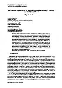
There are many medical image segmentation systems using k-means technique in detecting mass tumor in brain [15]. K-means technique is fast and simple, but it suffers from incomplete detection of tumor mainly if it is malignant tumor. On the other hand, other systems used fuzzy c-means technique because it retains more information from the original image. It can detect malignant tumor cells accurately [16], but these techniques are sensitive to noise and outliers and they take long execution time. Besides these systems, there is other systems used particle swarm optimization to segment tumor from brain images [17]. It may reach the optimal solution or near the optimal solution. It takes more computation time especially in color image segmentation. In our proposed medical image segmentation system, we get benefits from the last three techniques. As shown in Fig.1, the proposed medical image segmentation system consists of four main stages: pre-processing, clustering, tumor extraction and contouring, and validation. The main idea of doing the integration is to reduce number of iterations done by fuzzy c-means clustering technique. Of course, it minimizes execution time and gives qualitative results with KFCM (K means integrated with fuzzy c means) clustering. It also reduces computation time of particle swarm optimization to reach to the optimal clustering in KPSO (k means integrated with particle swarm optimization) clustering. The main stages of the proposed system will be discussed in more detail in the sequent subsections.
Fig. 1. The framework of the proposed medical image segmentation system
MRI Brain Tumor Segmentation System Based on Hybrid Clustering Techniques
3.1
405
Pre-processing Stage
This stage is implemented by applying a series of initial processing procedures on the image before any special purposes processing. The main purpose of this stage is to improve the image quality and removes the noise. Since, the brain images are more sensitive than other medical images; they should be of minimum noise and maximum quality. Therefore, this stage consists of de-noising and skull removal sub-stages. De-noising is important for medical images to be sharp, clear, and free of noise and artifacts. MRI images are normally corrupted by Gaussian and Poisson noise [18]. In this paper, we used median filter which is a nonlinear filter. It is often used in image processing to reduce salt and pepper noises [19]. It works by moving pixel by pixel through the image, replacing each value with the median value of neighboring pixels. The pattern of neighbors is called the "window", which slides pixel by pixel over the entire image. The median is calculated by first sorting all the pixel values from the window into numerical order, after that replacing the pixel being considered with the middle (median) pixel value. Median filtering is better than linear filtering for removing noise in the presence of edges [20]. On the other hand, image background doesn't usually contain any useful information but increasing processing time. So, removing background, skull, scalp, eyes, and all structures, which are not in interest, decrease the amount of used memory and increase the processing speed. Skull removed is done by using BSE (brain surface extractor) algorithm. It is used only with MRI images [21]. It filters the image to remove irregularities, detects edges in the image, and performs morphological erosions and brain isolation. It also performs surface cleanup and image masking. 3.2
Clustering Stage
By de-noising the MRI images and removing skulls, the images are fed to one of the proposed techniques: KFCM or KPSO. In case of supplying the image to the first technique KFCM we initialize cluster numbers K, max iterations, and termination parameter. The cluster centers are calculated by: mu= (1: k)*m/ (k+1)
(1)
Where mu are the initial means that can be calculated due to K the number of clusters and m=max (MRI image) +1. Then, assign each point to the nearest cluster center based on minimum distance and re-compute the new cluster centers. It repeats until some convergence criterion is met. Then, the resulting image can be clustered by initializing number of centroids (centroid of the cluster is the mean of all points in this cluster) equal to the number of k. This will reduce iterations and processing time. If initializing number of centroids differs from K number, it may increase time in some cases. Then, calculating the distance and updating membership and means values with determining the condition of closing. The output of the technique is the clustering image, execution time, and iteration. On the other hand, the free noise MRI images are fed to the second proposed approach (KPSO) by initializing cluster numbers k, population of particles, inertial weight value, and number of iterations. The algorithm
406
E.A. Abdel Maksoud, M. Elmogy, and R. Mokhtar Al-Awadi
follows the same steps of KFCM till determining the clusters means due to initial k. Each particle is updated by two best values. The first is the personal best (pbest) which is the best solution or fitness that has achieved so far by that particle. The second value is global best (gbest) which is the best value obtained so far by any particle in the neighborhood of that particle. Each particle modifies its position using the current positions, the current velocities, the distance between the current position and pbest, and the distance between the current position and the gbest. After that, the particle updates its velocity and positions until termination parameter which is the number of iterations. The output of the algorithm is a clustering image with optimal number of clusters, optimal clusters centroids, and computation time. 3.3
Extraction and Contouring Stage
In this stage, we used two segmentation methods: thresholding and active contour level set. Thresholding segmentation is intensity based segmentation. It is one of the important, simple, and popular segmentation techniques [22]. Thresholding segmentation technique converts a multilevel image into a binary image. It is used to extract or separate the objects in an image from the background. The segmenting image obtained from thresholding has the advantages of smaller storage space, fast processing speed, and ease manipulation [23]. The output of this stage is a segmented image with dark background and lighted object which is the brain tumor. On the other hand, the active contours have been used for image segmentation and boundary detection since the first introduction of snakes by Kass et al. [24]. The main idea is to start with initial boundary. Shapes are presented as closed curves, i.e. contours. It iteratively modifies them by applying shrink/expansion according to the constraints. An advantage of the active contours is that they partition an image into regions with continuous boundaries. So, we used level set to contour the boundary of tumor area or shape continuously after thresholding. Level set method is demonstrated in details by Lee et al. [25]. By using level set after thresholding, it gives user the resulting segmenting image of the original image with contoured tumor areas. 3.4
Validation Stage
In validation stage, the resulting segmenting images with the two proposed clustering techniques were compared to the ground truth as illustrated in experimental results. The results were evaluated by performance matrix which contains the precision and recall. Precision is the correct segmentation refers to the percentage of true positive, the number of pixels that belong to a cluster and are segmented into that cluster. Recall or sensitivity is defined as the number of true positives divided by the total number of elements that actually belong to the positive cluster [26]. The performance matrix will be illustrated in details in experimental results.
MRI Brain Tumor Segmentation System Based on Hybrid Clustering Techniques
3.5
407
Experimental Results
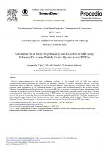
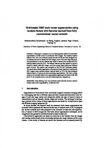
In order to check the performance of our two hybrid proposed clustering techniques in our proposed medical image segmentation system, we used three benchmark data sets. The first is the Digital Imaging and Communications in Medicine (DICOM) data set. DICOM consists of 21 images which contain brain tumors. It has no ground truth images for the contained images. The second data set is Brain Web data set. It contains simulated brain MRI data based on normal and multiple sclerosis (MS). This dataset consists of 152 images. The last data set is BRATS database from Multimodal Brain Tumor Segmentation. The data set consists of multi-contrast MRI scans and has ground truth images. This data set contains 81 images. In this section, we show the results of our two hybrid proposed clustering techniques obtained using real MRI brain images from the three different databases. This work was implemented using MATLAB 7.12.0 (R2011a). We run our experiments on a core i5/2.4 GHZ computer with 8 GB RAM and a NVEDIA/ (1 GB VRAM) VGA card. Table 1 and Table 2 demonstrate the results of applying the main four stages of our framework including our proposed techniques KFCM and KPSO on the three image data sets. Table 3, shows that EM (Expectation Maximization) like KM (K means) in accuracy but it takes longer time (T in seconds) than K Means. On the other hand, the mean shift clustering technique (MS) need to enter the parameters of bandwidth, threshold and output number of clusters K and time (T in seconds). It takes less processing time but it does not give accurate results as in DS2 when K=3.We observed that without skull removal, the processing time on all techniques was increased in DS1. On the contrary, when removing skull as in DS2 or using images with removed skull like in DS3, the processing time is reduced as shown in Table 3. In table 4, KFCM seems like FCM (Fuzzy C Means) in accuracy but KFCM take less processing time T than FCM with less iteration. From Table 5, we observed that KPSO seems like PSO (Particle Swarm Optimization) in accuracy but KPSO take less processing time than PSO. Table 6, 7 describe the performance matrix of K-means and expectation maximization. The results prove that expectation maximization may be like K-means in accuracy in the last two data sets (DS2 and DS3), but in first data set (DS1), KM accuracy is 85.7% where EM accuracy is 66.6%. From Table 6 and 8, we can observe that MS technique also seems to be the same as KM technique in performance matrix except in the second data set (DS2). Table 9 and 10 ensure that KFCM is more accurate than FCM and it is very clear in the results of first data set where KFCM accuracy is 90.50% but FCM accuracy is 85.7%. Table 11, 12 describe the performance matrix comparisons between particle swarm optimization PSO and the integration between k means and particle swarm optimization KPSO. The results prove that they are the same in accuracy, but PSO takes long time compared to KPSO in Table 5.The results showed that FCM takes longest execution time (T in seconds) in clustering, and then EM. After that, PSO takes the third level in execution time less than FCM and EM. KFCM is in the fourth level and KPSO in the fifth level. KM is in the sixth level and MS is in the last level.
408
E.A. Abdel Maksoud, M. Elmogy, and R. Mokhtar Al-Awadi
Table 1. The main stages of the proposed framework by using KFCM applied on three benchmark data sets Original mri
BSE
Median filter
KFCM
Threshold
Truth/ Normal
Level Set
No truth or normal images
NO skull removal
DS2
Ds1
DS
DS3
Already skull removed
Table 2. The main stages of the proposed framework by using KPSO applied on three benchmark data sets Original mri
BSE
Median filter
KPSO
Threshold
Truth/ Normal
Level Set
No truth or normal images
NO skull removal
DS2
Ds1
DS
DS3
Already skull removed
Table 3. The comparison between KM (K mean), EM (Expectation Maximization) and MS (Mean Shift) clustering algorithms DS
KM
EM
Ds1
K 9
T(s) 7.5
Ds2
4
1.76
Ds3
12
4.34
K 9
MS
T(s) 34.47
K 4
T(s) 0.35
4
8.00
3
0.29
12
32.06
5
0.47
MRI Brain Tumor Segmentation System Based on Hybrid Clustering Techniques
409
Table 4. The comparison between FCM (Fuzzy C means) and our proposed technique (KFCM) DS
59.52
Iteration No 8
12.87
19
15.92
4
5.18
14
6.89
3
3.46
DS1
Iteration No 51
DS2 DS3
FCM
T(s)
KFCM
T(s)
Table 5. The comparison between PSO (Particle Swarm Optimization) and our proposed technique (KPSO) DS
Iteration No 7
PSO
DS1
T(s)
Iteration No 9
KPSO
34.58
T(s) 9.23
DS2
25
58.75
25
8.42
DS3
4
6.26
4
6.01
Table 6. The performance metrics of KM Data Sets DS1 DS2 DS3
TP 85.7 96.7 95.06
TN 0 0 0
FP 0 0 0
FN 14.3 3.3 4.94
Accuracy 85.7 96.7 95.06
Precision 100 100 100
Recall 85.7 96.7 95.06
Table 7. The performance metrics of EM Data Sets DS1 DS2 DS3
TP 66.6 95.4 95.06
TN 0 0 0
FP
FN 0 33.4 0 4.6 0 4.94
Accuracy 66.6 95.4 95.06
Precision 100 100 100
Recall 66.6 95.4 95.06
Table 8. The performance matrices of MS Data Sets DS1 DS2 DS3
TP 85.7 96.05 95.06
TN
FP 0 0 0
0 0 0
FN 14.3 3.95 4.94
Accuracy 85.7 96.05 95.06
Precision 100 100 100
Recall 85.7 96.05 95.06
410
E.A. Abdel Maksoud, M. Elmogy, and R. Mokhtar Al-Awadi Table 9. The performance matrices of FCM Data Sets DS1 DS2 DS3
TP 85.7 100 100
TN 0 0 0
FP
FN 0 14.3 0 0 0 0
Accuracy 85.7 100 100
Precision 100 100 100
Recall 85.7 100 100
Table 10.The performance matrices of KFCM Data Sets TP DS1 90.5 DS2 100 DS3 100
TN 0 0 0
FP 0 0 0
FN 9.5 0 0
Accuracy 90.5 100 100
Precision 100 100 100
Recall 90.5 100 100
Table 11. The performance matrices of PSO Data Sets DS1 DS2 DS3
TP 95 100 100
TN FP 0 0 0 0 0 0
FN 5 0 0
Accuracy 95 100 100
Precision 100 100 100
Recall 95 100 100
Table 12. The performance matrices of KPSO Data Sets DS1 DS2 DS3
4
TP 95 100 100
TN FP FN 0 0 5 0 0 0 0 0 0
Accuracy Precision 95 100 100 100 100 100
Recall 95 100 100
Conclusion and Future Work
Image segmentation plays an important role in medical image. In this paper, we proposed a brain image segmentation system based on two different clustering techniques. The first is integration between fuzzy c means with k means which is called KFCM. The second is integration between particle swarm optimization and k means which is called KPSO. We applied the two clustering techniques on the three different data sets to detect the brain tumor. From experiments, we proved the effectiveness of our techniques in segmenting the brain tumor by comparing it with five state-of-theart algorithms: K-means, Expectation Maximization, Mean Shift, Fuzzy C means, and particle swarm optimization. The result of KPSO is very near to KFCM in accuracy and time but in first data set, the KPSO accuracy is 95% and KFCM is 90.5% and KPSO time is less than KFCM time. In future work, the 3D evaluation of the brain tumor detection using 3D slicer will be carried out. As well as to increase the efficiency of the segmentation process, an intensity adjustment process will provide more challenging and may allow us to refine our segmentation techniques to the MRI brain tumor segmentation, refer to [27].
MRI Brain Tumor Segmentation System Based on Hybrid Clustering Techniques
411
References 1. Bai, X., Wang, W.: Saliency-SVM: An automatic approach for image segmentation. J. Neur. Comput. 136, 243–255 (2014) 2. Patil, D.D., Deore, S.G.: Medical Image Segmentation: A Review. Int. J. Comp. Sci. Mob. Comput. 2(1), 22–27 (2013) 3. Dass, R., Priyanka, D.S.: Image segmentation techniques. Int. J. Electron. Commun. Technol. 3(1), 66–70 (2012) 4. Fazli, S., Ghiri, S.F.: A Novel Fuzzy C-Means Clustering with Hybrid Local and Non Local Spatial Information for Brain Magnetic Resonance Image Segmentation. J. Appl. Eng. 2(4), 40–46 (2014) 5. Patel, J., Doshi, K.: A Study of Segmentation Methods forDetection of Tumor in Brain MRI. Advance in Electronic and Electric Engineering 4(3), 279–284 (2014) 6. Leela, G.A., Kumari, H.M.V.: Morphological Approach for the Detection of Brain Tumour and Cancer Cells. J. Electron. Comput. Eng. Res. 2(1), 7–12 (2014) 7. Neshat, M., Yazdi, S.F., Yazdani, D., Sargolzaei, M.: A New Cooperative Algorithm Based on PSO and K-Means for Data Clustering. J. Comput. Sci. 8(2), 188–194 (2012) 8. Madhulatha, T.S.: An overview on clustering methods. IOSR. J. Eng. 2(4), 719–725 (2012) 9. Acharya, J., Gadhiya, S., Raviya, K.: Segmentation Techniques For Image Analysis: A review. Int. J. Comput. Sci. Mang. Res. 2(1), 1218–1221 (2013) 10. Jumb, V., Sohani, M., Shrivas, A.: Color Image Segmentation Using K-Means Clustering and Otsu’s Adaptive Thresholding. Int. J. Innov. Technol. Explor. Eng. 3(9), 72–76 (2014) 11. Kumar, K.S., Sivasangareswari, P.: Fuzzy C-Means Clustering with Local Information and KernelMetric for Image segmentation. Int. J. Adv. Res. Comput. Sci. Technol. 2(1), 95–99 (2014) 12. Abdul-Nasir, A.S., Mashor, M.Y., Mohamed, Z.: Colour Image Segmentation Approach for Detection of Malaria Parasites Using Various Colour Models and k-Means Clustering. J. WSEAS Transactions. Biol. Biomed. 10(1), 41–55 (2013) 13. Joseph, R.P., Singh, C.S., Manikandan, M.: brain tumor MRI image segmentation and detection in image processing. Int. J. Res. Eng. Technol. 3(1), 1–5 (2014) 14. Wang, X., Guo, Y., Liu, G.: Self-adaptive Particle Swarm Optimization Algorithm with Mutation Operation based on K-means. Advanced materials research. In: 2nd International Conference on Computer Science and Electronics Engineering, pp. 2194–2198. Atlantis Press, Paris (2013) 15. Mohan, P., Al, V., Shyamala, B.R., Kavitha, B.C.: Intelligent Based Brain Tumor Detection Using ACO. Int. J. Innov. Res. Comput. Commun. Eng. 1(9), 2143–2150 (2013) 16. Anandgaonkar, G., Sable, G.: Brain Tumor Detection and Identification from T1 Post Contrast MR Images Using Cluster Based Segmentation. Int. J. Sci. Res 3(4), 814–817 (2014) 17. Arulraj, M., Nakib, A., Cooren, Y., Siarry, P.: Multicriteria Image Thresholding Based on Multiobjective Particle Swarm Optimization. J. Applied Mathe. Sci. 8(3), 131–137 (2014) 18. Rodrigues, I., Sanches, J., Dias, J.: Denoising of Medical Images corrupted by Poisson Noise. In: 15th IEEE International Conference on Image Processing ICIP, pp. 1756–1759. IEEE Press, San Diego (2008) 19. Abinaya, K.S., Pandiselvi, T.: Brain tissue segmentation from magnitude resonance image using particle swarm optimization Algorithm. Int. J. Comput. Sci. Mob. Comput. 3(3), 404–408 (2014)
412
E.A. Abdel Maksoud, M. Elmogy, and R. Mokhtar Al-Awadi
20. Kumar, S.S., Jeyakumar, A.E., Vijeyakumar, K.N., Joel, N.K.: An adaptive threshold intensity range filter for removal of random value impulse noise in digital images. J. Theoretical Appl. Info. Technol. 59(1), 103–112 (2014) 21. Medical Medica limage processing analysis and visualization, http://mipav.cit.nih.gov/pubwiki/index.php/ Extract_Brain:_Extract_Brain_Surface_BSE 22. Narkhede, H.P.: Review of Image Segmentation Techniques. Int. J. Sci. Modern. Eng 1(8), 54–61 (2013) 23. Saini, R., Dutta, M.: Image Segmentation for Uneven Lighting Images using Adaptive Thresholding and Dynamic Window based on Incremental Window Growing Approach. Int. J. Compu. App. 56(13), 31–36 (2012) 24. Kass, M., Witkin, A., Terzopoulos, D.: Snakes: Active contour Models. Int. J. Compu. Vison 1(4), 321–331 (1988) 25. Lee, G.P.: Robust image segmentation using active contours: level set approaches. PhD, North Carolina State University (2005) 26. Dakua, P.S.: Use of chaos concept in medical image segmentation. J. Comput. Meth. Biomech. Biomed. Eng. Imag. Visua. 1(1), 28–36 (2013) 27. Own, H.S., Hassanien, A.E.: Rough Wavelet Hybrid Image Classification Scheme. Journal of Convergence Information Technology JCIT 3(4), 65–75 (2008)