in Elendt M4 medium without food. After 24 h, the larvae that did not move after disturbance with a blunt probe for five seconds were considered dead. Dashed ...
SLCO4A1. PRDM16. ADRB2. ELOVL3. DIO2. PGC1ACPT1B. 0. 5. 10. 15. Re la tive m. RNA. E xp re ssio n vehicle. Rosi. GQ-16 average ± SEM; *p
Md Shakir Hussain Haider, Wajihul Hasan Khan, Farah Deeba, Sher Ali, Anwar Ahmed, Irshad H Naqvi, Ravins Dohare,. Hytham A. Alsenaidy, Abdulrahman M.
K3,8,10,14R, the four lysines at K3, 8, 10, and 14 of endogenous Htz1 were mutated to ... Data are the average of at least three independent experiments ± SD.
Jul 4, 2018 - 29 Burwood Grove, Hayling Island, PO11 9DS. 15. Lot 11. 17 Rosslyn .... accessible for commuting across th
May 24, 2017 - particular lot will be offered for sale at Auction or whether it has .... Trigg House, Monks Brook, St Cr
May 24, 2017 - e) sign each sale memorandum; and f) treat a contract as repudiated if the buyer fails to sign a sale mem
B) Tree length=[ Ks (dog-mouse)+ Ks (human-rat)+ Ks (human-dog)+ Ks (rat-mouse)]/2. The figure below shows a plot of this tree length versus the human- ...
Determining correlation between infectious virus titre and real time RT-PCR copy number. Correlation of infectious virus titres as measured by TCID50 assay ...
Page 1. Figure S1 â Correlation between students' age and musicality scores.
SUPPLEMENTAL FIGURE LEGENDS. Figure S1. Mmp2 and Mmp9 are expressed in discrete populations within hemogenic regions. Related to Figure 1.
progressive allograft vasculopathy; (b) representative photomicrographs of elastin van Gieson stained paraffin sections depicting typical fibroproliferative arterial ...
Chryssa Kanellopoulou, Timothy Gilpatrick, Gokhul Kilaru, Patrick Burr, Cuong K. Nguyen, Aaron Morawski, Michael J. Lenardo, and Stefan A. Muljo ...
Quantibody arrays using Genepix Pro software. .... PBS and serum-free MesenCult® XF media (Stem Cell Technologies) was added, stimulated with 10 ng/ml TNF-α ..... Lee, R.H., Pulin, A.A., Seo, M.J., Kota, D.J., Ylostalo, J., Larson, B.L., ...
... involving the 3'Eκ and MiEκ bait across the Igk locus in pre-B, Immature B, DN and DP cells and their replicates. This matrix is denoted as M3 in the main text.
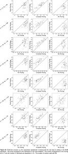
May 29, 2015 - Complete Pooling. No Pooling. Figure S1 Prediction accuracy rı (for populations represented in training set) for trait days to silking in the NAM.
ascidians suggests the presence of multiple paralogues (Wnt5a and Wnt5b) which originated in the. Stolidobranchia ... Despite the fact that the number of Wnt5 genes is the same in species of the Molgula ..... S12.g13523â¡. -. Wnt9 ..... KR, Eppig JT
The samples for this study were obtained from a patient with a primary diffuse gastric cancer tumor and metastases located in each ovary (Krukenberg tumors).
Poignard, Elise Landais, James C. Paulson, Ian A. Wilson, and Andrew B. Ward ... (A) C-terminally truncated (âCT) and full-length (FL) Env constructs ...
lilah-Seyfried, Thomas Scheper, Henning Kempf, Robert Zweigerdt, and .... Cultures were dissociated using Collagenase II (Worthington, Lakewood, USA) and ...
negative inputs of Nkx2.2, Olig2 and Nkx6.1. Related to Figures 5 and 6. Supplemental Tables (available as separate files). Table S1. Genes, their expression ...
Figure S1. Dendrogram and heatmap of the Pearson's correlation ...
Dendrogram and heatmap of the Pearson's correlation coefficient for human tissue gene expression (Log2FPKM). Only genes with FPKM>1 in at least one ...
Figure S1. Dendrogram and heatmap of the Pearson’s correlation coefficient for human tissue gene expression (Log2FPKM). Only genes with FPKM>1 in at least one sample were used for analysis. The cartilage samples are highlighted with a red frame.
Figure S2. Box plots showing cartilage-selective gene expression in cartilage (red) and noncartilage (blue) tissues. The thick horizontal line in each box represents the median gene expression value.
Figure S3. Bar graph showing RUNX2 expression in human tissues. Red bars indicate cartilage samples and blue bars indicate non-cartilage samples, respectively. Gene expression levels are shown as FPKM values.
Figure S4. Genome browser views of H3K4me1 and H3K27ac peaks along a COL2A1 and b COL11A1 loci. Chromosome coordinates are shown as black bars on top. Black arrows indicate the direction of transcription on a diagram of each gene below. CH, chondrocytes. AG, adrenal gland. LI, large intestine. SI, small intestine. MU, muscle. SC, spinal cord. ST, stomach. TH, thymus.
Figure S5. Genome browser views of H3K4me1, H3K4me3, H3K27ac peaks and RNA expression track along lncRNA loci (A) AC058791.1 and (B) RP11-261P24.2 in human chondrocytes.
Figure S6. Sox9 binds to the lncRNA 2610035D17Rik promoter and gene body in mouse rib chondrocytes. a Genome browser view of Sox9 binding along Sox9 and 2610035D17Rik loci on chromosome 11. b Shaded region in a was enlarged for a close view of Sox9 binding at the 2610035D17Rik promoter and first intron.