September 1, 2006 11:40 WSPC/147-MPLB
01172
Final Reading Brief Review
Modern Physics Letters B, Vol. 20, No. 20 (2006) 1217–1231 c World Scientific Publishing Company
COMPUTATIONAL MODELING OF TISSUE SELF-ASSEMBLY
ADRIAN NEAGU Department of Physics & Astronomy, University of Missouri-Columbia, Columbia, MO, 65211, USA Department of Biophysics and Medical Informatics, Victor Babes University of Medicine and Pharmacy Timisoara, P-ta Eftimie Murgu Nr. 2, 300041 Timisoara, Romania
[email protected] IOAN KOSZTIN∗ , KAROLY JAKAB and BOGDAN BARZ Department of Physics & Astronomy, University of Missouri-Columbia, Columbia, MO, 65211, USA ∗
[email protected] MONICA NEAGU Department of Biophysics and Medical Informatics, Victor Babes University of Medicine and Pharmacy Timisoara, P-ta Eftimie Murgu Nr. 2, 300041 Timisoara, Romania RICHARD JAMISON Department of Physics & Astronomy, University of Missouri-Columbia, Columbia, MO, 65211, USA GABOR FORGACS Department of Physics & Astronomy, Department of Biology, University of Missouri-Columbia, Columbia, MO, 65211, USA Received 7 August 2006 As a theoretical framework for understanding the self-assembly of living cells into tissues, Steinberg proposed the differential adhesion hypothesis (DAH) according to which a specific cell type possesses a specific adhesion apparatus that combined with cell motility leads to cell assemblies of various cell types in the lowest adhesive energy state. Experimental and theoretical efforts of four decades turned the DAH into a fundamental principle of developmental biology that has been validated both in vitro and in vivo. Based on computational models of cell sorting, we have developed a DAH-based lattice model for tissues in interaction with their environment and simulated biological selfassembly using the Monte Carlo method. The present brief review highlights results on specific morphogenetic processes with relevance to tissue engineering applications. Our own work is presented on the background of several decades of theoretical efforts aimed to model morphogenesis in living tissues. Simulations of systems involving about 10 5 cells have been performed on high-end personal computers with CPU times of the order of days. Studied processes include cell sorting, cell sheet formation, and the development 1217
September 1, 2006 11:40 WSPC/147-MPLB
1218
01172
Final Reading
A. Neagu et al. of endothelialized tubes from rings made of spheroids of two randomly intermixed cell types, when the medium in the interior of the tube was different from the external one. We conclude by noting that computer simulations based on mathematical models of living tissues yield useful guidelines for laboratory work and can catalyze the emergence of innovative technologies in tissue engineering. Keywords: Cell adhesion; differential adhesion hypothesis; tissue engineering.
1. Introduction Starting from the beginning of the 20th century, theoretical models of living tissues have evolved along two distinct conceptual lines. The first of these views considers the tissue as a set of discrete, interacting cells, whereas the other treats it as a continuum, and monitors cell densities instead of individual cells.18 Here we briefly describe a few of these models. The interested reader can find further details in the cited literature. The continuum approach, promoted by Murray and Oster, uses the methods of continuum mechanics and allows for modeling tissues built of realistic numbers of cells (see Ref. 14 and references therein). The distribution of cells of various types throughout the tissue is described in terms of their densities, whereas their morphogenetic rearrangements are treated as fluxes. The method has been applied for studying developmental morphogenesis, dermal wound healing, contraction, scar formation and vasculogenesis.18 The latter phenomenon may occur in vivo via two different mechanisms: (i) vasculogenesis, a term used for de novo vessel formation by the self-assembly of an endothelial cell population, and (ii) angiogenesis, the formation of capillary vessels by endothelial cell sprouting from preexisting vessels. (Endothelial cells represent the major component of small blood vessels and line the internal face of large ones. Besides endothelial cells, capillaries also feature attached pericytes, a cell type responsible for the stability of capillary vessels.) Growing large, vascularized organ replacements in the laboratory is one of the major challenges of tissue engineering. Therefore, understanding the intimate mechanisms of vasculogenesis is vital for building perfusable tissue constructs. Starting from a cell population randomly distributed on the planar surface of a homogeneous extracellular matrix, the model of Murray et al.14 predicts the emergence of interconnected filamentous structures of cells that are similar to a vascular network. The results are in good agreement with in vitro experiments on endothelial cells seeded on Matrigel.11 One of the most important principles of developmental biology, which inspired numerous discrete cell models, is the differential adhesion hypothesis (DAH). 20 It states that (i) cell adhesion corresponds to cell-type-dependent energies and (ii) the constituent cells of a tissue are motile enough to reach the lowest energy configuration. The DAH withstood the test of time, being confirmed by many experiments. This principle leads to a close analogy between true liquids and living tissues made of adhesive and motile cells, such as most embryonic and some artificial tissues.3
September 1, 2006 11:40 WSPC/147-MPLB
01172
Final Reading
Computational Modeling of Tissue Self-Assembly
1219
A considerable number of discrete cell models rely on DAH. Monte Carlo simulations of the large-N Potts model from statistical physics reproduced experimental findings regarding the segregation of different cell populations and the mutual engulfment of adjacent tissue fragments.5 In this model the tissue is represented on a lattice, each cell spans several lattice sites, and has a unique identification number; the average number of sites per cell is maintained around a target value via an elastic energy term containing a Lagrange multiplier. The simulations are based on the Metropolis algorithm, accounting for cell migration and shape changes in systems made of up to several thousand cells.4 These simulations suggested that cell motility may be ascribed to an effective, temperature-like parameter.2 Recent computational models of in vivo morphogenesis, besides DAH, also include chemical signaling, i.e. chemotaxis, cell differentiation and extracellular matrix production.12, 16 The process of culmination of the cellular slime mold Dictyostelium discoideum under the condition of scarce food supply, was simulated in two dimensions by combining the Glazier and Graner model4 with a set of partial differential equations able to describe cAMP signaling. The model is defined by parameters characteristic for the subcellular level and is able to predict phenomena that involve the self-organization of thousands of cells. In this respect, this model bears the potential to characterize the morphogenetic impact of genes whose function is elucidated at the subcellular level.12 Slime mold aggregation has also been described using a distinct, force-based, three-dimensional (3D) model, in which individual amoebae are treated as viscoelastic ellipsoids with type-dependent adhesion apparatus, intrinsic motility and cAMP-mediated signaling capacity.16 2. In Silico Tissue Engineering Tissue engineering (TE) is a rapidly developing field of biomedical research, which aims to repair, replace or regenerate damaged tissues. It exploits biological morphogenesis, a self-assembly process that gives birth to a large variety of structures in living systems. TE employs a number of techniques meant to engage cells into forming tissues10 (see also the web page http://www.nsf.gov/pubs/2004/nsf0450/). It is hard to overestimate the importance of the field, given that growing organs in vitro could solve the problem of transplantable organ shortage. TE evolved in close relationship with regenerative medicine, and proved successful in developing various functional organ modules. These may be used for in vivo tissue repair, may promote tissue regeneration, and can also be used for testing new drugs. The clinical success of TE depends on finding a suitable cell source, on optimizing scaffolds or hydrogels that support cell growth, differentiation and assembly, on building bioreactors able to provide physiological conditions for the engineered tissues, and, most importantly, on the development of techniques able to assure long-enough shelf life for the tissue construct to reach the patient.6
September 1, 2006 11:40 WSPC/147-MPLB
1220
01172
Final Reading
A. Neagu et al.
The success of the theoretical methods currently used to describe both in vivo and in vitro rearrangements of cell populations motivated recent attempts of modeling phenomena of interest in TE. In order to be efficient in screening alternative experimental designs and in offering hints for related laboratory studies, computational tissue engineering must properly account for the dynamics of cell populations in the presence of scaffolds and extracellular matrices that on the one hand guide cell behavior and, on the other hand, are subject to degradation and restructuring by cells. Several groups are presently engaged in this endeavor. For example, a recent computational model describes the movement of fibroblasts within an acellular dermal matrix of oriented fibers.18 The simulations predict the dynamics of matrix invasion as a function of various parameters, such as fiber size distribution, packing density, and matrix morphometry. The scaffold is generated by a random walk algorithm and the fibroblast movement along the fibers is described by a five-state Markovian process of directional change. The hopping of cells between nearby fibers is simulated using the Monte Carlo method. The simulations are remarkable also from the point of view of the computational platform they used, a versatile 3D modeling and animation package, MAYA (Alias, Toronto, ON, Canada; http://www.alias.com). It incorporates an onboard programming language, along with physics simulation, visualization, and animation engines. The computational model is meant as a supplement for experimental efforts to streamline the workflow of matrix design. In the context of bone and cartilage morphogenesis in vitro, hierarchical computational techniques have been employed to design anatomically shaped 3D scaffolds with controlled porosity and chemical composition via solid free-form fabrication. 7 The method proved useful for improving the mechanical properties of scaffolds and resulted in accelerated tissue formation. Computer simulations, serving as proof-of-concept in silico experiments, may also speed up the development of new technologies. As an example, we present results related to the modeling of artificial organs built from spheroids containing tens of thousands of cells. The self-assembly of these cell aggregates resembles the fusion of adjacent liquid drops, thus justifying the name of “bioink” given to the cell aggregates used in bioprinters. Their computer-controlled, layer-by-layer deposition into a supportive hydrogel (also referred to as “biopaper”) goes by the name of bioprinting.9, 13, 15 Model assumptions and parameter estimations are based on experiments. Simulations of similar systems starting from new initial configurations, with same or modified conditions, are used to optimize future experiments. In the following we describe the employed computer simulation method along with some of the obtained results. 3. A Lattice Model of Living Tissues We first build a lattice model of a system of living cells in a culture medium or an extracellular matrix, and then turn to simulate its evolution using the Metropolis
September 1, 2006 11:40 WSPC/147-MPLB
01172
Final Reading
Computational Modeling of Tissue Self-Assembly
1221
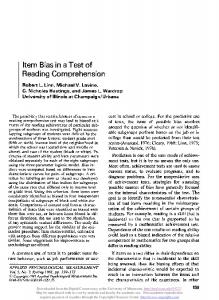
Fig. 1. A simplified, two-dimensional square-lattice model. Sites are occupied by cells (black), or volume elements of medium or extracellular matrix (light gray). A cell is considered to interact to the same extent with nearest or next-nearest neighbors. Cells interact with adjacent cells (light gray bond) and with the surrounding medium (dark gray bond). In order to avoid double-counting, only half of the depicted bonds are attributed to the given cell [see Eq. (1)].
Monte Carlo method. Our goal is to describe the self-assembly of cells within tissue constructs made of hundreds of thousands of cells. Therefore, in contrast to the Glazier–Graner model,4, 5 our program focuses on the types of particles present on each lattice site rather than following cell shape changes or monitoring the position of individual cells. For computational simplicity, we discretize the space and represent the biological system on a cubic lattice. Each lattice site is occupied either by a cell, or a similarsized volume element of the embedding medium. Figure 1 depicts the 2D version of the model that enables us to explain the significance of the terms in the total interaction energy: X E= [J(σi,j , σi,j+1 ) + J(σi,j , σi+1,j+1 ) + J(σi,j , σi+1,j ) + J(σi,j , σi+1,j−1 )] . (1) i,j
The occupancy of a given site,(i, j), is specified by a type index, σi,j , which can take two values, namely 0 for a medium (type 1) particle, and 1 for a cell (type 2) particle. A given cell interacts with its neighbors either directly, via cell adhesion molecules (e.g. cadherins) or indirectly by binding to extracellular matrix filaments via integrins. In our model adhesivities are associated to contact interaction energies also referred to as bond energies, which are determined by both the strength and dynamics of the involved chemical bonds. Each term on the right hand side of Eq. (1) can take the values J(0, 0) = − ε11 , J(1, 1) = − ε22 or J(1, 0) = J(0, 1) = − ε12 . The ε’s are positive quantities and represent the mechanical work needed to disrupt the corresponding bond.
September 1, 2006 11:40 WSPC/147-MPLB
1222
Final Reading
01172
A. Neagu et al.
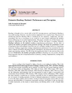
Fig. 2. Spontaneous rounding of an irregular tissue fragment made of CHO cells during 24 hours of incubation.
The second law of thermodynamics tells us that such a system will evolve towards the less structured, highest symmetry state that has maximum entropy. In the associated biological problem, however, we are dealing with an open system, so the principle of maximum entropy does not necessarily imply less structure in the emergent cellular pattern. That is why, for example, the differential adhesion hypothesis is not a direct consequence of the second law of thermodynamics. DAH has its origin in experiments like that of Fig. 2, showing that an irregular tissue fragment placed in a nonadhesive environment spontaneously rounds up, as a liquid droplet would.3 This experiment indicates that the rearrangement of cells is dictated by interfacial forces: the tissue rounds up in order to minimize the area exposed to the tissue culture medium. (A sphere is the geometrical object of smallest surface area for a given volume). The total interaction energy may be rewritten in terms of interfacial contributions. To this end, consider a configuration of N1 particles of type 1, out of which N1I are located on the 1–2 interface while the rest, N1B , reside in the bulk. A similar partitioning may be done for the type 2 particles as well. Thus, Eq. (1) can be rewritten as " # N1I N2I X X 1 B B (n1i1 ε11 + n2i1 ε12 ) + (n2i2 ε22 + n1i2 ε12 ) E = − N1 nn ε11 + N2 nn ε22 + 2 i =1 i =1 1
2
(2)
where the factor 1/2 cancels the double counting of interacting pairs. The dummy index i1 (i2 ) runs over interfacial particles of type 1 (2), whereas n1i1 (n2i1 ) stands for the number of type 1 (2) neighbors interacting with the type 1 particle labeled by i1 . A similar notation holds for the type 2 interfacial particles labeled by i2 . The simulations are performed for systems of finite size, such that cells are coaxed to move within the medium. With these boundary conditions, it is technically convenient to maintain an immobile medium layer on the frontier of the system. The contribution to the interaction energy coming from this layer is constant and, therefore, it can be discarded. Thus we are dealing with particles lying in
September 1, 2006 11:40 WSPC/147-MPLB
01172
Final Reading
Computational Modeling of Tissue Self-Assembly
1223
the interior of the system with nn significant neighbors (nn = 8 in two dimensions and nn = 26 in three dimensions); therefore n1i1 = nn − n2i1 and n2i2 = nn − n1i2 , which leads to " N1I X 1 B I B I n2i1 E = − (N1 + N1 )nn ε11 + (N2 + N2 )nn ε22 + (ε12 − ε11 ) 2 i =1 1
I
+ (ε12 − ε22 )
N2 X
i2 =1
#
n1i2 .
(3)
Both sums in Eq. (3) yield the total number of heterotypic bonds PN1I PN2I i1 =1 n2i1 = i2 =1 n1i2 = B12 . Indeed, the first is obtained by cumulating the numbers of type 2 particles in the significant neighborhood of each interfacial particle of type 1, whereas the second one is the sum of the numbers of type 1 particles around all type 2 particles from the 1–2 interface. Thus, the total adhesive interaction energy, both in 2D and 3D, becomes 1 1 E = γ12 B12 − N1 nn ε11 − N2 nn ε22 2 2
(4)
where B12 is the number of 1–2 bonds, directly proportional to the area of the interface, and γ12 =
ε11 + ε22 − ε12 2
(5)
is the interfacial tension parameter. During simulations that do not include cell proliferation, differentiation and death the last two terms on the right hand side of Eq. (4) are constant and, therefore, they may be omitted. Canonical Monte Carlo simulations using E = γ12 B12 yielded results in qualitative agreement with experiments on living tissue self-assembly.9 This expression is remarkable, since it does not depend on the strengths of all types of interactions, but only on their combination, γ12 . In the case of a complex tissue of several cell types and media, the total interaction energy, under the constraint of constant numbers of particles of each type, is given by E=
T X
γij · Bij ,
(6)
i,j =1 i