ORIGINAL ARTICLE
Force Requirements for Artificial Muscle to Create an Eyelid Blink With Eyelid Sling Craig W. Senders, MD; Travis T. Tollefson, MD; Shane Curtiss; AnnJoe Wong-Foy, PhD; Harsha Prahlad, PhD
Objective: To determine the force requirements, opti-
mal vector, and appropriate materials of a novel eyelid sling device that will be used to rehabilitate eyelid closure (blink) in congenital or acquired permanent facial paralysis with an artificial muscle. Methods: The force required to close the eyelids in
human cadavers (n = 6) were measured using a load cell system. The eyelid sling using either expanded polytetrafluoroethylene (ePTFE) or temporalis muscle fascia was implanted. The ideal vector of force and placement within the eyelid for a natural eyelid closure were compared. Results: The eyelid sling concept was successful at cre-
ating eyelid closure in a cadaver model using an upper eyelid sling attached to the distal tarsal plate. Less force was necessary to create eyelid closure using a tempora-
lis muscle fascia sling (627±128 mN) than for the ePTFE eyelid sling (1347±318 mN). Conclusions: The force and stroke required to close an
eyelid with the eyelid sling are well within the attainable range of the electroactive polymer artificial muscle (EPAM). This may allow the creation of a realistic and functional eyelid blink that is symmetric and synchronous with the contralateral, normally functioning blink. Future aims include consideration of different sling materials and development of both the EPAM device and an articulation between the EPAM and sling. The biocompatibility and durability studies of EPAM in a gerbil model are under way. The successful application of artificial muscle technology to create an eyelid blink would be the first of many potential applications. Arch Facial Plast Surg. 2010;12(1):30-36
E
LECTROACTIVE POLYMER ARtificial muscle (EPAM) is an emerging technology that has the potential to be used in rehabilitating facial movement in patients with paralysis. These electroactive polymers act like human muscles by expanding and contracting based on variable voltage input levels. Mimicry of
Video available online at www.archfacial.com
Author Affiliations: Departments of Otolaryngology–Head and Neck Surgery, Facial Plastic and Reconstructive Surgery, Cleft and Craniofacial Program (Drs Senders and Tollefson), and Orthopedics (Mr Curtis), University of California Davis Medical Center, Sacramento; and SRI International, Menlo Park, California (Drs Wong-Foy and Prahlad).
diaphragm movement in a canine model has shown promise and has led to the examination of reanimating smaller muscle groups, such as those responsible for eyelid closure or facial expression. Conventional treatment of permanent facial paralysis has included a variety of surgical procedures that endeavor to recreate the form and function of the facial musculature. Management of inadequate eyelid closure in facial paralysis seeks to protect the eye from corneal ulceration or blindness. This study seeks to develop the protocol and device design for human implanta-
(REPRINTED) ARCH FACIAL PLAST SURG/ VOL 12 (NO. 1), JAN/FEB 2010 30
tion of EPAM to reproducibly create a longlasting eyelid blink that will protect the eye and improve facial appearance. Permanent facial paralysis that results from congenital facial anomalies, facial trauma (eg, temporal bone), or oncologic surgical resection is a common and significant clinical problem. Currently, options for ocular protection in permanent facial paralysis include gold1 or platinum2 weight eyelid loading, lower eyelid repositioning (tarsal strip canthoplasty), tarsorhaphy, or muscle transfer such as local-regional (eg, temporalis muscle tendon transfer)3 or free tissue transfer (eg, gracilis).4 These options are limited by implant extrusion, donor site morbidity, and surgical candidacy. Herein, we describe a novel alternative method for eyelid rehabilitation in permanent facial paralysis. We propose that the eyelid sling mechanism (Figure 1) can create an eyelid blink when actuated by an artificial muscle.5 Electroactive polymer artificial muscle was developed at SRI International (Menlo Park, California) in the 1990s and has the potential to mimic a variety of biologic muscle activities if it can be successfully implanted in humans. To our WWW.ARCHFACIAL.COM
Downloaded from www.archfacial.com at Emory University, on February 9, 2010 ©2010 American Medical Association. All rights reserved.
A
B
#
Battery
II
§
‡
EPAM
¶ †
∗ Sling
Figure 1. Eyelid sling conceptual illustration with artificial muscle and power source design. Illustration of left upper eyelid sling attached to the electroactive polymer artificial muscle (EPAM) device (A) through an interpolation device that is implanted in the lateral orbital wall (B). The power supply and artificial muscle are implanted in the temporal fossa. An electroactive polymer device (*) includes an actuator (†) having an electroactive polymer (‡) in a biocompatible sheath (§). The electroactive polymer is attached the eyelid sling (㛳) and energized by a power supply (¶). A myoelectric sensor (#) is in electrical contact with the actuator to provide a signal from the contralateral orbicularis oculi muscle to coordinate eyelid blinking.
∗
A
B
†
‡
Figure 2. Mechanism of polymer expansion and contraction. A, Electroactive polymer (*) comprises an elastomeric dielectric polymer film (†) sandwiched between compliant electrodes (‡), as shown in Figure 1. B, When a DC voltage is applied to the electrodes, the elastomeric polymer expands in a plane perpendicular to the force due to the attraction between the electrodes. The elastomer polymer contracts back to the original shape with substantial force when the voltage differential across the electrodes is neutralized.
knowledge, there have been no published reports of applications of artificial muscle in humans or animals. ARTIFICIAL MUSCLE The use of an artificial muscle in a variety of human disorders has great potential. The characteristics of the ideal artificial muscle should be similar to those of biologic muscle. Biologic muscles are “optimized systems” that are relatively similar in all species and exhibit quick reaction times with large linear forces. Biologic muscle contraction, stimulated by electrochemical nerve conduction, is caused by chemically induced reversible hydrogen bonding between actin and myosin.6 In the field of robotics, a variety of actuators (motion-generating devices) have been developed, which include electromagnetic motors, piezoelectric ceramics, or shape memory alloys. The use of these devices is limited in humans owing to poor biocompatibility. The development of elec-
troactive polymer technology has created the potential for a vast array of applications ranging from precisely delayed-release drug delivery to powering hydraulic systems and as flat-panel speaker diaphragms.7 Electroactive polymer artificial muscle is composed of an inner core of a dielectric elastomer film (eg, silicone or acrylic) with a flexible electrode coating placed on either side (Figure 2). An electrostatic compressive force is then created by compressing the elastomer when voltage is applied to the 2 outer electrodes. The elastomer expands in area and becomes thinner as the voltage is applied. When the electrical current is released, the artificial muscle contracts back to its original shape, thus converting the electrostatic force into mechanical work.8 INDICATIONS Over 40 000 cases of facial paralysis are seen in the United States every year, with most of these being idiopathic (Bell
(REPRINTED) ARCH FACIAL PLAST SURG/ VOL 12 (NO. 1), JAN/FEB 2010 31
WWW.ARCHFACIAL.COM
Downloaded from www.archfacial.com at Emory University, on February 9, 2010 ©2010 American Medical Association. All rights reserved.
Upper eyelid loading with gold or platinum is a safe and effective method to correct paralytic lagophthalmos, so why do we need to pursue other options? Any alternative method to restore eyelid closure and function would need to be compared with gold weight eyelid loading in which a carefully selected weight (0.6-1.8 g) can be positioned into the upper eyelid pretarsal region to allow gravity to provide the force by which the eyelid can close. This is technically a straightforward, reversible technique with a remarkable success rate.1 The disadvantages are that there is a visible weight in the eyelid that can extrude or, if too heavy, induce visual field impairment with eyelid ptosis. Most important, eyelid closure relies on inhibition of the levator palpebral superioris to relax and on the force of gravity to allow the eyelid to close, thus producing a slow eyelid blink that is not synchronous with the contralateral normal eye (10-15 blinks per minute).
A
‡ †
∗ B §
‡ †
EYELID SLING CONCEPT ∗
Figure 3. Illustration of a sling. A, The sling (*) is mounted between a fixed mount point (on right) and the dielectric polymer (†) that contracts when the voltage is discontinued into the electrodes (‡). B, The sling flattens to mimic eyelid closure. Although we use an upper eyelid sling, this figure demonstrates a lower eyelid sling (§).
palsy) or virally induced.6 In the few cases of a facial paralysis in which the patients do not recover or the paralysis is permanent, a variety of surgical options can be used to maintain facial muscle tone, rehabilitate some function, and protect the globe from exposure keratopathy. These surgical options include facial nerve grafting, cable grafting (sural nerve), cross-face jump grafts, or anastomosis with other cranial nerves (hypoglossalfacial nerve). However, in many cases of permanent facial paralysis, nerve grafting may not be possible owing to (1) extensive tumor excision, including the facial nerve or distal facial muscles; (2) permanent facial nerve dysfunction after Bell palsy or herpes zoster palsy; or (3) congenitally absent facial nerve function with multiple other cranial neuropathies (Mobius syndrome). Management of paralytic lagophthalmos and lower eyelid ectropion (eg, tarsal strip canthoplasty, or temporary tarsorhaphy) is imperative. Immediate reconstruction options are considered when nerve grafting is not possible and include temporalis muscle transfer into the eyelids. Traditional static facial slings using tensor fascia lata or alloplastic materials (expanded polytetrafluoroethylene [ePTFE]; or cadaveric acellular dermis) are effective at midface suspension,9 while free tissue transfer (gracilis or serratus anterior) has shown good efficacy for restoring perioral movement10 and eyelid function.4 Elderly patients with permanent facial paralysis after oncologic resection may not be good candidates for major reconstructive procedures (eg, microvascular free tissue reconstruction) owing to multiple comorbidities.
In the concept development of the eyelid sling mechanism (Figure 3), we sought to create eyelid closure with an artificial sphincter mechanism that could mimic the innate orbicularis oculi muscle. The orbicularis oculi muscle has 2 major divisions: (1) the orbital and (2) the palpebral, which is further subdivided into the pretarsal and preseptal components. The pretarsal orbicularis is mostly responsible for the involuntary eyelid blink, while the preseptal contributes to voluntary eyelid closure (wink). The orbital component of the orbicularis oculi protects the globe in bright sunlight or adverse conditions by squeezing the eyelids. The coordinated eyelid blink is a balance of eyelid closure by the orbicularis oculi (innerverated by the facial nerve) and eyelid opening by the levator palpebrae superioris muscle (innervated by the oculomotor nerve).11 To develop the eyelid sling concept, we relied on an amalgam of techniques such as medial and lateral canthal reconstruction (eg, tarsal strip canthoplasty),12-14 gold weight eyelid loading, and concepts from free tissue transfer. The goal of the eyelid sling mechanism is to protect the eye and prevent corneal exposure. When lower eyelid muscle tone is lost in permanent facial paralysis, it results in midfacial drooping and loss of lower eyelid tone. For this reason, the lower eyelid is pulled away from the globe, which contributes to the associated paralytic lagophthalmos and ectropion.15 The medial punctum is also everted from the globe and creates the unfortunate condition of epiphora with dry eye symptoms in the same setting. A tarsal strip canthoplasty can reposition and shorten the lower eyelid to a more stable position over the inferior globe by suspending the lateral tarsal plate to the medial periosteum of the orbital rim and the superior horn of the lateral canthal tendon. An effective modification, lateral orbital wall anchoring, has allowed more permanent fixation of the lateral tarsal strip. A suture can be passed through drill holes in the lateral orbital wall to anchor the lateral canthal tendon (Figure 4). The overall goal of the project is to rehabilitate eyelid closure (blink) in congenital or acquired permanent facial paralysis using an eyelid sling that is actuated with
(REPRINTED) ARCH FACIAL PLAST SURG/ VOL 12 (NO. 1), JAN/FEB 2010 32
WWW.ARCHFACIAL.COM
Downloaded from www.archfacial.com at Emory University, on February 9, 2010 ©2010 American Medical Association. All rights reserved.
A
Figure 4. Intraoperative photograph demonstrating the lateral orbital wall drill holes that are used for suture suspension of lateral canthus for repair of paralytic lower eyelid extropion.
B
EPAM. This will create a realistic and functional eyelid blink that is symmetric and synchronous with the contralateral, normally functioning blink. To actuate the eyelid sling, we must develop the first successful human implantation of artificial muscle. Herein, we report on our force and vector analysis of the eyelid sling using cadaver experiments. METHODS The cadaveric dissections and experiments were conducted at the University of California, Davis, Anatomy Laboratory, with approval from the Donated Body Program. The eyelid sling mechanisms were implanted in 6 cadaver dissections of the orbit to analyze vector analysis and force requirements using a miniature tension-compression load cell (Omega Corp, Stanford, Connecticut) attached to a novel handheld device (Figure 5); this device was developed and validated in an associated laboratory by one of us (S.C). Data collection was completed using Daqview software (version 7.13.14; Iotech, Cleveland, Ohio) and an 8-channel Daqbook 2000E 16-bit data acquisition system connected to a DBK43a strain gage module (DBK434; Iotech). Force measurements were collected and analyzed using Excel 2004 software (Microsoft Corp, Redmond, Washington).
Figure 5. Handheld tension-compression load cell for acquiring force measurements. A, The load cell is attached to the eyelid sling. B, Force applied in the direction of the arrow creates eyelid closure.
EYELID SLING PROCEDURE The implantation began with an extended curvilinear incision similar to the supraciliary blepharoplasty incision (approximately 10 mm above the eyelash line) (video available at http:// www.archfacial.com). The orbit was injected with normal saline using a 28-gauge needle to attain physiologic globe volume and pressure (normal range, 10-21 mm Hg), measured with tonometry.16 We dissected through the pretarsal orbicularis to expose the tarsal plate and then medially through the orbicularis oculi to expose the medial orbital wall and the insertion sites (anterior and posterior lacrimal crests) of the 2 heads of the medial canthal tendon. Lacrimal probes were placed in the eyelid punctum to protect the canaliculi during dissection and placement of the medial orbital sling anchoring screw.14 We completed the lateral canthal dissection with an incision extending over the lateral orbital rim (similar to a lateral canthal anchoring procedure) (Figure 6). The lateral orbital periosteum was dissected free from the lateral orbital wall, and a malleable retractor protected the globe while the lateral orbitotomy was completed using a drill. The inferior division of the
Figure 6. Lateral canthal suture suspension of lower eyelid through the lateral orbital drill holes in a cadaver model. Note the temporalis fascia eyelid sling that extends from the medial canthal dissection and over the inferior-most upper eyelid tarsal plate.
lateral canthal tendon was resuspended to the lateral orbital wall through the drill holes. The position of the orbitotomy (at or above the Whitnall tubercle) and lateral canthal resuspension are performed to prevent the upper eyelid from overriding the lower eyelid.12,13 With the orbitotomy complete, the eyelid sling material (ePTFE or temporalis muscle fascia) was secured to the inferior one-third of the tarsal plate using 6-0 suture. The eyelid sling was inset to the medial orbital wall with a 1-mm titanium microscrew (3-mm length; Stryker Leibinger, Kalamazoo, Michigan).14 The ideal medial canthal anchoring position was tested by placement of the screw in (1) the frontal process of the maxillary bone and both (2) the anterior and (3) the posterior lacrimal crest. The gap between the medial
(REPRINTED) ARCH FACIAL PLAST SURG/ VOL 12 (NO. 1), JAN/FEB 2010 33
WWW.ARCHFACIAL.COM
Downloaded from www.archfacial.com at Emory University, on February 9, 2010 ©2010 American Medical Association. All rights reserved.
A
B
C
D
Figure 7. Cadaver dissection. A, Demonstration of the eyelid sling mechanism in the upper eyelid of the right eye (temporalis muscle fascia in right; expanded polytetrafluoroethylene in left) to allow direct comparison of force requirements. B, Right eyelid implanted temporalis fascial sling showing eyelid closure with application of posterior-lateral force. C, Expanded polytetrafluoroethylene in left eyelid. D, Eyelid closure with application of force to sling.
canthal eyelid and the globe was measured with a digital caliper. The lateral aspect of the sling was passed either over the lateral orbital rim or through the orbitotomy with exit into the temporal fossa. This was performed to mimic the proposed position of the artificial muscle device and a battery. The eyelid is closed by activating the eyelid sling mechanism with posteriolateral force.
EXPERIMENTS Two separate experiments were completed. First, the force required to close the eyelids was measured on 4 frozen (thawed) cadaver heads using the ePTFE as a sling. The specimen was positioned upright to simulate head position in an upright person. Force testing was completed with the sling either passed (1) over the lateral orbital wall or (2) through lateral orbital drill holes. Results were collected for each group and analyzed. Second, force measurements were obtained with a comparative study using fresh cadavers (n = 2). One eye was implanted with a temporalis muscle fascia sling, and an ePTFE sling was implanted in the contralateral eye (Figure 7). The temporalis muscle fascia graft (8 ⫻2 cm) was harvested with a standard scalp incision. The force measurements were recorded for 4 series of 6 eyelid closures on each side (temporalis fascia and ePTFE) and compared with a t test. RESULTS
We completed the vector of pull and suspension of the medial and lateral aspects of the sling in the initial 3 eyelid dissections. The most natural eyelid position in relation to the globe was noted when the medial canthal an-
choring screw was placed in the posterior lacrimal crest (0 mm from the globe) when compared with the frontal process of the maxillary bone (5 mm) and in the anterior lacrimal crest (3 mm). The most natural eyelid closing was seen when the eyelid sling was secured at the most inferior border of the tarsal plate instead of the more cephalad. The ideal position for the sling to pass over the lateral orbital wall was 1 to 3 mm inferior to the Whitnall tubercle. The first 2 specimens required approximately 1500 to 1800 mN of force to close the eyelids using just the upper eyelid sling actuation. The force requirement for closing the eyelid with the temporalis fascia sling (627±128 mN) was less than for the ePTFE eyelid sling (1347 mN± 318 mN; P ⬍ .05) (Figure 8). COMMENT
Our first priority was to determine if the stroke and load requirements of the eyelid sling mechanism were within the specifications of the artificial muscle (EPAM). As reported in our previous publication,5 eyelid sling placement in both the upper and lower eyelid created eyelid closure, but an unnatural lower eyelid movement was noted. The eyelid sling mechanism concept was altered to use only an upper eyelid material. Ideally, the artificial muscle force requirements could be reduced from the initial measurements we made using frozen cadaver specimens (ePTFE sling; 1.5-1.8 N). By using fresh cadaver specimens and adding the lateral canthal anchoring procedure, we were able to decrease the force requirements for the ePTFE sling (to
(REPRINTED) ARCH FACIAL PLAST SURG/ VOL 12 (NO. 1), JAN/FEB 2010 34
WWW.ARCHFACIAL.COM
Downloaded from www.archfacial.com at Emory University, on February 9, 2010 ©2010 American Medical Association. All rights reserved.
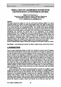
1800 1600 1400 1200
Force, mN
1.3±0.3 N). We considered using an autograft such as tensor fascia lata or temporalis fascia to reduce friction and enhance biocompatibility of the sling material in the upper eyelid. Substituting a temporalis muscle fascia sling markedly reduced the force requirements (to 627±128 mN) for complete eyelid closure in direct comparison with the ePTFE sling. We developed the eyelid sling device so that on contraction of an artificial muscle device, an ellipticalshaped sling would pull the 2 slings toward one another, creating eyelid closure. Contracting the EPAM can pull the eyelid sling design to straighten a curvilinear sling. Initially, we proposed 2 slings (for the upper and lower eyelids). The eyelid sling’s anatomic positioning was designed to mimic the position of the pretarsal orbicularis oculi muscle (which is responsible for involuntary blink). The preferred upper eyelid sling position (similar to that of a gold weight implant) is on the tarsal plate as inferior as possible on the tarsal plate, immediately superior to the ciliary margin. The upper eyelid would evert abnormally when the eyelid sling was secured onto a more cephalic tarsal plate position. In facial paralysis, nasolacrimal dysfunction is exacerbated when the pretarsal orbicularis oculi muscles fail to create a negative pressure that draws tears through the eyelid punctum and into the nasolacrimal system. We ultimately chose to position the eyelid sling anchored to the medial orbital wall to protect the nasolacrimal system and establish proper medial canthal positioning against the globe. The complex medial canthal anatomy includes the pretarsal orbicularis oculi, which divides into a superficial limb and inserts 4 mm anterior to the anterior lacrimal crest, and a deep limb, inserting posterior to the posterior lacrimal crest and enveloping the superior and inferior lacrimal canaliculi. We first chose the frontal process of the maxilla as an anchor site owing to its thickness for screw purchase, but a “medial gaposis”17 between the eyelid and globe was unacceptable. The medial canthal position relative to the globe was best when the self-drilling screw secured the sling in the thickened posterior lacrimal crest, with care to not injure the lacrimal sac. The inferior and superior lacrimal punctum were cannulated for identification and preservation during dissection. The stroke requirements of the proposed artificial muscle device were within the range of available EPAM technology. The displacement of the eyelid sling that was required for complete eyelid closure was 3 mm when both a lower and upper eyelid sling was used, and 6 mm with the solitary upper eyelid sling.5 Compared with the eyelid sling mechanism, a gold weight implant (1.2 g) uses the force of gravity on the gold weight (⬍0.1 N) to close the eyelid. The greater force requirements for the sling mechanism are due to both the sling material’s elasticity and friction of the sling material on the lateral orbital rim. Eyelid closure in the cadaver specimens is also different than in vivo owing to the deficiency of recoil in the connective tissue (canthal tendons and superior transverse ligament).18 One of the major limitations of this study is the assumption that this cadaver model can represent a similar situation in vivo in which fibrosis and wound
1000 800 600 400 200 0 Fascia
ePTFE
Figure 8. Force requirements to create eyelid closure using expanded polytetrafluoroethylene (ePTFE) or temporalis muscle fascia in the eyelid sling design. Error bars indicate standard deviations.
contracture will be involved. The next phase of the project involves creating an articulation between EPAM and the sling material that will minimize the amount of force required from the artificial muscle device by minimizing friction, and maximizing the stroke of the sling mechanism. Currently, the evaluation of the biocompatibility and durability of EPAM in a gerbil model are promising. Successful long-term EPAM function in an animal model will allow application of the technology to create other facial expressions. Other than eyelid closure, we seek to develop human implantation of artificial muscle to animate the perioral area (smile) in acquired or congenital facial paralysis (eg, Mobius sequence19). In conclusion, the eyelid sling concept was successful at creating eyelid closure in a cadaver model. The stroke requirements of the proposed artificial muscle device were within the range of available EPAM technology. When we tested the upper eyelid sling mechanism, less force was necessary to create eyelid closure using a temporalis muscle fascia sling than an ePTFE sling. Future aims include consideration of different sling materials and development of both the EPAM device and the articulation component, which is planned for the lateral orbital rim. Biocompatibility and durability studies of EPAM in a gerbil model are under way. The successful application of artificial muscle technology to create an eyelid blink would be the first of many potential applications. Accepted for Publication: June 29, 2009. Correspondence: Travis T. Tollefson, MD, Department of Otolaryngology–Head and Neck Surgery, Facial Plastic and Reconstructive Surgery, Cleft and Craniofacial Program, University of California, Davis Medical Center, 2521 Stockton Blvd, Ste 7200, Sacramento, CA 95817 (
[email protected]). Author Contributions: Study concept and design: Senders, Tollefson, Curtiss, Prahlad, and Wong-Foy. Acquisition of data: Senders and Tollefson. Analysis and interpretation of data: Senders and Tollefson. Drafting of the manuscript: Senders and Tollefson. Critical revision of the manuscript for important intellectual content: Senders, Tollefson, Curtiss, Prahlad, and Wong-Foy. Statistical analysis: Tollefson and Curtiss. Obtained funding: Tollef-
(REPRINTED) ARCH FACIAL PLAST SURG/ VOL 12 (NO. 1), JAN/FEB 2010 35
WWW.ARCHFACIAL.COM
Downloaded from www.archfacial.com at Emory University, on February 9, 2010 ©2010 American Medical Association. All rights reserved.
son. Administrative, technical, and material support: Tollefson, Prahlad, and Wong-Foy. Study supervision: Tollefson, Curtiss, and Wong-Foy. Financial Disclosure: Drs Senders and Tollefson have applied for a patent for “Greater Eyelid Closure With an Eyelid Sling” jointly with the University of California, Davis, and SRI International. Funding/Support: This project was supported by a grant from the American Academy of Facial Plastic and Reconstructive Surgery Bernstein grant and completed with the collaboration of SRI International. Online-Only Material: A video is available at http://www .archfacial.com. Additional Contributions: Erica Whitney, BA, provided expert technical editing. We also thank the individuals who donated their bodies and tissues for the advancement of education and research, which were provided by the University of California Anatomical Materials Programs. REFERENCES 1. Jobe RP. A technique for lid loading in the management of the lagophthalmos of facial palsy. Plast Reconstr Surg. 1974;53(1):29-32. 2. Berghaus A, Neumann K, Schrom T. The platinum chain: a new upper-lid implant for facial palsy. Arch Facial Plast Surg. 2003;5(2):166-170. 3. Byrne PJ, Kim M, Boahene K, Millar J, Moe K. Temporalis tendon transfer as part of a comprehensive approach to facial reanimation. Arch Facial Plast Surg. 2007;9(4):234-241.
4. Frey M, Giovanoli P, Tzou CHJ, Kropf N, Friedl S. Dynamic reconstruction of eye closure by muscle transposition or functional muscle transplantation in facial palsy. Plast Reconstr Surg. 2004;114(4):865-875. 5. Tollefson TT, Senders CW. Restoration of eyelid closure in facial paralysis using artificial muscle. Laryngoscope. 2007;117(11):1907-1911. 6. Bar-Cohen Y. Electroactive Polymer (EAP) Actuators as Artificial Muscles: Reality, Potential, and Challenges. 2nd ed. Bellingham, WA: SPIE Press; 2004:8. 7. Ashley S. Artificial muscles. Sci Am. 2003;289(4):52-59. 8. Pelrine R, Kornbluh R, Pei Q, Joseph J. High-speed electrically actuated elastomers with strain greater than 100%. Science. 2000;287(5454):836-839. 9. Tate JR, Tollefson TT. Advances in facial reanimation. Curr Opin Otolaryngol Head Neck Surg. 2006;14(4):242-248. 10. Chuang DC, Mardini S, Lin S, Chen HC. Free proximal gracilis muscle and its skin paddle compound flap transplantation for complex facial paralysis. Plast Reconstr Surg. 2004;113(1):126-135. 11. Helmchen C, Rambold H. The eyelid and its contribution to eye movements. In: Straube A, Buttner U, eds. Neuro-Opthalmology: Developments in Ophthalmology. Vol 40. Basel, Switzerland: Karger; 2007:110-131. 12. McCord CD, Ford DT, Hanna K, Hester TR, Codner MA, Nahai F. Lateral canthal anchoring: special situations. Plast Reconstr Surg. 2005;116(4):1149-1157. 13. Moe KS, Linder T. The lateral transorbital canthopexy for correction and prevention of ectropion: report of a procedure, grading system, and outcome study. Arch Facial Plast Surg. 2000;2(1):9-15. 14. Moe KS, Kao CH. Precaruncular medial canthopexy. Arch Facial Plast Surg. 2005; 7(4):244-250. 15. May M. Eye reanimation techniques. In: May M, Schaitkin BM, eds. The Facial Nerve. 2nd ed. New York, NY: Thieme Medical Publishers; 2000:701-715. 16. Riordan-Eva P, Whitcher J. Vaughan and Asbury’s General Ophthalmology. New York, NY: McGraw-Hill; 2007:36. 17. May M. Prospective evaluation of eyelid function with gold weight implant and lower eyelid shortening for facial paralysis. Arch Facial Plast Surg. 2002;4(1):60-61. 18. Evinger C, Manning KA, Sibony PA. Eyelid movement: mechanisms and normal data. Invest Ophthalmol Vis Sci. 1991;32(2):387-400. 19. Mobius PJ. Uber angeboren doppelseitige abducens-facialis-lahmung. Munch Med Wochenschr. 1888;35:91-94.
Announcement Visit www.archfacial.com. As an individual subscriber, references are more useful to you. References within the text are linked to the reference list, and vice versa, for easy browsing and navigation. Journal references are linked whenever possible to at least 1 of 3 locations to permit accessing the abstract and/or full text: (1) references to articles published in journals posted on HighWire are linked to free full text of articles (shown as FULL TEXT); (2) references to MEDLINE-indexed articles are linked to the free PUBMED abstract (PUBMED); and (3) references to articles published in journals that participate in CrossRef or ISI’s Web of Science link to the full text (CrossRef or ISI). Articles linked through CrossRef or ISI may require you to log in for access.
(REPRINTED) ARCH FACIAL PLAST SURG/ VOL 12 (NO. 1), JAN/FEB 2010 36
WWW.ARCHFACIAL.COM
Downloaded from www.archfacial.com at Emory University, on February 9, 2010 ©2010 American Medical Association. All rights reserved.