IEEE REVIEWS IN BIOMEDICAL ENGINEERING, VOL. 5, 2012
15
Formal Design Methods for Reliable Computer-Aided Diagnosis: A Review Oliver Faust, U. Rajendra Acharya, and Toshiyo Tamura Methodological Review
Abstract—Physiological signals, medical images, and biosystems can be used to access the health of a subject and they can support clinicians by improving the diagnosis for treatment purposes. Computer-aided diagnosis (CAD) in healthcare applications can help in automated decision making, visualization and extraction of hidden complex features to aid in the clinical diagnosis. These CAD systems focus on improving the quality of patient care with a minimum of fault due to device failures. In this paper, we argue that a formal and model driven design methodology can lead to systems which meet this requirement. Modeling is not new to CAD, but modeling for systems design is less explored. Therefore, we discuss selected systems design techniques and provide a more concrete design example on computer-aided diagnosis and automated decision making. Index Terms—Algorithms, artificial neural network (ANN), computer-aided diagnosis, formal methods, infrastructure, reliability, safety critical, systems engineering.
I. NOMENCLATURE ANN
Artificial neural network.
AR
Autoregressive.
BA
Bayesian averaging.
BCI
Brain computer interface.
BPSO
Binary particle swarm optimization.
BSN
Biomedical sensor network.
CAD
Computer-aided diagnosis.
CD
Correlation dimension.
CEUS
Contrast enhanced ultrasound.
CLDA
Clustering linear discriminant analysis algorithm.
CSP
Communicating sequential processes.
CT
Computed tomography.
DR
Diabetic retinopathy.
DWT
Discrete wavelet transform.
ECG
Electroencephalogrphy.
EEG
Electroencephalograph.
GMM
Gaussian mixture model.
HOS
Higher order spectra.
IR
Infrared.
K-NN
K-Nearest neighbor.
LLE
Largest Lyapunov exponet.
MC
Clustered microcalcifications.
MRI
Magnetic resonance imaging.
PCA
Principal component analysis.
PNN
Probabilistic neural network.
QoS
Quality of service.
RBF
Radial basis function.
ROC
Receiver operating characteristic.
SNN
Spiking neural network.
SVM
Support vector machine. II. INTRODUCTION
I
Manuscript received August 15, 2011; revised October 16, 2011; accepted January 02, 2012. Date of publication January 23, 2012; date of current version December 06, 2012. O. Faust is with the Department of Electrical and Computer Engineering, Ngee Ann Polytechnic University, Singapore (e-mail:
[email protected]). U. R. Acharya is with Ngee Ann Polytechnic University, Singapore. T. Tamura is with Medical System Engineering, Chiba University, Chiba, Japan. Digital Object Identifier 10.1109/RBME.2012.2184750
T is well recognized that as humans get older, they are more likely to depend on biomedical systems for their wellbeing. In recent years this has sparked rapid development and widespread deployment of biomedical systems. These systems have progressed from single purpose island systems to massively networked health care systems with personal health records [1], [2]. Another fact, which documents the rapid progress, is that these networks distribute an ever-increasing amount of biomedical data [3], [4]. Therefore, our society is more and more dependent on a technology which gets more complex. Computer-aided diagnosis (CAD) helps physicians to cope with this complexity by providing computer output based on quantitative analysis of biomedical data [5], [6]. Therefore, CAD has become one of the major research subjects in medical imaging and diagnostic radiology [7], [8]. The basic concept of CAD is to provide a computer output which serves as a “second opinion” to assist biomedical data analysis [9]. Hence, for the development of a successful CAD scheme it is necessary to develop computer algorithms and investigate how useful the computer output would
1937-3333/$31.00 © 2012 IEEE
16
be for the diagnoses, how to quantify the benefits of the computer output as well as how to maximize the effect of the computer output on the diagnoses [10]. Thus, large-scale observer performance studies using a reliable methodology, such as receiver operating characteristic (ROC) analysis [11] are as important as the development of computer algorithms in the field of CAD. From these diverse requirements it is understandable that research and development of CAD has involved a team effort by investigators with different backgrounds such as physicists, radiologists, computer scientists, engineers, psychologists and statisticians [12]. The concept of CAD is broad, therefore it can be applied to both imaging modalities, and biosignals [13], [14]. However, the majority of CAD schemes developed in the past include the detection of breast lesions on mammograms [15]–[18], the detection of lung nodules in chest radiographs [19], [20] and thoracic computed tomography (CT) [21]–[23], as well as the detection of polyps in CT colonography [24]–[26]. However, there is a downside of relying too much on machine-based decision making, because human decisions are more reliable when compared to machine generated decisions [27]. In Section II, we show that the need for reliability is clearly recognized on both algorithmic and systemic levels, by reviewing algorithms used in CAD systems. However, there is a gap which opens up during systems design. This gap results from the fact that the demand for reliability impacts on the system requirements, because the reliability requirement is more difficult to achieve for complex systems. In this case, the word difficulty expresses the increase in resources or cost in general to meet the requirement. Over time, the increase in system complexity coupled with similar levels of reliability has caused an exponential rise in the difficulty to meet the system requirements. In general, high reliability systems need extensive modeling during the design phase. Therefore, it is quite natural for biomedical systems to employ information modeling. Examples for modeling in biomedical system design are given in Section III. Despite the widespread use of modeming, Kent et al. argue that biomedical information system designers regard modeling as low priority [28]. Designers rush to design and implement hardware and software solutions only after brief assessments of domain requirements. Although this process results in a rapid development cycle, the system usually does not or not completely satisfy the user needs and the developers are forced to reprogram certain aspects of the system. More fundamentally, projects with a rapid development cycle follow an evolutionary design methodology [29], [30] which makes it difficult to cope with a steep requirement increase. The reason for this slow progress comes from the fact that this evolutionary approach relies on progress which is made from one system generation to another. Unfortunately, even with a rapid development, the product or system lifecycle is still counted in years instead of month or weeks, therefore evolutionary progress takes time. But, with the exponential rise in difficulty the requirements (or demand) outpaces the rate of progress achievable with evolutionary system design methodologies. Therefore, all solutions to this fundamental problem require a paradigm shift. Having established the need for a paradigm shift we need to look for solutions from other areas, else there is a danger of
IEEE REVIEWS IN BIOMEDICAL ENGINEERING, VOL. 5, 2012
reinventing the wheel. Engineers deal with systems all the time, therefore it is likely that there are engineering solutions for this problem. In biomedical engineering the problem is that we may have to build highly complex systems which have to be very reliable. Aerospace engineers are frequently confronted with similar situations. They have to build flying machines which carry humans and goods through the third dimension from point A to point B. In 1961, point A was Cape Canaveral here on earth and point B was the southern Sea of Tranquility on the moon. The Apollo program was setup to build a physical solution for this aerospace problem. The engineers, involved in this project, did not experience an exponential growth in requirements, they saw the requirements jump to another level. This jump was fuelled by the same factors which currently drive the requirements for biomedical systems: technical complexity and reliability. Therefore, it is natural to investigate how the engineers, during that time, overcame these massive obstacles. To start with, they had some of the brightest minds working on this project. However, there is a limit of what even the brightest minds can do without appropriate support structures. These support structures came in the form of design methodologies. More specifically, the Apollo program used an engineering method called systems engineering [31]. Many experts believe that the consequent application of systems engineering principals lead to ultimate success. In Section IV, we elaborate on design methods for biomedical systems with a focus on the systems engineering methodology. In this review paper we adopt the position that formal and model driven design approaches can improve the reliability of CAD systems. We support our decision to advocate systems engineering as a possible solution for the problem of the exponential rise in the difficulty to meet the system requirements by discussing a practical example. For this example, we have extended the systems engineering design methodology with formal modeling. The resulting framework was used as design concept for an embedded decision making system. In Section V, we sketch out our design approach by establishing the need for such a decision making system. Lastly, specification refinement and implementation are described. This paper is written in the form of an argument, which culminates by showing a formal specification for a biomedical information processing system. Therefore, we close this paper directly with the conclusion in Section VI. III. RELIABILITY OF COMPUTER-AIDED DIAGNOSIS SYSTEMS We start the discussion of reliability for CAD systems by reasoning about the need for this requirement. Fox and Thomson argue that traditional clinical practices are proving unsustainable (e.g., overuse of drugs and investigations, waiting lists measured in years), and the cost of medical services continues to rise inexorably. In addition, high profile media exposures of clinical errors and increases in litigation following North American trends are forcing many European governments and healthcare agencies to acknowledge that their services have structural problems and that the Quality of Service (QoS) needs to increase [32].
FAUST et al.: FORMAL DESIGN METHODS FOR RELIABLE COMPUTER-AIDED DIAGNOSIS
A major aspect of system quality is reliability which means one aspect of improving the QoS is to make heath care systems, such as CADs, more reliable. This aspect is well understood in the biomedical research community. There is no shortage on projects which incorporate reliability as one of their goals. In the next sections we review CAD algorithms in terms of their claims on reliability. A. Algorithms Used in Computer-Aided Diagnosis An algorithm is a finite list of instructions for calculating a function [33]. Reliability of algorithms is normally related to the function itself and not to the machine executing the instructions. To be specific, algorithm designers require their algorithm to perform the functionality reliable in some statistical sense. In most cases the reliability assessment is supported by experimental results which yield the required data for statistical analysis. For example, an algorithm for visual positioning of previously refined regions of interest on microscopic slides is assessed in terms of its tolerance to possible variations in visual appearance due to slide rotations, scaling and illumination changes [34]. Other algorithms are assessed based on their ability to perform in well-known scenarios [35], [36]. This leads to statements like: Algorithm accuracy and robustness were demonstrated on two tissue-mimicking phantoms, subjected to controlled amount of angular deviation and the proposed method shows a great reliability in the prediction of these phantoms [37]. Measures for reliability are sensitivity, specificity and positive predictive value. These reliability measures are of particular importance for classification algorithms. Soda and Iannello worked on the aggregation of classifiers for staining pattern recognition in antinuclear autoantibodies analysis. In terms of reliability, their contribution was a framework, where a novel parameter measures the reliability of the final classification [38]. In their work on detection of multiple sclerosis classification, the experimenters claim that the use of multiple stimulation patterns appears to improve the reliability of the algorithm [39]. Authors have used auditory stimulus optimization with feedback, where optimized algorithms showed a higher reliability [40]. These classification systems can be used in CAD systems. Such systems must be reliable, because human wellbeing and even human life directly depends on the correct functionality of these systems. Therefore, researchers constantly thrive to improve the reliability of the algorithms used to construct these systems [41]–[44]. Reliability in the communication of data analysis results, from medical personal to patient, is also an important topic. Researchers in biomedical sciences thrive to make the results as accessible as possible [45]. The reliability of measurements is also an important topic in biomedical science. In general, biomedical data is noisy, riddled with artifacts and dependent on the particular measurement setup. Data, such as magnetic resonance imaging (MRI) physiological signals, can be analyzed and assessed for their reliability based on unsupervised clustering methods [46]. Sun et al. present numerical methods and workstation for the quantitative analysis of real-time myocardial contrast echocardiography. They have illustrated a range of clinical studies to indicate the effectiveness of the system and the reliability of the
17
methods [47]. Even with rather reliable data, one major requirement for algorithms was that the results must be more reliable than the input data [48]. Katouzian et al. have described the challenges in atherosclerotic plaque characterization with intravascular ultrasound. For this characterization system they have explored the reliability of both the training dataset and the recognition algorithm complexity [49]. A support vector machine (SVM) system for the characterization of clustered microcalcifications using mammograms was proposed [50]. The microcalcifications regions were segmented using edge detection and morphological methods to extract features based on shape, texture, and statistical values. The SVM classifier with radial basis function (RBF) kernel presented 97% accuracy, and that with polynomial kernel gave 95% accuracy. Automatic detection of clustered microcalcifications (MCs) in digitized mammograms was proposed [51]. The pixels of MC were detected and grouped into MC objects using a multilayer feedforward neural network classifier. Seventeen statistical features were extracted from the MC objects and fed to an Adaboost with SVM classifier yielded 89.55% mean true positive detection rate. Gene expression was used to distinguish benign and malignant thyroid carcinoma. The immunohistochemistry correctly classified 90.6% of fine-needle aspirations and 85% of follicular thyroid adenomas [52]. Fluorescent scanning was used to classify benign and malignant thyroid carcinoma and obtained an accuracy of 90% [53]. Acharya et al. have extracted relevant discrete wavelet transform (DWT) and texture features from the contrast enhanced ultrasound (CEUS) thyroid images [45]. These features coupled with K-nearest neighbor (K-NN) resulted in the classification (benign and malignant) accuracy of 98.9%, a sensitivity of 98%, and a specificity of 99.8%. Texture and motion pattern features were extracted from carotid atherosclerosis images and fed to fuzzy C-means classifier for classification into symptomatic and asymptomatic plaques [54]. The experimenters were able to classify 74% of plaques based solely on texture features, 79% of plaques were correctly classified based entirely on motion features and 84% were correctly classified using a combination of motion and texture features. The texture features, coupled with SVM classifiers, were used for the automated identification of symptomatic and asymptomatic plaque images [55]. The classification accuracy was 82.4% for a SVM classifier with RBF kernel. A clinical trial is under way to examine the impact of metabolic control over time in children with type 1 diabetes, as compared to standard insulin treatments due to the infusion of autologous cord blood stem cells [56]. The results showed that an infusion of cord blood stem cell was safe. Furthermore, this technique may provide some slowing of the loss of insulin production in children with type 1 diabetes. Biomaterials or new drugs must be biocompatible, only then they can be used in a clinical setting [57]. For example, a defective heart valve which is replaced by a mechanical valve implant. This valve is coated with pyrolytic carbon and secured to the surrounding tissue with a mesh of woven fabric called Dacron (du Pont’s trade name for polyethylene terephthalate). The mesh allows for the body’s tissue to grow while incorporating the valve [58].
18
In olden days, telemedicine was made possible using telephone and radio; recently, these techniques have been supplemented with video telephony, advanced diagnostic methods are supported by distributed client/server applications and telemedical devices to aid in-home care [59]. Recent developments in mobile collaboration technology have benefitted from using hand-held mobile devices which allow healthcare professionals in multiple locations to view and monitor patient issues as if they were in the same room [60]. Karimi et al. have presented a technique which uses wavelet analysis and artificial neural network (ANN) for analyzing heart sounds to detect coronary artery disease [61]. They detected normal classes with 90% accuracy and coronary artery disease classes with 85% accuracy. A combination of uncertainty methods (fuzzy and probabilistic) was used in the diagnosis of coronary artery disease using electroencephalography (ECG) stress signals, and the experimenters observed an accuracy of around 80% [62]. Binary particle swarm optimization (BPSO) and genetic algorithm techniques were used to extract features from exercise stress testing data and obtained 81.46% accuracy in coronary artery disease detection using SVM classifier [63]. Babaoglu et al. have also used principal component analysis (PCA) for dimension reduction on the same dataset and obtained an accuracy of 79.71% with the SVM classifier [64]. Wavelet-chaos-neural network methodology was used for classification of healthy, ictal, and interictal electroencephalograph (EEG) classes [65]. Wavelets were used to decompose the EEG signals into delta, theta, alpha, beta, and gamma sub-bands. Standard deviation, correlation dimension (CD), and largest Lyapunov exponent (LLE) were extracted from the subbands. A spiking neural network (SNN) was developed using three training algorithms (SpikeProp, QuickProp, and RProp). With the RProp algorithm, the SNN classifier obtained an accuracy of 92.5%, using the mixed-band feature space consisting of nine parameters (CD and LLE from wavelet coefficients). Also, they obtained an accuracy of 96.7% using the Levenberg-Marquardt backpropagation neural network and a mixed-band feature space consisting of nine parameters [66]. Lyapunov features were used in a recurrent neural network (RNN) for classifying the three classes with an efficiency of more than 96% [67]. Higher order spectra (HOS) features were found to be significant enough for differentiating normal, interictal and epileptic EEG signals [68]. On using the HOS features in Gaussian mixture model (GMM) and SVM classifier, they obtained accuracies of 93.11% and 92.67%, respectively [69]. A Clustering Linear Discriminant Analysis algorithm (CLDA) was used to decode hand movement directions from a small number of training trials for magnetoencephalography-based brain computer interface (BCIs) [70]. CLDA starts with a spectral clustering algorithm which automatically partitions the BCI features into several groups where the within-group correlation was maximized and the between-group correlation was minimized. The results show an average accuracy of 87% in decoding of four directions for single movement. The design of spring-loaded crutches was optimized for reliability by choosing an appropriate spring stiffness based on their dynamic characteristics [71].
IEEE REVIEWS IN BIOMEDICAL ENGINEERING, VOL. 5, 2012
Acoustic attenuation coefficients, wavelet coefficients and autoregressive (AR) model coefficients were used as features for a Bayes classifier to classify steatosis and normal livers [72]. The results show very high sensitivity ( 95%), specificity ( 95%), and accuracy ( 95%). Using only the AR model coefficients, the experimenters obtained accuracy, sensitivity, and specificity dropped to 90%, 87%, and 95%, respectively. Three types of liver images, namely, normal, hepatoma, and cirrhosis, were classified using fractal feature vector based on M-band wavelet transform [73]. The results show that a hierarchical classifier was least 96.7% accurate in classifying normal and abnormal liver images. Variables, such as age, menopausal status, maximum tumor diameter, tumor volume, locularity, presence of papillary projections, presence of random echogenicity, presence of analyzable blood flow velocity waveforms, peak systolic velocity, time-averaged maximum velocity, pulsatility index, and resistance index were obtained from 52 benign and 15 malignant transvaginal B-mode ultrasonography images [74]. These features coupled with back propagation method, obtained sensitivity and specificity of 100% and 98.1% respectively. Zimmer et al. have proposed an automatic analysis of benign and malignant ovarian cancer ultrasound images by quantification of grey-level intensity variations (mean, standard deviation, etc.) [75]. They obtained a low accuracy of 70% for tumors containing solid portions. Advanced image processing, data mining techniques, and computer simulations are used to improve the diagnostic accuracy in healthcare systems, such as ophthalmology [76]. Glaucoma and diabetic retinopathy (DR) are common eye diseases which may cause vision loss, if not diagnosed early enough [77]. Thermography using infrared (IR) imaging is an effective noninvasive imaging technique that is widely used in the medical field. This technique detects temperature changes in vascular tissues, hence, it provides an instrument with which to study ocular surface temperature and ocular diseases [78]. IV. MODELING Modeling is a significant part of research, education and practice in biomedical and health informatics [79]. To be specific, mathematical dependency modeling aims to produce reliable results which are, in some sense, relevant in the solution of a practical problem [80]. Despite widespread applications in biomedical research, the role of models and modeling is often controversial and ill understood. It is usual to find that fundamental definitions, axioms, and postulates used in the modeling process have become implicit assumptions. It is essential to have a clear vision of the fundamental principles of modeling [81]. In biomedical engineering, modeling is driven by the understanding of physical phenomena and the modeling environment capabilities [82]. For example, biomedical phenomena can be modeled in terms of metabolomics [83]–[86], genomics [87]–[90], and proteomics [91]–[96]. In these fields, very complex models have been used to understand the behavior at a decreasing scale: from metabolomics, which is focused on the system, to proteomics, which is focused on the proteins, to genomics, which is focused on genes. In general, the decreasing
FAUST et al.: FORMAL DESIGN METHODS FOR RELIABLE COMPUTER-AIDED DIAGNOSIS
scale was associated with more model data and modeling algorithms with higher computational complexity [97]. Therefore, the processing requirements for the modeling environment increased [98]. We explore modeling for biomedical system design from three different perspectives: formal modeling, biomedical informatics and processing platforms. Each of these domains has requirements and expectations on the model. A. Formal Modeling Computer simulation enables system developers to execute a model of an actual or theoretical system with a computer and analyze the execution output. Peleg et al. have used Petri net tools to study systems behavior. The systems were represented using three kinds of biomedical models: a biological workflow model used to represent biological processes, and two different computer-interpretable models of health care processes derived from clinical guidelines [99]. A typical example of formal modeling is the mathematical formulation of working-memory capacity limits proposed on the key assumption of mutual interference between items in working memory [100]. Tarakanov and Dasgupta have presented a mathematical model based on the features of antigen, antibody bindings in the immune system [101]. Hakman and Groth have designed a system which integrates quantitative simulation with symbolic reasoning techniques, under the control of a user interface management system, using a relational database management system for data storage and interprocess communication [102]. Lyons and Arbib have used port automata to model sensor based robotics. This formal model ensured consistency and well-definedness, and it facilitates plan verification as well as automatic plan generation [103]. Bernot et al. have provided a computer science formal approach to treat temporal properties of biological regulatory networks, expressed in computational tree logic [104]. Jetley et al. have used formal methods to model medical device reviews [105]. Arney et al. have based their development of a patient-controlled analgesia infusion pump on formal methods [106]. B. Biomedical Informatics Biomedical informatics is the science of information as applied to or studied in the context of biomedicine [107]. The underlying scientific principles are beginning to be identified and defined, educators are increasingly acknowledging the growing importance of computer science for physicians, and the technology itself is growing at rates that make the future of the field both unbounded and impossible to predict [108]. However, a general problem with all software systems is the almost limitless state space of such systems. In order to create awareness and to manage this huge state space, biomedical information uses modeling. Maojo et al. have provided an overview of current approaches related to biomedical informatics, GRID, and genomic medicine, particularly in Europe [109]. Objects play a major role in both database and artificial intelligence research [110]. Hakman and Groth have addressed the issue of modeling objects with a new object-oriented biomedical continuous system modeling language. With object-oriented approaches like this, complex models were structured as multilevel, multicomponent
19
model hierarchies [111]. Another application area for biomedical informatics is prediction and reaction of current and future bioterrorist attacks. The challenge is that a comprehensive and timely response requires data acquisition, threat detection, and response infrastructure with unprecedented scope in time and space [112]. C. Processing Platforms Telemedicine has emerged as a new health care field. This new field offers health care providers, professionals and patients a plethora of opportunities to respond to social and geographical inequalities in health care provision [113]. Manolokos et al. have proposed a parallel processing system for biomedical signal processing that is optimal in terms of total execution time for multiple pipelined data blocks [114]. Bajaj et al. have implemented a computational environment to produce libraries of executable, combinable and customizable computer models of natural and synthetic biosystems, which provide a supporting framework for the predictive understanding of both structure and behavior. This was achieved by multiscale geometric modeling and multiphysics simulations [115]. A biomedical sensor network (BSN) is a small-size sensor network for medical applications, that may contain tens of sensor nodes. A formal model was used to validate and tune the temporal configuration parameters of a BSN in order to meet desired QoS requirements on network connectivity, packet delivery ratio and end-to-end delay [116]. V. DESIGN METHODS According to Alan Turing, computers are universal machines [117], therefore these devices can be used to provide various forms of assistance to clinicians, such as better clinical records, timely prompts and reminders, and assistance in following care pathways. A number of well-known centers, particularly in North America, have pioneered such systems, in some cases over decades [118]. Their faith was justified, because objective benefits of computer support on both clinical and cost effectiveness are emerging [119]. From this perspective it is only logical to scale these systems up and harvest the rewards on a larger scale. However, scale is a problem, design methods which simply scale up existing systems almost always fail, especially for complex networked systems which are composed from independent entities. Well-thought-out design methodologies are required to translate the process made by biomedical sciences into benefits for people. There is no shortage in very specifically targeted design methods for biomedical systems. For example, Shah et al. have used an object-oriented design methodology to design and develop a software system in a modular fashion [120]. Fox and Thomson have described both development and application of a unified technology for clinical decision support and disease management. Their work was based on logic engineering, a distinct design and development methodology which combines concepts from knowledge engineering, logic programming, and software engineering [32]. All these projects were done by domain level experts and outstanding researchers. However, all the literature they produced just documents the project design which was executed. This is
20
IEEE REVIEWS IN BIOMEDICAL ENGINEERING, VOL. 5, 2012
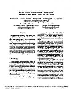
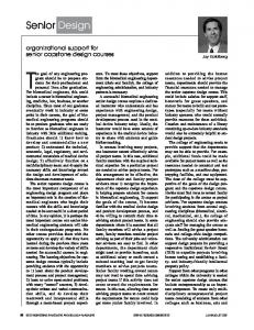
a problem, because the research papers do not document the thought process which led the authors to adopt a specific way of designing a system. That means, lots of implicit knowledge is required to design biomedical systems successfully and the design process takes a considerate amount of creative energy. Now, domain level expertise is always required, however the energy spend on thinking about the design process can be reduced. The way to achieve this reduction is by following well proven and standardized design methods. The following sections introduce systems engineering as a well proven and successful design methodology. A. Systems Engineering Systems engineering is a methodical, disciplined approach for design, realization, technical management, operations, and retirement of a system [121]. Systems engineering takes into account all steps necessary to create a system. This leads to reliable and safe systems, because negligence in any of the design steps leads to weaker and therefore less trustworthy systems. The design and development of safety critical information systems can benefit from adopting the full systems engineering approach, particularly with regard to bringing a human-centered perspective to the formulation of system requirements and the configuration of effective user interfaces [122]. This helps us to design medical information systems which have to provide a reliable link and good coordination between hospital departments [123]. As with all structuring methods, systems engineering does not hold the solution to all health care problems. Issues that typically arise upon the introduction of systems engineering include the readiness of the organization to embrace the new approach and specifying the degree of formalization that is appropriate for a given project, what metrics one should use to characterize the human factors challenges that are inherent in a given system and what software tools can be adopted to facilitate the systems engineering processes [124], [125]. Nevertheless, systems engineering can be applied to a wide range of engineering problems. For example, Diez et al. used the systems engineering methodology to develop computer-supported learning systems [126]. Palanisamy and Selvan have proposed a novel method for identifying relevant subspaces for data mining using fuzzy entropy and perform clustering [127]. Their theories and algorithms were evaluated through experiments which were designed according to the systems engineering method. The next section introduces the design of an ANN classification system. We use this design to introduce systems engineering in greater detail and we put forward formal methods as a useful extension in the specification refinement step. VI. FORMAL METHODS IN EMBEDDED DECISION MAKING AND ABSTRACT EXAMPLE The embedded decision making system design illustrates some of the ideas, which were developed above, in a more practical setting. We follow the systems engineering meta model, shown in Fig. 1, to design the system. The systems engineering meta model describes an iterative process where both ideas about and understanding of the problem get more and more concrete. The needs definition is still speculative and
Fig. 1. Systems engineering lifecycle model.
it is the phase where ideas get thrown around. At the end of speculative stage a group of experts decides whether or not the project progresses to the requirement capturing phase. Requirements define what the system is expected to achieve and they should be obtained as a result of informed discussion [128, ch. 1]. Therefore, field specific research work and the associated mathematical models constitute some of the system requirements. Analyzing field specific research answers questions about the work context. To be specific, it answers the questions like: “What can be achieved?” and “What are the most promising approaches to solve a problem in practice?” It is not uncommon that a specific need translates into a requirement which is impossible meet. Therefore, the requirements have to be verified against the needs and the needs have to adjusted if they were overambitious. However, adjusting the need always brings the whole project into question. That means, once the need has been adjusted, there must be a decision on whether or not the project will continue. Once the requirements have been captured, the project moves into the specification stage. The specification must be expressed as formally as possible, because only a high degree of formalism leads us to an unambiguous system description [129], [130]. Such a system description is important, because the specification is discussed by both engineering and management groups. The systems engineering model is constructive, the ideas of iteration and looping processes hold also for the relationship between specification and requirements. To be specific, the specification has to be validated against the requirements. With the specification we establish how we build the system, in the implementation we actually go about building it. In the best case the implementation is just an automated or mechanical translation of the specification. However, most of the time the specification just describes the high level architecture and not the detailed processes which make up the system functionality. Therefore, lots of handcrafting is necessary in the translation process. Once the implementation is done, we still do not have a product. The design process is only complete after product verification. This step is particularly important for high reliability and safety critical systems, such as biomedical products, because product verification involves use and failure case testing. In the validation loop we check whether or not we have built the right system, because this step checks the implementation against the requirements. If and only if the implementation passes all tests, will we have a product which can be released.
FAUST et al.: FORMAL DESIGN METHODS FOR RELIABLE COMPUTER-AIDED DIAGNOSIS
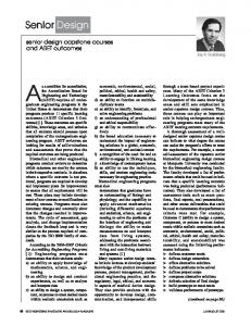
The focus of this abstract example is set to needs definition, requirements capturing and specification refinement. The example shows that systems engineering combined with formal models improves our ability to reason about the design. This reasoning breeds understanding and understanding is what it takes to build reliable systems which are functional, reliable and safe. A. Need Definition Empirical science dictates the need for data collection in order to learn about specific physical phenomena. Biomedical science is no exception, with ever more sensitive sensors an increasing amount of data is acquired in medical information systems. In the past, the organization of this data was still driven by the data source, this did not support the cognitive processes of physicians [131]. Therefore, new methods to visualize patient medical records are becoming imperative in order to assist physicians with clinical tasks and medical decision-making. This is a crucial result of the need definition process: we have a clearly defined problem. In all subsequent discussions we will not go beyond this problem. The next step in the need definition is to focus on and to reason about the specific application area. To cope with the sheer data volume methods like data mining and knowledge representation are needed [132]. However, these techniques do not go far enough, because they just extract relevant information from the data. Nevertheless, this is an important prerequisite for disease diagnosis [133]. The most advanced form of help for physicians is CAD. For example, accurate and reliable decision making in oncological prognosis can help in the planning of suitable surgery and therapy, and generally, improve patient management through the different stages of the disease [134]. Böröczky et al. have proposed a feature subset selection method based on genetic algorithms to improve the performance of false positive reduction in lung nodule CAD [135]. In this step we have narrowed down the application area and the next step is to focus on state of the art techniques for CAD. The diagnosis itself can be modeled as a decision making process. Any method of data analysis, intended to support the clinical decision-making process, should meet several criteria: it should capture clinically relevant features, be computationally feasible, and provide easily interpretable results [136]. Movement of health care delivery towards the primary care sector and the home setting can result in substantial benefits in terms of health outcome, social provision, cost effectiveness and resource utilization. In order to gain maximum benefit from such a distributed approach, it is necessary to ensure that there is a full and proper understanding of the roles of all those involved in health care delivery as well as making available the best possible support for clinical decision making [137]. More specifically, machine learning algorithms can provide predictions complemented with valid confidence measures. In medical diagnosis, such predictions are highly desirable, as medical experts can gain additional information for each machine diagnosis. A risk assessment in each prediction can play an important role for medical decision making, in which the outcome can be critical for the patients [138]. Ji et al. have proposed a novel team-based intelligent agent software system approach
21
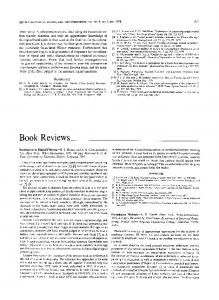
Fig. 2. Block diagram of a general decision making system.
for proactively monitoring and detecting potential ADRs of interest using electronic patient records [139]. Fig. 2 shows a typical block diagram of a medical decision making system. Data acquisition, preprocessing and feature extraction steps are data dependent. Classification methods are more general, i.e., classification algorithms can be used for a wide range of feature vectors . The example we want to introduce in this section should be as general as possible, therefore we focus on the classification method. There is a wide range of classification algorithms available [140], hence the main task in the need definition step is to establish what algorithm is needed. Medical decision making can be regarded as a process, combining both analytical cognition and intuition. It involves reasoning within complex causal models of multiple concepts, usually described by uncertain, imprecise, and/or incomplete information. Iakovidis and Papageorgiou have proposed a novel approach based on cognitive maps and intuitionistic fuzzy logic [141]. Kusiak and Law have proposed a data mining algorithm for extraction of robust rules that can be used in clinical practice for better understanding and prevention of unwanted medical events [142]. Güler and Übeyli have proposed a multiclass SVM with the error-correcting output codes for the multiclass EEG signals classification problem. The probabilistic neural network (PNN) and multilayer perceptron neural network were also tested and benchmarked for their performance on the classification of the EEG signals [143]. Bayesian averaging (BA) over ensembles of decision models allows technologists to evaluate the uncertainty of decisions, this is of crucial importance for safety critical applications, such as medical diagnostics [144]. Exarchos et al. have used association rules for the automated detection and classification of transient events in EEG recordings [145]. The expanded disability status scale has been the most widely used measure of disability in multiple sclerosis clinical trials. Automatic EDSS (AEDSS) is an expert system designed to overcome this problem. It constrains the neurologist to follow precise reasoning steps, enhancing EDSS reliability [146]. ANN is among the oldest machine classification algorithms [147], [148]. It mimics both functionality and structure of a human brain. Therefore, it is widely used in medical decision making [149]–[152]. Blood product transfusion is a financial concern for hospitals and patients. This study evaluates the efficacy of using ANN to predict the transfusion requirements of trauma patients using readily available information [153], [154]. Weight-elimination neural networks were applied to coronary surgery mortality prediction. The objective was to assess the effectiveness of the weight-elimination cost function in improving classification performance of ANNs and to observe how changing the a priori distribution of the training set affects network performance [155]. ANN-based techniques have been used for monitoring dangerous infections [156], predicting high-risk preterm birth [157], and epileptic EEG
22
IEEE REVIEWS IN BIOMEDICAL ENGINEERING, VOL. 5, 2012
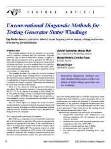
Fig. 4. Neuron process model which is defined in (3) and (4) and used in (5) to build the ANN.
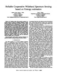
Fig. 3. General ANN network diagram which gives a graphical representation of (5).
detection [158]. Shin et al. have developed an automatic classification system for cough sounds to symptoms which indicate abnormal health conditions [159]. The ANN model used energy cepstral coefficients obtained by filter banks based on human auditory characteristics as input parameters. A novel neural-network model for deriving standard 12-lead ECGs from serial three-lead ECGs: application to self-care [160]. Tzallas et al. used ANN classification to extract epileptic seizure events from EEG signals [161]. The wide range of applications and the pervasive use of ANN led us to adopt this algorithm as basis for the system design. In general, and this example is no exception, the need definition is a highly speculative process and a result is reached by a panel of experts. Nevertheless, this need definition forms the bedrock of the design. All subsequent design steps aim to realize a system which fulfills these needs. B. Requirements In the previous section we established the need for an ANN classification system. Hence, the first requirement is that the system, to be designed, must operate according to the ANN theory. In the need definition we focused on biomedical information processing problems. From the discussion in Section II it necessarily follows that the system must be reliable and safe, this constitutes the second requirement. In the need definition we have fixed the application area to biomedical signal processing, but the application itself was left open. Hence, the ANN implementation must be as generic as possible. Fig. 3 shows the network diagram of a general ANN system [162, ch. 1]. The system assumes an -dimensional input vector which is the result of the feature extraction step, as shown in Fig. 2. The process network is modeled as an array of neurons which are arranged in layers. The neurons in layer are fully connected to the neurons the adjacent layers ( and ). The network structure can be further refined by the programming of the neurons itself. In general, the system requirements are not complete, because a specific processing platform is not defined. However, a discussion on processing platforms and processing requirements, such as real-time, is not within the scope of this review paper. For the purpose of this paper, the requirements are sufficient to proceed to the specification.
C. Specification In this section we introduce formal methods for biomedical systems design. By using formal methods we respond to both safety and reliability requirements [163], [164]. We have chosen Communicating Sequential Processes (CSP) as the modeling language. CSP is a process algebra which was invented by Hoare [165], [166] and which has by now over 30 years of solid research behind it [167], [168]. In a practical setting, CSP has been used to model percolation effects for large scale mesh networks [169]. Sputh et al. used CSP to model hardware and software for in situ remote monitoring systems [170]. The CSP part of the ANN algorithm model defines the network structure. There is ample literature which describes the ANN functionality in terms of algebraic equations [171]–[173], but these equations do not specify how to construct processing networks which execute the algorithm instructions. Without a formal language it is difficult to reason about the way this network structure is implemented. This discussion is important, because it allows engineers to communicate their ideas to the management team and potential architectural problems can be found in an early design stage. To model the system in CSP we follow the process network diagram shown in Fig. 3. First we define a neuron as a process. Fig. 4 shows a diagram which depicts the inputs and outputs of this process. Block or network diagrams, such as the ones shown in Figs. 3 and 4, fail to communicate the internal functionality of the components they show. Therefore, it is impossible to extract the systemic information from such diagrams. For example, it is impossible to reason about the sequence of events based on a diagram alone. A much stronger method is to define all necessary functionality in a formal but abstract way. Mathematical descriptions offer the required clarity and logical consistency. Before we proceed with the mathematical description of the process shown in Fig. 4, there is some housekeeping necessary. Equation (1) defines three sets which are used in the formal model of the ANN functionality: from to column
(1)
The connections between the neurons are modeled as channels. Equation (2) defines as an channel array: (2)
FAUST et al.: FORMAL DESIGN METHODS FOR RELIABLE COMPUTER-AIDED DIAGNOSIS
The process models the communication behavior of the neuron. The parameters and indicate the neuron position in terms of row and column. The variable is used internally by the process to keep track of the messages which were received. Equation (3) defines that the neuron consumes a data value from all connected channels before it progresses to :
(3) Equation (4) defines the communication functionality of . To be specific, outputs data on all connected output channels.
(4) After defining the process, which represents the individual neuron, we assemble the process network shown in Fig. 3. Equation (5) combines the individual layers with the interleave operator and the resulting layers are connected with the parallel operator . MAIN
(5)
The MAIN process network was subjected to extensive testing and formal model checking. Through testing we gained insights in the communication within the ANN process network. Through formal model checking we established that the model is livelock- and deadlock-free [174]. These are two important properties for parallel systems. Only rigorous proofs, such as model checking, ensure that the model works reliably. The implementation is just a translation of the formal model into language constructs for the processing platform. With the right tool support this translation is almost mechanical, therefore it is not a part of this paper. VII. CONCLUSION The progress made by biomedical science is enormous, because it is fueled by the rapid development of computer systems and more ingenious algorithms for signal analysis. There is a need or social obligation to bring this progress to the people. One of the fundamental problems is that in order for a biomedical system to be useful, it must be complex. This is usually referred to as the enormous complexity of practical systems. Com-
23
puter-aided diagnosis systems are no exceptions to this rule. In recent years novel algorithms in data mining, signal and image processing have been developed to aid the clinicians in making faster and efficient diagnosis. In the first section of this paper we show that this complexity is coupled with a demand for reliability. To express the impact of complexity and reliability on the system requirements we have introduced the term difficulty. The term refers to the resources needed or cost to meet the requirement. We argue that the difficulty increases exponentially with the complexity under the assumption that the same reliability is required. To cope with this increasing system complexity it is necessary to move away from evolutionary system design and towards system-based development. Evolutionary systems design makes only progress, i.e., copes with the complexity problem, from one revision to the next. In contrast, systems-based development incorporates feedback loops within the (systems engineering) design methodology. Therefore, it can cope with the complexity problem much better. One of the cornerstones of modern design is modeling. We established that this fact also holds for computer-aided diagnosis systems in Section III. However, modeling is most of the time not enough to meet the system requirements. A methodical design approach is needed to create complex computer-aided diagnosis systems which are reliable. This leads us to formal and model driven designs. A prominent example for formal design is systems engineering. Systems engineering defines a meta model which needs to be adopted to the application domain. Systems engineering is applicable to a wide range of biomedical projects. When it is employed, it will guide both technical and management teams to see the bigger picture, i.e., providing the holistic view. This is especially important for the engineering team, because engineers are usually bogged down by a great deal of technical detail and they (sometimes) miss the bigger picture. Therefore, each management decision, which impacts on their work, is seen as an intrusion and usually causes resistance. Systems engineering aims to minimize this resistance by providing adequate structures for information exchange between the different teams. This makes the development process flexible and ultimately this enables the project to cope with the inherent complexity problem of modern biomedical systems. The main thesis of this review paper is that the combination of formal modeling and systems engineering has the capability to improve the reliability of diagnosis systems. By reviewing scientific literature we have established that reliability is a central requirement for computer-aided diagnosis systems and that reliability can be established through modeling. The main contribution of this review is to work out the relationship between modeling and reliability for computer-aided diagnosis. We adopt the position that the relationship between modeling and reliability can only be understood from a design perspective. To be more specific, structured design methodologies, such as systems engineering, help us to understand and achieve reliability. We are aware that this review is by far not sufficient to achieve an adoption of this methods, it constitutes only a small step towards this goal. More work is needed to address the difficulties that arise from the design of complex systems which must be reliable. To be specific, research on large scale biomedical systems must involve formalizing the design methodology.
24
IEEE REVIEWS IN BIOMEDICAL ENGINEERING, VOL. 5, 2012
REFERENCES [1] N. Archer, U. Fevrier-Thomas, C. Lokker, K. A. McKibbon, and S. E. Straus, “Personal health records: A scoping review,” J. Amer. Med. Informatics Assoc. vol. 18, no. 4, pp. 515–522, 2011, 10.1136/amiajnl-2011-000105 [Online]. Available: http://jamia.bmj.com/content/18/4/515.abstract, arXiv:http://jamia.bmj.com/content/18/4/515. full.pdf+html [2] D. Detmer, M. Bloomrosen, B. Raymond, and P. Tang, “Integrated personal health records: Transformative tools for consumer-centric care,” BMC Med. Informatics Decision Making, vol. 8, no. 1, pp. 1–45, 2008, 10.1186/1472-6947-8-45 [Online]. Available: http://www.biomedcentral.com/1472-6947/8/45 [3] J. S. Duncan and N. Ayache, “Medical image analysis: Progress over two decades and the challenges ahead,” IEEE Trans. Pattern Anal. Machine Intell., vol. 22, no. 1, pp. 85–106, Jan. 2000. [4] A. Burgun and O. Bodenreider, “Accessing and integrating data and knowledge for biomedical research,” Yearbook Med. Informatics pp. 91–101, 2008 [Online]. Available: http://www.ncbi.nlm.nih.gov/ pubmed/18660883 [5] K. Doi, “Computer-aided diagnosis in medical imaging: Historical review, current status and future potential,” Computerized Med. Imaging Graphics, vol. 31, no. 4–5, pp. 198–211, 2007. [6] B. J. Erickson and B. Bartholmai, “Computer-aided detection and diagnosis at the start of the third millennium,” J. Digital Imaging, vol. 15, pp. 59–68, 2002. [7] K. Doi, “Current status and future potential of computer-aided diagnosis in medical imaging,” Brit. J. Radiol., vol. 78, no. suppl_1, pp. S3–S19, 2005. [8] K. Doi, M. Giger, R. Nishikawa, K. Hoffmann, H. MacMahon, and R. Schmidt, “Potential usefulness of digital imaging in clinical diagnostic radiology: Computer-aided diagnosis,” J. Digital Imaging, vol. 8, pp. 2–7, 1995. [9] H. P. Chan, K. Doi, S. Galhotra, C. J. Vyborny, H. MacMahon, and P. M. Jokich, “Image feature analysis and computer-aided diagnosis in digital radiography. I. Automated detection of microcalcifications in mammography,” Med. Phys., vol. 14, no. 4, pp. 538–548, 1987. [10] M. Das, G. Mühlenbruch, A. H. Mahnken, T. G. Flohr, L. Gündel, S. Stanzel, T. Kraus, R. W. Günther, and J. E. Wildberger, “Small pulmonary nodules: Effect of two computer-aided detection systems on radiologist performance,” Radiology, vol. 241, no. 2, pp. 564–571, Nov. 2006. [11] L. Monnier-Cholley, F. Carrat, B. P. Cholley, J.-M. Tubiana, and L. Arrivé, “Detection of lung cancer on radiographs: Receiver operating characteristic analyses of radiologists’, pulmonologists’, and anesthesiologists’ performance,” Radiology, vol. 233, no. 3, pp. 799–805, 2004. [12] B. Van Ginneken, B. M. Ter Haar Romeny, and M. A. Viergever, “Computer-aided diagnosis in chest radiography: A survey,” IEEE Trans. Med. Imaging, vol. 20, no. 12, pp. 1228–1241, Dec. 2001. [13] O. Faust, U. R. Acharya, L. C. Min, and B. H. C. Sputh, “Automatic identification of epileptic and background EEG signals using frequency domain parameters,” Int. J. Neural Syst., vol. 20, no. 2, pp. 159–176, 2010. [14] Y. Zhu, S. Williams, and R. Zwiggelaar, “Computer technology in detection and staging of prostate carcinoma: A review,” Med. Image Anal., vol. 10, no. 2, pp. 178–199, 2006. [15] D. A. Berry, K. A. Cronin, S. K. Plevritis, D. G. Fryback, L. Clarke, M. Zelen, J. S. Mandelblatt, A. Y. Yakovlev, J. D. F. Habbema, and E. J. Feuer, “Effect of screening and adjuvant therapy on mortality from breast cancer,” New Engl. J. Medicine, vol. 353, no. 17, pp. 1784–1792, 2005. [16] R. A. Smith, D. Saslow, K. A. Sawyer, W. Burke, M. E. Costanza, W. P. Evans, R. S. Foster, E. Hendrick, H. J. Eyre, and S. Sener, “American cancer society guidelines for breast cancer screening: Update 2003,” CA: Cancer J. Clinicians, vol. 53, no. 3, pp. 141–169, 2003. [17] E. D. Pisano, C. Gatsonis, E. Hendrick, M. Yaffe, J. K. Baum, S. Acharyya, E. F. Conant, L. L. Fajardo, L. Bassett, C. D’Orsi, R. Jong, and M. Rebner, “Diagnostic performance of digital versus film mammography for breast-cancer screening,” New Engl. J. Medicine, vol. 353, no. 17, pp. 1773–1783, 2005. [18] L. Ma, E. Fishell, B. Wright, W. Hanna, S. Allan, and N. F. Boyd, “Case-control study of factors associated with failure to detect breast cancer by mammography,” J. Nat. Cancer Inst., vol. 84, no. 10, pp. 781–785, 1992. [19] L. Monnier-Cholley, H. MacMahon, S. Katsuragawa, J. Morishita, T. Ishida, and K. Doi, “Computer-aided diagnosis for detection of interstitial opacities on chest radiographs,” Amer. J. Roentgenol., vol. 171, no. 6, pp. 1651–1656, 1998.
[20] J. Shiraishi, H. Abe, R. Engelmann, M. Aoyama, H. MacMahon, and K. Doi, “Computer-aided diagnosis to distinguish benign from malignant solitary pulmonary nodules on radiographs: Roc analysis of radiologists’ performance-initial experience,” Radiology, vol. 227, no. 2, pp. 469–474, 2003. [21] S. G. Armato, F. Li, M. L. Giger, H. MacMahon, S. Sone, and K. Doi, “Lung cancer: Performance of automated lung nodule detection applied to cancers missed in a CT screening program,” Radiology, vol. 225, no. 3, pp. 685–692, 2002. [22] P. A. Hein, L. D. Krug, V. C. Romano, S. Kandel, B. Hamm, and P. Rogalla, “Computer-aided detection in computed tomography colonography with full fecal tagging: Comparison of standalone performance of 3 automated polyp detection systems,” Canadian Assoc. Radiologists J., vol. 61, no. 2, pp. 102–108, 2010. [23] J. Näppi and K. Nagata, “Sources of false positives in computer-assisted CT colonography,” Abdominal Imaging, vol. 36, pp. 153–164, 2011. [24] H. Yoshida, J. Näppi, P. MacEneaney, D. T. Rubin, and A. H. Dachman, “Computer-aided diagnosis scheme for detection of polyps at CT colonography,” Radiographics, vol. 22, no. 4, pp. 963–979, 2002. [25] H. Yoshida, Y. Masutani, P. MacEneaney, D. T. Rubin, and A. H. Dachman, “Computerized detection of colonic polyps at CT colonography on the basis of volumetric features: Pilot study,” Radiology, vol. 222, no. 2, pp. 327–336, 2002. [26] H. Yoshida and J. Nappi, “Three-dimensional computer-aided diagnosis scheme for detection of colonic polyps,” IEEE Trans. Med. Imaging, vol. 20, no. 12, pp. 1261–1274, Dec. 2001. [27] F. Li, R. Engelmann, C. E. Metz, K. Doi, and H. MacMahon, “Lung cancers missed on chest radiographs: Results obtained with a commercial computer-aided detection program,” Radiology, vol. 246, no. 1, pp. 273–280, Jan. 2008. [28] J. Kent, S. Hoo, and S. T. C. Wong, “Information system modeling for biomedical imaging applications,” in Proc. Medical Imaging 1999: PACS Design and Evaluation: Eng. Clinical Issues, G. J. Blaine and S. C. Horii, Eds., 1999, vol. 3662, pp. 202–208, SPIE. [29] K.-S. Lee and K. Lee, “Framework of an evolutionary design system incorporating design information and history,” Computers Industry, vol. 44, no. 3, pp. 205–227, 2001. [30] C. Haubelt, J. Falk, J. Keinert, T. Schlichter, M. Streubühr, A. Deyhle, A. Hadert, and J. Teich, “A systemic-based design methodology for digital signal processing systems,” EURASIP J. Embedded Syst., vol. 2007, pp. 15–15, 2007. [31] AIAA Space 2006 Conf., Societal Impacts of the Apollo Program. [32] J. Fox and R. Thomson, “Decision support and disease management: A logic engineering approach,” IEEE Trans. Inform. Technol. Biomedicine, vol. 2, no. 4, pp. 217–228, Apr. 1998. [33] A. V. Aho and J. E. Hopcroft, The Design and Analysis of Computer Algorithms, 1st ed. Boston, MA: Addison-Wesley Longman, 1974. [34] G. Begelman, M. Lifshits, and E. Rivlin, “Visual positioning of previously defined ROIs on microscopic slides,” IEEE Trans. Inform. Technol. Biomedicine, vol. 10, no. 1, pp. 42–50, Jan. 2006. [35] O. Faust, U. R. Acharya, E. Ng, K.-H. Ng, and J. S. Suri, “Algorithms for the automated detection of diabetic retinopathy using digital fundus images: A review,” J. Med. Syst. 2010, 1-1310.1007/s10916-010-9454-7 [Online]. Available: http://dx. doi.org/10.1007/s10916-010-9454-7 [36] V. S. Sree, E. Y. Ng, U. R. Acharya, and O. Faust, “Breast imaging: A survey,” World J. Clin. Oncol., vol. 2, no. 4, pp. 171–178, 2011. [37] M. G. Danilouchkine, F. Mastik, and A. F. W. van der Steen, “Accuracy in prediction of catheter rotation in ivus with feature-based optical flow: A phantom study,” IEEE Trans. Inform. Technol. Biomedicine, vol. 12, no. 3, pp. 356–365, Mar. 2008. [38] P. Soda and G. Iannello, “Aggregation of classifiers for staining pattern recognition in antinuclear autoantibodies analysis,” IEEE Trans. Inform. Technol. Biomedicine, vol. 13, no. 3, pp. 322–329, Mar. 2009. [39] T. J. Dasey and E. Micheli-Tzanakou, “Detection of multiple sclerosis with visual evoked potentials—An unsupervised computational intelligence system,” IEEE Trans. Inform. Technol. Biomedicine, vol. 4, no. 3, pp. 216–224, Mar. 2000. [40] M. Anderson and E. Micheli-Tzanakou, “Auditory stimulus optimization with feedback from fuzzy clustering of neuronal responses,” IEEE Trans. Inform. Technol. Biomedicine, vol. 6, no. 2, pp. 159–170, Feb. 2002. [41] C. Lu, A. Devos, J. A. K. Suykens, C. Arus, and S. Van Huffel, “Bagging linear sparse Bayesian learning models for variable selection in cancer diagnosis,” IEEE Trans. Inform. Technol. Biomedicine, vol. 11, no. 3, pp. 338–347, Mar. 2007.
FAUST et al.: FORMAL DESIGN METHODS FOR RELIABLE COMPUTER-AIDED DIAGNOSIS
[42] C. Wittke, J. Mayer, and F. Schweiggert, “On the classification of prostate carcinoma with methods from spatial statistics,” IEEE Trans. Inform. Technol. Biomedicine, vol. 11, no. 4, pp. 406–414, Apr. 2007. [43] H. Kim, R. F. Yazicioglu, P. Merken, C. Van Hoof, and H.-J. Yoo, “ECG signal compression and classification algorithm with quad level vector for ecg holter system,” IEEE Trans. Inform. Technol. Biomedicine, vol. 14, no. 1, pp. 93–100, Jan. 2010. [44] Q. Zhu, H. Cui, K. Cao, and W. C. Chan, “Algorithmic fusion of gene expression profiling for diffuse large b-cell lymphoma outcome prediction,” IEEE Trans. Inform. Technol. Biomedicine, vol. 8, no. 2, pp. 79–88, Feb. 2004. [45] U. R. Acharya, O. Faust, S. V. Sree, F. Molinari, R. Garberoglio, and J. S. Suri, “Cost-effective and non-invasive automated benign and malignant thyroid lesion classification in 3D contrast-enhanced ultrasound using combination of wavelets and textures: A class of thyroscan,” Algorithms Technol. Cancer Res. Treat., vol. 10, pp. 371–380, 2011. [46] A. Meyer-Baese, O. Lange, A. Wismueller, and M. K. Hurdal, “Analysis of dynamic susceptibility contrast MRI time series based on unsupervised clustering methods,” IEEE Trans. Inform. Technol. Biomedicine, vol. 11, no. 5, pp. 563–573, May 2007. [47] F. R. Sun, M. Q. Zhang, X. B. Jia, X. J. Wang, G. H. Yao, and Y. Zhang, “Numerical methods and workstation for the quantitative analysis of real-time myocardial contrast echocardiography,” IEEE Trans. Inform. Technol. Biomedicine, vol. 14, no. 5, pp. 1204–1210, May 2010. [48] F. Veronesi, C. Corsi, E. G. Caiani, A. Sarti, and C. Lamberti, “Tracking of left ventricular long axis from real-time three-dimensional echocardiography using optical flow techniques,” IEEE Trans. Inform. Technol. Biomedicine, vol. 10, no. 1, pp. 174–181, Jan. 2006. [49] A. Katouzian, S. Sathyanarayana, B. Baseri, E. E. Konofagou, and S. Carlier, “Challenges in atherosclerotic plaque characterization with intravascular ultrasound (IVUS): From data collection to classification,” IEEE Trans. Inform. Technol. Biomedicine, vol. 12, no. 3, pp. 315–327, Mar. 2008. [50] S. Singh, V. Kumar, H. K. Verma, and D. Singh, “SVM based system for classification of microcalcifications in digital mammograms,” in Proc. 28th Annu. Int. Conf. IEEE Eng. Medicine Biology Soc. EMBS ’06, 2006, pp. 4747–4750. [51] F. Dehghan, H. Abrishami-Moghaddam, and M. Giti, “Automatic detection of clustered microcalcifications in digital mammograms: Study on applying adaboost with svm-based component classifiers,” in Proc. 30th Annu. Int. Conf. IEEE Eng. Medicine Biology Soc., 2008, pp. 4789–4792. [52] J. M. Cerutti, R. Delcelo, M. J. Amadei, C. Nakabashi, R. M. B. Maciel, B. Peterson, J. Shoemaker, and G. J. Riggins, “A preoperative diagnostic test that distinguishes benign from malignant thyroid carcinoma based on gene expression,” J. Clinical Investigation, vol. 113, no. 8, pp. 1234–1242, 2004. [53] J. A. Patton, J. W. Hollifield, A. B. Brill, G. S. Lee, and D. D. Patton, “Differentiation between malignant and benign solitary thyroid nodules by fluorescent scanning,” J. Nucl. Medicine, vol. 17, no. 1, pp. 17–21, 1976. [54] J. Stoitsis, S. Golemati, K. S. Nikita, and A. N. Nicolaides, “Characterization of carotid atherosclerosis based on motion and texture features and clustering using fuzzy c-means,” in Proc. 26th Annu. Int. Conf. IEEE Eng. Medicine Biology Soc., 2004, vol. 1, pp. 1407–1410. [55] U. R. Acharya, O. Faust, A. Alvin, S. Sree, F. Molinari, L. Saba, A. Nicolaides, and J. Suri, “Symptomatic vs. asymptomatic plaque classification in carotid ultrasound,” J. Med. Syst., pp. 1–11, 2011. [56] M. J. Haller, H.-L. Viener, C. Wasserfall, T. Brusko, M. A. Atkinson, and D. A. Schatz, “Autologous umbilical cord blood infusion for type 1 diabetes,” Experimental Hematol., vol. 36, no. 6, pp. 710–715, 2008. [57] M. A. Meyers, P.-Y. Chen, M. I. Lopez, Y. Seki, and A. Y. M. Lin, “Biological materials: A materials science approach,” J. Mechanical Behavior Biomedical Materials, vol. 4, no. 5, pp. 626–657, 2011. [58] T. Brown, Chemistry: The Central Science: A Broad Perspective. Sydney, Australia: Pearson, 2007. [59] I. Sachpazidis, Image and Medical Data Communication Protocols for Telemedicine and Teleradiology Oct. 2008. [60] C. V. Haaff, “Virtually on-sight,” Just for Canadian Doctors, vol. 22, pp. 22–22, 2009. [61] M. Karimi, R. Amirfattahi, S. Sadri, and S. A. Marvasti, “Noninvasive detection and classification of coronary artery occlusions using wavelet analysis of heart sounds with neural networks,” in Proc. 3rd IEE Int. Seminar Medical Applicat. Signal Processing, 2005, pp. 117–120, Ref. 2005-1119. [62] S. Arafat, M. Dohrmann, and M. Skubic, “Classification of coronary artery disease stress ECGS using uncertainty modeling,” in Proc. ICSC Congr. Computational Intelligence Methods Applications, 2005.
25
[63] I. Babaoğlu, O. Findik, and M. Bayrak, “Effects of principle component analysis on assessment of coronary artery diseases using support vector machine,” Expert Syst. Appl., vol. 37, pp. 2182–2185, 2010. [64] I. Babaoglu, O. Findik, and E. Ílker, “A comparison of feature selection models utilizing binary particle swarm optimization and genetic algorithm in determining coronary artery disease using support vector machine,” Expert Syst. Appl., vol. 37, pp. 3177–3183, 2010. [65] S. Ghosh-Dastidar and H. Adeli, “Improved spiking neural networks for eeg classification and epilepsy and seizure detection,” Integr. Comput.-Aided Eng., vol. 14, no. 3, pp. 187–212, 2007. [66] S. Ghosh-Dastidar, H. Adeli, and N. Dadmehr, “Mixed-band waveletchaos-neural network methodology for epilepsy and epileptic seizure detection,” IEEE Trans. Biomed. Eng., vol. 54, no. 9, pp. 1545–1551, Sep. 2007. [67] N. F. Güler, E. D. Übeyli, and I. Güler, “Recurrent neural networks employing Lyapunov exponents for EEG signals classification,” Expert Syst. Applicat., vol. 29, no. 3, pp. 506–514, 2005. [68] K. C. Chua, V. Chandran, U. R. Acharya, and C. M. Lim, “Automatic identification of epileptic electroencephalography signals using higher-order spectra,” Proc. Inst. Mechanical Engineers, Part H: J. Eng. Medicine, vol. 223, no. 4, pp. 485–495, 2009. [69] K. C. Chua, V. Chandran, U. R. Acharya, and C. M. Lim, “Analysis of epileptic EEG signals using higher order spectra,” J. Med. Eng. Technol., vol. 33, no. 1, pp. 42–50, 2009. [70] J. Zhang, G. Sudre, X. Li, W. Wang, D. Weber, and A. Bagic, “Clustering linear discriminant analysis for MEG-based brain computer interfaces,” IEEE Trans. Neural Syst. Rehabil. Eng., vol. 19, to be published. [71] G. Liu, S.-Q. Xie, and Y. Zhang, “Optimization of spring-loaded crutches via boundary value problem,” IEEE Trans. Neural Syst. Rehab. Eng., vol. 19, no. 1, pp. 64–70, Jan. 2011. [72] R. Ribeiro and J. A. Sanches, “Fatty liver characterization and classification by ultrasound,” in Proc. 4th Iberian Conf. Pattern Recognition Image Analysis, Berlin, Germany, 2009, pp. 354–361, Springer-Verlag. [73] W.-L. Lee, Y.-C. Chen, and K.-S. Hsieh, “Ultrasonic liver tissues classification by fractal feature vector based on m-band wavelet transform,” IEEE Trans. Med. Imaging, vol. 22, no. 3, pp. 382–392, Mar. 2003. [74] A. Tailor, D. Jurkovic, T. H. Bourne, W. P. Collins, and S. Campbell, “Sonographic prediction of malignancy in adnexal masses using an artificial neural network,” BJOG: Int. J. Obstetrics Gynaecol., vol. 106, no. 1, pp. 21–30, 1999. [75] Y. Zimmer, R. Tepper, and S. Akselrod, “An automatic approach for morphological analysis and malignancy evaluation of ovarian masses using b-scans,” Ultrasound Medicine Biol., vol. 29, no. 11, pp. 1561–1570, 2003. [76] J.. Nayak, P. S. Bhat, U. R. Acharya, C. M. Lim, and M. Gupta, “Automated identification of different stages of diabetic retinopathy using digital fundus images,” J. Med. Syst. USA, vol. 32, no. 2, pp. 107–115, 2008. [77] U. R. Acharya, S. Dua, X. Du, S. V. Sree, and C. K. Chua, “Automated diagnosis of glaucoma using texture and higher order spectra features,” IEEE Trans. Inform. Technol. Biomedicine, vol. 15, no. 5, pp. 449–455, May 2011. [78] J. H. Tan, E. Y. K. Ng, U. R. Acharya, and C. Chee, “Infrared thermography on ocular surface temperature: A review,” Infrared Phys. Technol., vol. 52, no. 4, pp. 97–108, 2009. [79] A. Hasman and R. Haux, “Modeling in biomedical informatics—An exploratory analysis: Part 2,” Int. J. Med. Informatics, vol. 76, no. 2–3, pp. 96–102, 2007. [80] M. Sapir, “Formalization of induction logic in biomedical research,” in Proc. 4th Int. Symp. Robotics Automation ISRA’2004, Queretaro, Mexico, 2004, pp. 1–8. [81] T. F. Massoud, G. J. Hademenos, W. L. Young, E. Gao, J. Pile-Spellman, and F. Viñuela, “Principles and philosophy of modeling in biomedical research,” FASEB J., vol. 12, no. 3, pp. 275–285, 1998. [82] K. S. Burrowes, A. J. Swan, N. J. Warren, and M. H. Tawhai, “Towards a virtual lung: Multi-scale, multi-physics modelling of the pulmonary system,” Philosophical Trans. Roy. Soc. A: Mathematical, Physical Eng. Sci., vol. 366, no. 1879, pp. 3247–3263, 2008. [83] O. Fiehn, “Metabolomics—The link between genotypes and phenotypes,” Plant Molecular Biol., vol. 48, pp. 155–171, 2002. [84] J. Hugenholtz, W. Sybesma, M. Nierop Groot, W. Wisselink, V. Ladero, K. Burgess, D. van Sinderen, J.-C. Piard, G. Eggink, E. Smid, G. Savoy, F. Sesma, T. Jansen, P. Hols, and M. Kleerebezem, “Metabolic engineering of lactic acid bacteria for the production of nutraceuticals,” Antonie van Leeuwenhoek, vol. 82, pp. 217–235, 2002, 10.1023/A:1020608304886.
26
[85] I. Mierau and M. Kleerebezem, “10 years of the nisin-controlled gene expression system (nice) in lactococcus lactis,” Applied Microbiol. Biotechnol., vol. 68, pp. 705–717, 2005. [86] P. Droste, M. Weitzel, and W. Wiechert, “Visual exploration of isotope labeling networks in 3D,” Bioprocess Biosyst. Eng., vol. 31, pp. 227–239, 2008. [87] O. Morozova and M. A. Marra, “Applications of next-generation sequencing technologies in functional genomics,” Genomics, vol. 92, no. 5, pp. 255–264, 2008. [88] P. Lamesch, N. Li, S. Milstein, C. Fan, T. Hao, G. Szabo, Z. Hu, K. Venkatesan, G. Bethel, P. Martin, J. Rogers, S. Lawlor, S. McLaren, A. Dricot, H. Borick, M. E. Cusick, J. Vandenhaute, I. Dunham, D. E. Hill, and M. Vidal, “horfeome v3.1: A resource of human open reading frames representing over 10,000 human genes,” Genomics, vol. 89, no. 3, pp. 307–315, 2007. [89] H. T. Tsang, J. W. Connell, S. E. Brown, A. Thompson, E. Reid, and C. M. Sanderson, “A systematic analysis of human chmp protein interactions: Additional mit domain-containing proteins bind to multiple components of the human escrt iii complex,” Genomics, vol. 88, no. 3, pp. 333–346, 2006. [90] E. Pettersson, J. Lundeberg, and A. Ahmadian, “Generations of sequencing technologies,” Genomics, vol. 93, no. 2, pp. 105–111, 2009. [91] R. M. Twyman, Principles of Proteomics. Oxford, U.K.: Taylor and Francis, 2004, Advanced text. [92] A. K. Konopka, “Surrogacy theory and models of convoluted organic systems,” PROTEOMICS, vol. 7, no. 6, pp. 846–856, 2007. [93] A. W. Dowsey, J. A. English, F. Lisacek, J. S. Morris, G.-Z. Yang, and M. J. Dunn, “Image analysis tools and emerging algorithms for expression proteomics,” PROTEOMICS, vol. 10, no. 23, pp. 4226–4257, 2010. [94] A. Tuukkanen, B. Huang, A. Henschel, F. Stewart, and M. Schroeder, “Structural modeling of histone methyltransferase complex set1c from saccharomyces cerevisiae using constraint-based docking,” PROTEOMICS, vol. 10, no. 23, pp. 4186–4195, 2010. [95] P. Tsakanikas and E. S. Manolakos, “Protein spot detection and quantification in 2-de gel images using machine-learning methods,” PROTEOMICS, vol. 11, no. 10, pp. 2038–2050, 2011. [96] E. W. Deutsch, L. Mendoza, D. Shteynberg, T. Farrah, H. Lam, N. Tasman, Z. Sun, E. Nilsson, B. Pratt, B. Prazen, J. K. Eng, D. B. Martin, A. I. Nesvizhskii, and R. Aebersold, “A guided tour of the trans-proteomic pipeline,” PROTEOMICS, vol. 10, no. 6, pp. 1150–1159, 2010. [97] F. Ortega, K. Sameith, N. Turan, R. Compton, V. Trevino, M. Vannucci, and F. Falciani, “Models and computational strategies linking physiological response to molecular networks from large-scale data,” Philosophical Trans. Roy. Soc. A: Mathematical, Phys. Eng. Sci., vol. 366, no. 1878, pp. 3067–3089, 2008. [98] D. J. Doorly, D. J. Taylor, A. M. Gambaruto, R. C. Schroter, and N. Tolley, “Nasal architecture: Form and flow,” Philosophical Trans. Roy. Soc. A: Math., Phys. Eng. Sc., vol. 366, no. 1879, pp. 3225–3246, 2008. [99] M. Peleg, S. Tu, A. Manindroo, and R. B. Altman, “Modeling and analyzing biomedical processes using workflow/Petri net models and tools,” Medinfo, vol. 11, pt. 1, pp. 74–78, 2004. [100] K. Oberauer and R. Kliegl, “A formal model of capacity limits in working memory,” J. Memory Language, vol. 55, no. 4, pp. 601–626, 2006, Special Issue on Memory Models. [101] A. Tarakanov and D. Dasgupta, “A formal model of an artificial immune system,” Biosystems, vol. 55, no. 1–3, pp. 151–158, 2000. [102] M. Hakman and T. Groth, “Kbsim: A system for interactive knowledge-based simulation,” Computer Methods Programs in Biomedicine, vol. 34, no. 2–3, pp. 91–113, 1991. [103] D. M. Lyons and M. A. Arbib, “A formal model of computation for sensory-based robotics,” IEEE Trans. Robot. Automat., vol. 5, no. 3, pp. 280–293, Mar. 1989. [104] G. Bernot, J.-P. Comet, A. Richard, and J. Guespin, “Application of formal methods to biological regulatory networks: Extending Thomas’ asynchronous logical approach with temporal logic,” J. Theoretical Biol., vol. 229, no. 3, pp. 339–347, 2004. [105] R. Jetley, S. Purushothaman Iyer, and P. Jones, “A formal methods approach to medical device review,” Computer, vol. 39, no. 4, pp. 61–67, 2006, 10.1109/MC.2006.113. [106] D. Arney, R. Jetley, P. Jones, I. Lee, and O. Sokolsky, “Formal methods based development of a pca infusion pump reference model: Generic infusion pump (GIP) project,” in Proc. Joint Workshop High Confidence Medical Devices, Software, and Systems and Medical Device Plug-and-Play Interoperability, HCMDSS-MDPNP ’07, Washington, DC, 2007, pp. 23–33, http://dx.doi.org/10.1109/ HCMDSS-MDPnP.2007.36 [Online]. Available: http://dx.doi.org/ 10.1109/HCMDSS-MDPnP.2007.36, IEEE Computer Society
IEEE REVIEWS IN BIOMEDICAL ENGINEERING, VOL. 5, 2012
[107] E. V. Bernstam, J. W. Smith, and T. R. Johnson, “What is biomedical informatics?,” J. Biomed. Informatics, vol. 43, no. 1, pp. 104–110, 2010. [108] E. H. Shortliffe, “The science of biomedical computing,” Informatics Health Social Care, vol. 9, no. 3–4, pp. 185–193, 1984. [109] V. Maojo and M. N. Tsiknakis, “Biomedical informatics and healthgrids: A European perspective—Past and current efforts and projects in the synergy of bionformatics and medical informatics,” IEEE Eng. Medicine Biol. Mag., vol. 26, no. 3, pp. 34–41, 2007. [110] T. Barsalou, An object-based architecture for biomedical expert database systems, knowledge systems AI Lab., Tech. Rep. KSL-88-60, 1988. [111] M. Hakman and T. Groth, “Object-oriented biomedical system modelling—The language,” Computer Methods Programs Biomedicine, vol. 60, no. 3, pp. 153–181, 1999. [112] I. S. Kohane, “The contributions of biomedical informatics to the fight against bioterrorism,” J. Amer. Med. Informatics Assoc., vol. 9, no. 2, pp. 116–119, 2002. [113] C. May and N. T. Ellis, “When protocols fail: Technical evaluation, biomedical knowledge, and the social production of ‘facts’ about a telemedicine clinic,” Social Sci. Medicine, vol. 53, no. 8, pp. 989–1002, 2001. [114] E. S. Manolakos, H. M. Stellakis, and D. H. Brooks, “Parallel processing for biomedical signal processing,” Computer, vol. 24, pp. 33–43, 1991. [115] C. Bajaj, A. DiCarlo, and A. Paoluzzi, “Proto-plasm: Parallel language for adaptive and scalable modelling of biosystems,” Philosophical Trans. Roy. Soc. A: Mathematical, Physical Eng. Sci., vol. 366, no. 1878, pp. 3045–3065, 2008. [116] S. Tschirner, L. Xuedong, and W. Yi, “Model-based validation of QoS properties of biomedical sensor networks,” in Proc. 8th ACM Int. Conf. Embedded Software, EMSOFT ’08, New York, 2008, pp. 69–78, ACM. [117] A. M. Turing, “On computable numbers, with an application to the entscheidungsproblem,” Proc. London Math. Soc., vol. 42, pp. 230–265, 1936. [118] M. E. Johnston, K. B. Langton, R. B. Haynes, and A. Mathieu, “Effects of computer-based clinical decision support systems on clinician performance and patient outcome: A critical appraisal of research,” Ann. Intern. Med., vol. 120, no. 2, pp. 135–142, 1994. [119] R. S. Evans, S. L. Pestotnik, D. C. Classen, T. P. Clemmer, L. K. Weaver, J. F. Orme, J. F. Lloyd, and J. P. Burke, “A computer-assisted management program for antibiotics and other antiinfective agents,” New Engl. J. Medicine, vol. 338, no. 4, pp. 232–238, 1998. [120] P. J. Shah, R. Martinez, and B. P. Zeigler, “Design, analysis, and implementation of a telemedicine remote consultation and diagnosis session playback using discrete event system specification,” IEEE Trans. Information Technol. Biomedicine, vol. 1, no. 3, pp. 179–188, Mar. 1997. [121] “NASA Systems Engineering Handbook,” NASA, 1995, (Ed.). [122] G. M. Samaras and R. L. Horst, “A systems engineering perspective on the human-centered design of health information systems,” J. Biomed. Informatics, vol. 38, no. 1, pp. 61–74, 2005, Human-Centered Computing in Health Information Systems. Part 1: Analysis and Design. [123] S. R. Levin, R. Dittus, D. Aronsky, M. B. Weinger, J. Han, J. Boord, and D. France, “Optimizing cardiology capacity to reduce emergency department boarding: A systems engineering approach,” Amer. Heart J., vol. 156, no. 6, pp. 1202–1209, 2008. [124] L. T. Kohn, J. Corrigan, and M. S. Donaldson, To Err is Human: Building a Safer Health System. Washington, DC: National Academy, 2000. [125] M. S. Bogner, Misadventures in Health Care: Inside Stories. Hillsdale, NJ: Lawrence Erlbaum, 2000. [126] D. Diez, C. Fernandez, and D. J. Manuel, “A systems engineering analysis method for the development of reusable computer-supported learning systems,” Interdisciplinary J. Knowledge Learning Objects, vol. 4, pp. 243–257, 2008. [127] C. Palanisamy and S. Selvan, “Efficient subspace clustering for higher dimensional data using fuzzy entropy,” J. Syst. Sci. Syst. Eng., vol. 18, no. 1, pp. 95–110, 2009. [128] Systems Management College Department of Defense, System Engineering Fundamentals, 2001. [129] Z. Song, Z. Ji, J.-G. Maa, B. H. C. Sputh, U. R. Acharya, and O. Faust, “A systematic approach to embedded biomedical decision making,” Computer Methods Programs in Biomedicine, 2011, to be published. [130] O. Faust, R. Shetty, V. S. Sree, S. Acharya, U. R. Acharya, E. Ng, C. Poo, and J. S. Suri, “Towards the systematic development of medical networking technology,” J. Med. Syst. 2010, 1-1510.1007/s10916-009-9420-4 [Online]. Available: http://dx.doi.org/10.1007/s10916-009-9420-4
FAUST et al.: FORMAL DESIGN METHODS FOR RELIABLE COMPUTER-AIDED DIAGNOSIS
[131] A. A. Bui, D. R. Aberle, and H. Kangarloo, “Timeline: Visualizing integrated patient records,” IEEE Trans. Inform. Technol. Biomedicine, vol. 11, no. 4, pp. 462–473, Apr. 2007. [132] A. Kusiak, J. A. Kern, K. H. Kernstine, and B. T. L. Tseng, “Autonomous decision-making: A data mining approach,” IEEE Trans. Inform. Technol. Biomedicine, vol. 4, no. 2, pp. 274–284, Feb. 2000. [133] J. M. Cauvin, C. L. Guillou, B. Solaiman, M. Robaszkiewicz, P. L. Beux, and C. Roux, “Computer-assisted diagnosis system in digestive endoscopy,” IEEE Trans. Inform. Technol. Biomedicine, vol. 7, no. 2, pp. 256–262, Feb. 2003. [134] H. Seker, R. N. G. Naguib, M. O. Odetayo, and D. Petrovic, “A fuzzy logic based-method for prognostic decision making in breast and prostate cancers,” IEEE Trans. Inform. Technol. Biomedicine, vol. 7, no. 1, pp. 114–122, Jan. 2003. [135] L. Böröczky, L. Zhao, and K. P. Lee, “Feature subset selection for improving the performance of false positive reduction in lung nodule cad,” IEEE Trans. Inform. Technol. Biomedicine, vol. 10, no. 3, pp. 504–511, Mar. 2006. [136] K. Marsolo, M. D. Twa, M. A. Bullimore, and S. Parthasarathy, “Spatial modeling and classification of corneal shape,” IEEE Trans. Inform. Technol. Biomedicine, vol. 11, no. 2, pp. 203–212, Feb. 2007. [137] E. R. Carson, D. G. Cramp, A. Morgan, and A. V. Roudsari, “Clinical decision support, systems methodology, and telemedicine: Their role in the management of chronic disease,” IEEE Trans. Inform. Technol. Biomedicine, vol. 3, no. 1, pp. 80–88, Jan. 1998. [138] A. Lambrou, H. Papadopoulos, and A. Gammerman, “Reliable confidence measures for medical diagnosis with evolutionary algorithms,” IEEE Trans. Inform. Technol. Biomedicine, vol. 15, no. 1, pp. 93–99, Jan. 2011. [139] Y. Ji, H. Ying, M. S. Farber, J. Yen, P. Dews, R. E. Miller, and R. M. Massanari, “A distributed, collaborative intelligent agent system approach for proactive postmarketing drug safety surveillance,” IEEE Trans. Inform. Technol. Biomedicine, vol. 14, no. 4, pp. 826–837, Apr. 2010. [140] T.-S. Lim, W.-Y. Loh, and Y.-S. Shih, “A comparison of prediction accuracy, complexity, and training time of thirty-three old and new classification algorithms,” Machine Learning, vol. 40, pp. 203–228, 2000. [141] D. K. Iakovidis and E. Papageorgiou, “Intuitionistic fuzzy cognitive maps for medical decision making,” IEEE Trans. Inform. Technol. Biomedicine, vol. 15, no. 1, pp. 100–107, Jan. 2011. [142] A. Kusiak, I. H. Law, and D. MacDonald, II, “The g-algorithm for extraction of robust decision rules children’s postoperative intra-atrial arrhythmia case study,” IEEE Trans. Inform. Technol. Biomedicine, vol. 5, no. 2, pp. 225–235, Feb. 2001. [143] I. Güler and E. D. Übeyli, “Multiclass support vector machines for EEG-signals classification,” IEEE Trans. Inform. Technol. Biomedicine, vol. 11, no. 1, pp. 117–126, Jan. 2007. [144] V. Schetinin, J. E. Fieldsend, D. Partridge, T. J. Coats, W. J. Krzanowski, R. M. Everson, T. C. Bailey, and A. Hernandez, “Confident interpretation of bayesian decision tree ensembles for clinical applications,” IEEE Trans. Inform. Technol. Biomedicine, vol. 11, no. 2, pp. 312–319, Feb. 2007. [145] T. P. Exarchos, A. T. Tzallas, D. I. Fotiadis, S. Konitsiotis, and S. Giannopoulos, “EEG transient event detection and classification using association rules,” IEEE Trans. Inform. Technol. Biomedicine, vol. 10, no. 3, pp. 451–457, Mar. 2006. [146] M. Gaspari, D. Saletti, C. Scandellari, and S. Stecchi, “Refining an automatic edss scoring expert system for routine clinical use in multiple sclerosis,” IEEE Trans. Inform. Technol. Biomedicine, vol. 13, no. 4, pp. 501–511, Apr. 2009. [147] L. N. Kanal, “Perceptron,” in Encyclopedia Computer Science. Chichester, U.K.: Wiley, 2003, pp. 1383–1385. [148] F. Rosenblatt, The Perceptron—A Perceiving and Recognizing Automaton Cornell Aeronautical Lab., Rep. 85-460-1. [149] K. Ashizawa, T. Ishida, H. MacMahon, C. J. Vyborny, S. Katsuragawa, and K. Doi, “Artificial neural networks in chest radiography: Application to the differential diagnosis of interstitial lung disease,” Academic Radiol., vol. 6, no. 1, pp. 2–9, 1999. [150] H. Abe, K. Ashizawa, S. Katsuragawa, H. MacMahon, and K. Doi, “Use of an artificial neural network to determine the diagnostic value of specific clinical and radiologic parameters in the diagnosis of interstitial lung disease on chest radiographs,” Academic Radiol., vol. 9, no. 1, pp. 13–17, 2002. [151] P. B. Snow, D. M. Rodvold, and J. M. Brandt, “Artificial neural networks in clinical urology,” Urology, vol. 54, no. 5, pp. 787–790, 1999.
27
[152] H. Abe, K. Ashizawa, F. Li, N. Matsuyama, A. Fukushima, J. Shiraishi, H. MacMahon, and K. Doi, “Artificial neural networks (anns) for differential diagnosis of interstitial lung disease: Results of a simulation test with actual clinical cases,” Academic Radiol., vol. 11, no. 1, pp. 29–37, 2004. [153] S. Walczak, “Artificial neural network medical decision support tool: Predicting transfusion requirements of ER patients,” IEEE Trans. Inform. Technol. Biomedicine, vol. 9, no. 3, pp. 468–474, Mar. 2005. [154] R. M. Golden, “Artificial neural networks: Neurocomputation,” in Int. Encyclopedia Social Behavioral Sci., N. J. Smelser and P. B. Baltes, Eds. Oxford, U.K.: Pergamon, 2001, pp. 806–811. [155] C. M. Ennett and M. Frize, “Weight-elimination neural networks applied to coronary surgery mortality prediction,” IEEE Trans. Inform. Technol. Biomedicine, vol. 7, no. 2, pp. 86–92, Feb. 2003. [156] E. Lamma, P. Mello, A. Nanetti, F. Riguzzi, S. Storari, and G. Valastro, “Artificial intelligence techniques for monitoring dangerous infections,” IEEE Trans. Inform. Technol. Biomedicine, vol. 10, no. 1, pp. 143–155, Jan. 2006. [157] C. Catley, M. Frize, C. R. Walker, and D. C. Petriu, “Predicting high-risk preterm birth using artificial neural networks,” IEEE Trans. Inform. Technol. Biomedicine, vol. 10, no. 3, pp. 540–549, Mar. 2006. [158] V. Srinivasan, C. Eswaran, and N. Sriraam, “Approximate entropybased epileptic EEG detection using artificial neural networks,” IEEE Trans. Inform. Technol. Biomedicine, vol. 11, no. 3, pp. 288–295, Mar. 2007. [159] S.-H. Shin, T. Hashimoto, and S. Hatano, “Automatic detection system for cough sounds as a symptom of abnormal health condition,” IEEE Trans. Inform. Technol. Biomedicine, vol. 13, no. 4, pp. 486–493, Apr. 2009. [160] H. Atoui, J. Fayn, and P. Rubel, “A novel neural-network model for deriving standard 12-lead ECGS from serial three-lead ECGS: Application to self-care,” IEEE Trans. Inform. Technol. Biomedicine, vol. 14, no. 3, pp. 883–890, Mar. 2010. [161] A. T. Tzallas, M. G. Tsipouras, and D. I. Fotiadis, “Epileptic seizure detection in EEGS using time frequency analysis,” IEEE Trans. Inform. Technol. Biomedicine, vol. 13, no. 5, pp. 703–710, May 2009. [162] Artificial Neural Networks—Methodological Advances and Biomedical Applications, K. Suzuki, Ed. Rijeka, Croatia: InTech, 2011. [163] A. Hall, “Realising the benefits of formal methods,” J. UCS, vol. 13, no. 5, pp. 669–678, 2007. [164] A. Hall, “What does industry need from formal specification techniques?,” in Proc. Second IEEE Workshop Industrial Strength Formal Specification Techniques, WIFT ’98, Washington, DC, 1998, p. 2, IEEE Computer Soc. [165] C. A. R. Hoare, “Communicating sequential processes,” Commun. ACM, vol. 21, no. 8, pp. 666–677, 1978. [166] C. A. R. Hoare, Communicating Sequential Processes, 1st ed. Upper Saddle River, NJ: Prentice Hall, 1978. [167] A. W. Roscoe, C. A. R. Hoare, and B. Richard, The Theory and Practice of Concurrency. Upper Saddle River, NJ: Prentice Hall PTR, 1997. [168] C. B. Jones, A. W. Roscoe, and K. R. Wood, Reflections on the Work of C. A. R. Hoare, 1st ed. New York: Springer, 2010. [169] O. Faust, B. Sputh, and A. R. Allen, “A study of percolation phenomena in process networks,” in Communicating Process Architectures 2006, F. R. M. Barnes, J. M. Kerridge, and P. H. Welch, Eds., 2006, pp. 109–121. [170] B. Sputh, O. Faust, and A. R. Allen, “A versatile hardware-software platform for in-situ monitoring systems,” in Communicating Process Architectures, A. A. McEwan, S. Schneider, W. Ifill, and P. Welch, Eds., 2007. [171] T. R. Harris, J. D. Bransford, and S. P. Brophy, “Roles for learning sciences and learning technologies in biomedical engineering education: A review of recent advances,” Annu. Rev. Biomedical Eng., vol. 4, no. 1, pp. 29–48, 2002. [172] R. Nayak, L. Jain, and B. Ting, “Artificial neural networks in biomedical engineering: A review,” in Proc. Asia-Pacific Conf. Advance Computation, 2001. [173] P. J. G. Lisboa, “A review of evidence of health benefit from artificial neural networks in medical intervention,” Neural Networks, vol. 15, no. 1, pp. 11–39, 2002. [174] N. Kaveh and W. Emmerich, “Deadlock detection in distribution object systems,” in Proc. 8th Eur. Software Eng. Conf. Held Jointly With 9th ACM SIGSOFT Int. Symp. Found. Software Eng., ESEC/FSE-9, New York, 2001, pp. 44–51.
28
IEEE REVIEWS IN BIOMEDICAL ENGINEERING, VOL. 5, 2012
Oliver Faust completed his first degree in Germany at a private university of applied science, in 2001, then received the Ph.D. degree from the University of Aberdeen, Scotland, U.K., in 2006. He worked for two years as a Postdoctoral Researcher at the same institution. Subsequently, he moved to Belgium to work for one year in a private company as a Hardware Researcher. Currently, he is Visiting faculty at Ngee Ann Polytechnic University, Singapore. There he pursues his research interests of digital communication, nonlinear and cognitive signal processing.
U. Rajendra Acharya received the Ph.D. degree from National Institute of Technology Karnataka, Surathkal, India, and the D.Engg. from Chiba University, Japan. He is Visiting faculty in Ngee Ann Polytechnic, Singapore, Adjunct faculty in Singapore Institute of Technology—University of Glasgow, Singapore, Associate faculty in SIM University, Singapore, and Adjunct faculty in Manipal Institute of Technology, Manipal, India. He has published more than 240 papers, in refereed international SCI-IF journals (145), international conference proceedings (41), textbook chapters (62), books (11) with h-index of 19 (Google Scholar). His major interests are in biomedical signal processing, bio-imaging, data mining, visualization and biophysics for better healthcare design, delivery and therapy. Dr. Acharya is on the editorial board of many journals and served as Guest Editor for many journals.
Toshiyo Tamura received the Ph.D. degree from Tokyo Medical and Dental University, in 1980. He is currently a Professor in the Department of Medical System Engineering, Graduate School of Engineering, Chiba University. His research interests include biomedical instrumentation, bio-signal processing, telemedicine telecare and home care technology. Dr. Tamura serves as the Editor-in-Chief of the Japanese Journal of Medical Electronics and Biological Engineering.