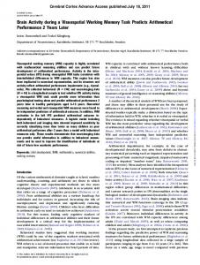
Frontal brain activation during a Working Memory task: a timedomain fNIRS study E. Moltenia,*, G. Basellia, A. M. Bianchia, M. Caffinib, D. Continib, L. Spinellib, A. Torricellib, S. Ceruttia, R. Cubeddub a Politecnico di Milano, Dipartimento di Bioingegneria, IIT unit, via Bonardi 3, I-20133 Milan, Italy b IIT, ULTRAS-INFM-CNR and IFN-CNR, Politecnico di Milano Dipartimento di Fisica, Piazza Leonardo da Vinci 32, I-20133 Milan, Italy *
[email protected]
ABSTRACT We evaluated frontal brain activation during a working memory task with graded levels of difficulty in a group of 19 healthy subjects, by means of time-resolved fNIRS technique. Brain activation was computed, and was then separated into a “block-related” and a “tonic” components. Load-related increases of blood oxygenation were studied for the four different levels of task difficulty. Generalized Linear Models were applied to the data in order to explore the metabolic processes occurring during the mental effort and, possibly, their involvement in short term memorization. Results attest the presence of a persistent attentional-related metabolic activity, superimposed to a task-related mnemonic contribution. Moreover, a systemic component probably deriving from the extra-cerebral capillary bed was detected. Keywords: near infrared spectroscopy, optical imaging, time-resolved imaging, working memory, sustained attention
1. INTRODUCTION It has been shown that brain activity is associated with focal cortical hyperemia, that is, a functionally induced focal cortical hyper-oxygenation. This focal activation has a vascular origin: it sets in whenever an increase in regional cerebral blood flow (rCBF) is not coupled to a correspondingly large increase in cerebral metabolic rate of oxygen consumption (CMRO2) [1]. Monitoring of brain functions based on the vascular response has a longstanding tradition [2]. However, the advent of powerful noninvasive technology, most notably positron emission tomography (PET) and functional magnetic resonance imaging (fMRI) made vascular imaging a cornerstone for the research on functional anatomy of the human cerebral cortex [3]-[4]. The generally close spatial and temporal coupling between the neuronal excitation/inhibition and changes in regional cerebral blood flow (rCBF) made these neuroimaging techniques especially suitable for the investigation of cognitive faculties, and measures of local cortical hemodynamic change can now be used as global indices of change in neural activity, thus providing functional maps of the cerebral cortex. Though crucial to any imaging approach and increasingly subject to research, the mechanisms of neurovascular coupling are far from being fully understood. A multimodal approach to the study of neurovascular dynamics, together with an attempt of integrated interpretation of the data will hopefully help to refine our knowledge about this issue. Among the available neuroimaging techniques, optical methods are able to directly detect focal hyper-oxygenation in response to a stimulus, when measuring over the correspondingly activated cortical area [5]. It follows that such methods provide relevant parameters which help us to investigate the origin of the widely used ‘blood oxygenation-dependent’ contrast (BOLD) in fMRI [6]. Specifically, functional near-infrared spectroscopy (fNIRS) is the elective tool for the quantitative investigation of the regulatory mechanisms underlying hemodynamic response to functional stimulation. By using specific wavelengths of light, injected at the scalp, fNIRS can noninvasively assess the dynamics of oxygenated, deoxygenated and total hemoglobin concentration ([oxy-Hb], [deoxy-Hb] and [tot-Hb]) during task-related brain activity [7]. Although it has been demonstrated that fNIRS is useful for studying higher cognitive functions, up to now, only few surveys investigated working memory functions with fNIRS.
Most biological tissues, bone included, are relatively transparent to near-infrared light (700-900 nm). In this optical window the absorption due to oxy-Hb and deoxy-Hb is higher than that of water. This fact makes transcranial measurements through the intact skull and scalp feasible [7]. A limitation of NIRS technique may reside in the fact that signals contain contributions from both the brain and the overlying tissues. Therefore, a proper instrumental design, combined with advanced methods of data analysis, must be applied to differentiate intracerebral from extracerebral contributions [8]. Time-resolved NIRS is based on the photons time of flight measurement, which naturally discriminates between superficial and deeper structures: on average, photons detected after long times of flight probe deeper tissue layers than early photons. In doing so, a separation of surface and deepness contributions is made feasible. Working memory has been the subject of numerous studies in recent years and continues to be one of the most widely researched cognitive functions. It consists of a brain system for temporarily storage, transfer and management of a representation of information. It is made up of several components and supports active maintenance of information as well as executive control processes. Its activation is required to carry out complex cognitive tasks such as learning, reasoning, and comprehension [9], as working memory is involved in the selection, initiation, and termination of information-processing functions such as encoding, storing, and retrieving data. Psychophysiology demonstrated sustained activity in the prefrontal cortex (PFC) while information is held in mind. Several studies attest sustained prefrontal and parietal activity during the delays of working memory tasks when information is held on-line [10]. Visual working memory involves the concerted activity of a distributed neural system, including posterior areas in visual cortex and anterior areas in prefrontal cortex. Within visual cortex, ventral stream areas are selectively involved in object vision, whereas dorsal stream areas are involved in spatial vision. This domain specificity appears to extend forward into prefrontal cortex, with ventralateral areas involved mainly in working memory for objects and dorsolateral areas involved mainly in working memory for spatial locations. Generally, the increase of workload in memory tasks requires additional mental resources and the recruitment of supplementary specialized networks; as a result, an increase of activation is observed. Many imaging studies supply evidence that the PFC is quantitatively more activated if the working memory load is increased, suggesting that executive function is involved for highly demanding working memory tasks [11]. Attentional networks are related to the PFC as well. Then, focusing attention to and remembering the task-relevant stimuli should increase the activity in distributed areas of the PFC [12]. It follows that working memory and attention both rely on cortical networks in the PFC, resulting in (1) a strong interaction between working memory and selective attention and (2) a crucial role of working memory in reducing distraction by maintaining the prioritization of relevant information. Impairments in working memory are found in several clinical disorders in which prefrontal and parietal cortices are damaged, such as after stroke, traumatic brain injury and in attention-deficit/hyperactivity disorder (ADHD). The vast majority of research on working memory has been carried out solely using behavioral experiments. Many behavioral studies with normal subjects can be found in literature, which were applied as well to brain-injured patients primarily concerned with functional dissociation: these works had little focus on the specific brain regions involved. In the past few years, however, a line of research has emerged that involves studying working memory at both behavioral and neural levels. This research uses neuroimaging techniques [13]-[14] to determine which brain areas are active when people perform various working-memory tasks, and to resolve upon which cognitive functions are mediated by those areas. The time scale of working memory is typically on the order of seconds. It is therefore difficult with PET, which integrates activity over 1-2 min, to precisely analyze the time course of memory in a working memory task. Up to now, fMRI (functional Magnetic Resonance) technique has provided us with useful information regarding the activation and metabolism of the brain areas involved in working memory tasks in both healthy subjects and patients. This approach to working memory places novel constraints on cognitive theories of working memory, and it offers new proposals about how cognitive processes are implemented in brain circuitry. In our experiment we tested working memory for verbal stimuli (letters), holding spatial locations constant. The “nback” task at four levels of difficulty was used: subjects viewed a continuous sequence of stimuli, deciding for each stimulus whether it matched the stimulus shown n stimuli earlier in the sequence, thus generating a specific response pattern. The task combines maintenance as well as active manipulation, i.e., executive processes, because of the necessity to continuously encode, update, and discard the information held in working memory with the presentation of each new stimulus. The value of n is regarded as related to memory load. Thus, by varying working memory load, it was possible to identify areas of brain activity related to working memory function.
The aim of the present study was to evaluate the vascular response correlated to neural activity within a working memory n-back task by means of near-infrared spectroscopy in a population of healthy volunteers.
2. MATERIALS AND METHODS 2.1 Time-resolved functional Near-Infrared Spectroscopy A couple of pulsed diode lasers, operating at 690 nm and 820 nm, with 80 MHz repetition rate and 1 mW average power (PDL, Picoquant GmbH, Germany), are used as light sources. The laser heads are connected to multimode graded index fibers (50/125 μm) by means of a home-made coupler which combines a neutral density attenuator (J54-082, Edmund OptiK GmbH; Germany), with variable attenuation in the range 0-40 dB, and a standard FC fiber optics coupler. Multimode graded index optical fibers (50/125 μm) with different lengths and a 2x2 fused fiber optic splitter (VISNIR5050, OZ Optics, Canada) are used to time multiplexing the laser pulses at the different wavelengths, and to create two independent channels. In each channel a 1x9 fiber optic switch (F-SM19, PiezoJena GmbH, Germany) creates up to 9 independent sources or injection points, therefore 18 sources are available. Four parallel detection chains accomplish acquisition of time-resolved reflectance curves. Each chain consists of a compact 4-channel photomultiplier (PMT) with a multialkali surface (R5900-20-M4, Hamamatsu Photonics, Japan), a routing electronics (HRT-41, Becker&Hickl, Germany), a fast amplifier (HFAC-26, Becker&Hickl, Germany) and a PC board (SPC130, Becker&Hickl, Germany) for time-correlated single photon counting (TCSPC). The parallel use of the four detection and acquisition lines enables a total of 16 independent detectors. Fiber optic bundles, 1.5 m-length with a distal end 90° bended and NA = 0.5 (LLR 3/1500-SIK-90°, Loptek GmbH, Germany), are used for light collection from tissue. The system is controlled by a personal computer (Pentium IV 3.5 GHz, 2 Gb RAM), which hosts the acquisition boards and it is used for data storage. A home made software, written in C language in the LabWindows/CVI environment (National Instruments, TX), is used to control the instrument. The software is interfaced to a micro-controller unit (dsPIC30F6014, Microchip Technology Inc., AZ) which is used for the hardware control of the instrumentation. In particular, the micro-controller unit generates trigger signals for the synchronization of data acquisition by the TCSPC boards and of the fiber optic switches. Typically, by means of the micro-controller unit, the sources can be activated sequentially in any desired sequence. The instrument response function (IRF) obtained by filling all the propagating modes of the bundle has a FWHM of approximately 500 ps. A detailed description and characterization of the system can be found in [15]. The system is also interfaced with dedicated software (Presentation, Neurobehavioral Systems Inc, Albany, CA) for precise stimulus delivery and experimental control program. For each wavelength λ a reference time-resolved fNIRS curve R0(t, λ) is derived by averaging the curves corresponding to an initial resting period (typically 20 s). Then, at each recording time T during the experiment, changes in the absorption coefficient are derived (Nomura, 1997) as
Δμ a (λ , T ) = −
1 ⎛ R(t ; λ ; T ) ⎞ ⎟, ln⎜ vt ⎜⎝ R0 (t ; λ ) ⎟⎠
(1)
where v is the speed of light in the medium, t is the arrival time of photons, and R(t, λ,T) is the time-resolved fNIRS curve at the recording time T. To enhance the contribution from deep layers and to remove possible disturbances caused by superficial layers, a correction method based on the use of late time windows (t = 1750-2500 ps) was also applied [16]. Taking the assumption that O2Hb and HHb are the main chromophores contributing to absorption, their concentrations changes are then derived by Lambert-Beer law. 2.2 Cognitive “n-back” task Subjects performed a variant of the n-back task using letters of the English alphabet as memoranda [10]. Four levels of memory load (0-, 1-, 2-, and 3-back) were presented, according to an increasing criteria. For each difficulty modality,
computerized lists of 30 stimuli were constructed and then presented in a pseudorandom order, making up a total of 240 intermingled upper- and lowercase letters shown to the subject. A block of a single condition lasted 60 s, and consisted of 30 stimuli (2 s inter-stimulus intervals). At the end of the first four conditions, subjects saw the word REST for 90 s (providing a rest period), before the same four conditions, in scrambled order, began. Globally, the task was made up of 17 blocks: 8 activation periods and 6 inter-trial rests and 3 major rests (initial, middle and final). Subjects responded to each stimulus presentation by pressing left mouse button for target stimuli and right button for non-targets. In the 0-back condition, the letter “A” was specified as the target stimulus in the instructions at the beginning of a block. In the 1-back condition, the target was any stimulus identical to the immediately preceding one. In the 2-back and 3-back conditions, the target was any stimulus identical to the stimulus presented two or three preceding trials, respectively. Subject performance during recording was monitored in terms of reaction time (RT) and accuracy (number of target letters identified correctly).
Fig. 1. n-back task is shown. In the 0-back condition (top left), letter “A” is the target stimulus. In the 1-back condition (top right), the target is any alphabetical letter identical to the immediately preceding one. In the 2-back and 3-back conditions, the target is any stimulus identical to the one presented two or three previous trials, respectively.
2.3 Data processing From the original data of [oxy-Hb] and [deoxy-Hb], the components of interest were extracted by using a digital filter. The choice was that of 9th order digital Chebyshev filter with the stop-band 30 decibels down. Transition frequency was selected as the inverse of the period of the task modulation, i.e. ft=0,0167 Hz. Then, a “low frequency” and a “high frequency” components were extracted from the original data. Time series of [oxy-Hb] and [deoxy-Hb] values for any given channel were modeled as a linear combination of L regressors (known functions) plus an error term: Y = X β + ε
(T ×1)
(T ×L ) ( L×1) (T ×1)
(2)
Where X is the design matrix (TxL matrix), each column is a regressor and T is the number of time points. β is the vector of the unknown parameters, one for each regressor, weighing their importance in modeling the original signal. Errors ε were assumed to have a normal distribution with zero mean and covariance σ2V. The ordinary least squares (OLS) estimation of the parameters was calculated:
βˆ = (X T X ) X T Y −1
(3)
Estimation of the error covariance component was performed in two steps; first the correlation matrix V was evaluated using the restricted maximum likelihood (ReML) procedure (iterative algorithm); then the variance was calculated, for each time series, using the estimation:
σˆ 2 =
eˆT eˆ trace(RV )
(4)
R being the residual forming matrix. Hypotheses on predictor variables could then be tested constructing the F statistic: ⎛⎜ βˆ i F=⎝
( )
var βˆi ⎞⎟ ⎠ L
2
(5)
Within the analysis protocol, a good trade-off between statistical reliability and temporal resolution was found by analyzing data with constant regressors. The task was modeled as a series of 17 consecutive boxcars that represented an equal number of activation periods, chosen accordingly to the different activities performed during the experiment. Brain activation was expected to yield an [oxy-Hb] increase or a [deoxy-Hb] decrease (positive and negative F-value in (5), respectively). Two-way repeated-measures ANOVA with time window (factor 1: ‘Time’) and channel (factor 2: ‘Channel’) as factors was run for low frequency signals, while two-way repeated-measures ANOVA with condition (factor 1: ‘Condition’) and channel (factor 2: ‘Channel’) as factors was run for high frequency components. 2.4 Subjects
Nineteen healthy volunteers took part in the present study, with a mean age of 27.15 years (SD 2.01 years, range 23-30 years). Seventeen subjects were right handed. All volunteers were native Italian speakers and were not paid for their participation; all of them were male. They all had normal vision and had no history of psychiatric disorders. The participants were screened thoroughly for neurological symptoms: they all did not show neuro-psychological illness; cognitive level, attentive capability and memory skills were in a normal state. Written informed consent was obtained from all volunteers after the examination and test procedure had been explained.
3. RESULTS 3.1 Behavioral results
The 19 subjects committed an average of 1,58 errors (SD=0,69) in the n-back task, thus falling in the normality range. On average, they committed 0,77 errors during the 0-back condition, 1,28 errors during the 1-back, 2,14 during the 2back and 2,15 during the 3-back. They answered correctly to the target in 511 ms (SD=36). More specifically, reaction times on targets were 464 ms for the 0-back condition, 508 for the 1-back, 540 ms for the 2-back and 535 ms for the 3back. 3.2 fNIRS results
The analysis of fNIR measures was performed after obtaining variations of oxygenated ([oxy-Hb]) and deoxygenated ([deoxy-Hb]) hemoglobin through a modified Beer-Lambert law. As an example, figure 3 shows the time course of [oxyHb] (red line) and [deoxy-Hb] (blue line) for all subjects and all channels (see Fig.2 for channels identification).
Fig. 2. Position of probes on the frontal head
Fig. 3. Time course of [oxy-Hb] (red line) and [deoxy-Hb] (blue line). Bold lines mark averages over the population of the study (all channels). Thin lines mark standard deviation.
Results emerging from time-resolved NIRS acquisitions show that variations of [oxy-Hb] remained positive in the PFC, over both the hemispheres and during all the test. Activation level increased during the first half of the test, reaching a peak in the central part of the test; then, it remained stable during the second part of the task, with a final and further increase in the last recovery period. In addition to the large, persistent changes described hitherto, transient and less prominent changes were observed time locked to the active blocks presentation. A slight hemispheric lateralization was observed, with a prevalence of activation on the right side, i.e., right PFC showed faintly higher [oxy-Hb] increases than the left one. Variations of [deoxy-Hb] globally stayed in a range of negative values throughout the test. Right channels showed an initial decrease and an ensuing increase in value, which moved towards the restoration of the rest value; left channels exhibited an initial decrease of [deoxy-Hb] as well, with a delayed recovery. In general, we found higher variations of [oxy-Hb] and [deoxy-Hb] over the right frontal area. After [oxy-Hb] and [deoxy-Hb] time courses were plotted, two components were separated from the metabolic response generated by the task. The first one is a “low frequency” component, significant of the general arousal level due to subject’s involvement in the cognitive challenge; the second is a “high frequency” oscillatory component, most probably modulated by the block presentation pattern and representative of the cerebral activity which is more specifically linked to the working memory effort. The two contributions were separately considered, with reference to the performance of the cognitive task.
Fig. 4. Grand average time course of low frequency component during the working memory task. [oxy-Hb] variations to the left and [deoxy-Hb] to the right.
Fig. 5. Cortical activation during working memory task detected by time resolved NIRS. The activation maps are obtained from F-statistic values, assessed by a second level analysis of the GLM model. [oxy-Hb] values are presented. Each map is relative to a different regressor. Arrows indicate rest periods. Task progression: from top to bottom.
Fig. 6. Deactivation maps obtained from second level analysis of the GLM model. Data for [deoxy-Hb] are presented. Arrows indicate rest periods. Columns show the task progression from top to bottom.
In figure 4 the grand average time course of low frequency component is presented. As the task starts, a strong increase in cerebral activation is noticed, lasting for the first half of the test. The central rest block, lasting one minute and a half, allows a partial recovery of the metabolic value: an important drop toward less activated level is evident. The second part of the test arouses a second metabolic adjustment. Repeated-measures ANOVA test was conducted on [deoxy-Hb]. A strong effect of time (p < 0.001) and of time-bychannel interaction (p < 0.001) emerged, while a main effect of channel was not observed (p > 0.05). The effect of time is paramount: the task generated a strong hemodynamic response throughout its entire duration, presenting a momentary decrease only in correspondence of the central rest period. Standard deviation increased with time within both parts of the task, thus testifying a greater uniformity of behavior among subjects during the “early” response to the task (and then a more pronounced inter-subject variability for the long-term response). Repeated-measures ANOVA test conducted on [oxy-Hb] attested a significant main effect of time and of time-by-channel interaction (p0.05). After extracting the low-frequency component of activation, GLM statistics were applied to the data, in order to identify frontal brain areas which showed statistically significant signal changes in response to the task stimulation. Time courses of [oxy-Hb] and of [deoxy-Hb] variations were analyzed: a peak of activation was found at the third block of working memory task, during the first half of the test. This level was, more or less, maintained during the following task blocks. Further, the above mentioned activation peak was observed during the middle baseline period, followed by a slight decrease afterwards (positive peak for [oxy-Hb], negative for [deoxy-Hb] ). Statistical maps were then calculated for the analyzed group of volunteers using the F-value parameters. Response of the prefrontal cortex to task presentation is given in figures 5 and 6, for [oxy-Hb] and [deoxy-Hb] respectively. The temporal trend noted above is confirmed by these activity maps. The task originated a strong metabolic response, leading to an increasing difference between parameters values registered during the first half of the test and during the baseline period. Central rest showed the most deviant values. The second part of the test was characterized by a strong, but steady, activation. Again, the final recovery period presented an apparently large activation. Modifications are nearly uniform over the PFC, attesting a general involvement on the frontal lobe due to this task. The only exception lies in a slightly right lateralization of [oxy-Hb] during the second part of the task. “High frequency” signals were then analyzed. Results seems to suggest that high frequency activity underwent a modulation induced by the task presentation sequence, thus following the alternation of task/rest blocks. Due to this characteristic, this component should be the one which mostly represents the real cortical activity related to the working memory effort. In figure 7 time courses of [oxy-Hb] and [deoxy-Hb] are presented. [deoxy-Hb] is greatly reduced during task blocks and recovers to baseline values during the various rest periods. Further, the second part of the test exhibits a
less intense hemodynamic response. Time-by-channel interaction attests a different distribution of cerebral activation over the PFC during different periods of the task.
Fig. 7. Grand average time course of high frequency component during a working memory task (channel 1). [oxy-Hb] (left) and [deoxy-Hb] (right) are plotted. The accordance between task presentation and parameter oscillation is well intuitable.
Statistical analysis was performed after having derived, for each channel and subject, a vector of only eight numbers, the values being the average of the parameters over the different task blocks provided by the experiment. Results for [deoxyHb] revealed a significant main effect of condition and condition-by-channel interaction (p0.05) was found. Repeated-measures ANOVA test conducted on [oxy-Hb] attested a significant main effect of condition and of condition-by-channel interaction (p0.05). The main effect of time is interpretable with the predominant increase in [oxy-Hb] levels during task blocks with respect to the baseline and the rest periods. The factor ‘Channel’ had no influence. GLM approach was newly applied to the data, epoched in accordance with the above mentioned statistical analysis. Regressors representing the aforementioned segmentation of the task were organized in the form of a contrast matrix. Results of the GLM F-statistic were obtained for both [oxy-Hb] and [deoxy-Hb]. In both cases, the rest periods present very low values of F-level, while active periods keep high. The preponderant activation of the first half of the test with respect to the second is also confirmed. Further, data present a higher variability among channels during the first part of the task, bearing out the time-by-channel interaction reported above. Figures 8 and 9 show the pseudocolor representation of the F-statistic values of [oxy-Hb] and [deoxy-Hb]. These values were obtained through the resolution of the complete model for the [oxy-Hb] first (Fig.8), and then for the [deoxy-Hb] (Fig.9), both at group level. Each map represents the distribution associated to a specific regressor. Rest periods present no activation and uniform distribution; regressor n°2, representative of the first half of the test (blocks n°1 to 4, task periods only), shows a strong activation ([deoxy-Hb] decrease) considerably lateralized over the right hemisphere. Regressor n°3, representative of the working memory blocks during the second part of the test, also shows an activation, though weaker and more uniform than that recorded during the early stage of the task, over the prefrontal cortex and without evident spatial localization. Decreased activity during the second part of the task and the absence of response during the rest periods are confirmed. The [oxy-Hb] level increases, as expected, with the test; maximum values are observed within the active task periods, during both parts of the test. Spatial distribution shows a mild lateralization to the right. Analysis were carried out in order to attest the dependence of activation on memory load, or rather on the difficulty of the test. A one way repeated-measures ANOVA was run, setting ‘memory load’ as factor. Results confirm that no influence of memory load is present (p>0.05). GLM was finally ran for superficial data (Fig. 10 and 11), that is, for signals obtained from “early” photons, detected after short times of flight. Superficial data were high-pass filtered first, by means of the Chebyshev filter described above. Then GLM F-statistic was applied. The same eight regressors previously used for the “high frequency” signals were included in the analysis. Maps revealed a faint activation, probably coming from the capillary bed belonging to the skin. Activation was found throughout the whole first half of the test: during both the initial rest and task blocks n°1 to 4. A poor (almost absent) lateralization was detected.
Fig. 8. F-statistic values of [oxy-Hb], expressing cortical activation during activity blocks (“high frequency”). Average values on the entire group. Columns show the task progression from top to bottom.
Fig. 9. F-statistic values of [deoxy-Hb]. Average values on the entire group. Columns show the task progression from top to bottom.
Fig. 10. F-statistic values of [oxy-Hb], expressing blood sprinkling in the skin during both task blocks and rests. Values on the entire group. Columns show the task progression from top to bottom.
Fig. 11. F-statistic values of [deoxy-Hb], expressing blood sprinkling in the skin during both task blocks and rests. Values on the entire group. Columns show the task progression from top to bottom.
4. DISCUSSION During the last decade, some researchers applied NIRS instrumentation to the study of working memory for both physiological and pathological situations. The employment of NIRS for the investigation of cognitive processes relies on three basic assumptions: (1) A selective change in some cognitive function—e.g., briefly remembering verbal information—is mediated by changes in neural activity in a region or in several selective regions of the brain; (2) A change in regional neural activity is accompanied by a change in oxygenation and/or deoxygenation and/or blood flow values to that region; (3) Changes in regional blood flow may be monitored by monitoring the sum of oxygenation and deoxygenation values in the blood stream using NIRS, if the hematocritus is constant. Besides, due to the great number of working memory tasks and the even larger number of their possible implementations, results in this field are not well established and under continuous improvement. We used the n-back task [10], which requires coding the stored letters with respect to their temporal position and constantly changing these temporal codes as new letters are presented. In n-back task, by varying the value of n, i.e. memory load, it is possible to
examine the effects of activations in different neural areas. A large number of results has been already reported in the neuroimaging literature for this test [3],[17], and evidences proved that, during the task, (1) subjects’ performance declines continuously with the increase of task load; (2) there are correlations between behavioral performance and activation measures; (3) continuous increases in memory load lead to continuous increases in activation in a fixed number of memory areas; and (4) continuous increases in memory load lead to new areas being recruited for the task. In our work, we have been following the goal of strengthen the knowledge of cerebral metabolism during working memory effort by studying, with a multi-channel time-of-flight NIRS, the hemodynamic response of cerebral cortex when engaged in a working memory n-back task. Moreover, signals were separated into “early photons” and “late photons” contributions, at the purpose to isolate intra-cortical and extra-cortical information and to perform a more refined interpretation of our results. Within the circuitry for verbal working memory, there is a separation between components that underlie storage and those that mediate rehearsal of information. Regions in the posterior parietal cortex (particularly in the left hemisphere) seem to be involved in storage, whereas frontal regions appear to mediate rehearsal. In contrast to storage and rehearsal functions, the processes that operate on the contents of working memory (executive processes) appear to be mediated by the dorsolateral prefrontal cortex (PFC). In particular, this region is activated when subjects have to temporally code the contents of working memory rather than just store the materials [13]. Temporal coding may not be needed in 0-back and 1-back conditions. According to Ungerlieder & al. [18], right prefrontal activity is prevalently associated with the simple, icon-like image of the visual stimulus, whereas left prefrontal activity is associated with a stimulus representation that is more symbolic, and, possibly, verbal. It should be taken into consideration, anyway, that laterality effects in visual memory may be influenced by a variety of factors, such as memory set size, retention interval length, and item familiarity, all of which may affect the extent to which subjects engage in symbolic or verbal encoding and rehearsal. Behavioural data presented above evidenced a prevalence of commission errors, occurred on target letters. Commissions on no-target letters and omissions for both target and no-target were quite rare. A direct correlation between load of the test and number of errors was observed, even though standard deviations underlined a notable inter-subject variability. Similar correlations were observed for reaction times, which tended to lengthen with workload, both for commissions errors and correct responses. In concordance with prior studies [19]-[20] we found a significant increase in [O2Hb] for the hemodynamic response of prefrontal neural tissue to cognitive stimuli (in this case a mnemonic task). Moreover, a decrease in [deoxy-Hb] values in the lateral PFC was observed during task performance. These two phenomena are intended to reflect the occurrence of rCBF increases in response to neuronal activation elicited by the test. The magnitude of the increase in [oxy-Hb] - typically two or three times that of the decrease in [deoxy-Hb] – was confirmed in our study as well. During the inter-trial rest, in the middle part of the test, a further increase in [oxy-Hb] was noted, and this fact has been associated to an uncoupling effect: the cessation of oxygen consumption is faster than the reduction in blood flow inside the cortex, and this is at the origin of [oxy-Hb] increase. Changes of [deoxy-Hb] affected channels less than [oxy-Hb] and were tendentially right lateralized. The increase of working memory load requires additional mental resources and the recruitment of supplementary specialized networks, especially over the prefrontal area [13]. On the other hand, it is assumed that the capacity of the cognitive system is limited. We predicted that cortical activity would increase in proportion to the working memory demands, i.e., we expected a relationship between activity and working memory load, especially in the PFC. Indeed, the pattern of activation should not differ quantitatively and qualitatively in most areas (i.e., no increasing activation and no recruitment of additional areas with increasing working memory demands) with the exception of the dorsolateral PFC where, by comparing the 1-back with the 2-back condition, an increase in cortical activation should be expected, due to novel task demands (updating and rehearsal). Jaeggi & al. [14] found load-dependent activation changes in the dorsolateral PFC. The percentage signal change continuously increased from the 1-back to the 3-back condition. The relationship between activation in PFC and working memory load was found to yield a monotous increase, although not with a strictly linear proportionality. The present work confirmed the augmentation of PFC activation along the first half of the test and showed some correlation to workload, though not statistically significant. After the middle rest, activation was restored during the second half of the test and even lasted over the final rest period, showing some sort of “residual hyperemia”. Lateralization effects were observed in our study, with [deoxy-Hb] changes mainly concentrated over the right PFC and [oxy-Hb] changes spanning also over the right frontal pole. Lateralized activation was observed in some other works using the n-back task [14],[21] but lateralization during working memory task is a topic still largely debated and controversial.
In addition, our study with time-resolved NIRS demonstrated that, during a working memory test, the mental operation which correctly performs the task is composed of at least two different mental processes: we observed and separately analyzed two kinds of temporal responses, characterized by different dynamics and associable to different psychological aspects: the “low” and “high” frequency responses. “High frequency” response has been interpreted as tightly connected to the temporization of task presentation, i.e. it was found to follow the alternation of activity/rest blocks during the experiment. This working memory-related process reflected the block-design of the experiment, showing the onset of a metabolic response any time an active block was presented, and the fading of the same response during the following rest blocks. No effect of memory load was found on this component, and even the 0-back condition showed a cerebral activation. These results are in accordance with D’esposito et al [4], who reported a block-related activity as well. An increase in memory load may sometimes be accompanied by a new task requirement (engagement of an additional cognitive faculty), which can lead to a marked increase in the activation of an area. A growing number of studies showed interactions between attention and working memory [22]. While working memory processes connected with strategies of maintenance of the stimulus are associated with activation of the left PFC [19], working memory tasks which require subvocal rehearsal are apparently linked with the ventrolateral PFC. Perhaps different parts of this prefrontal region subserve distinct functions, or rather these functions have some component in common, and this common element is what activates dorsolateral PFC. The component could be a metacognitive process that monitors operations performed on working memory, or a process responsible for the maintenance of the goal structure needed to guide processing in any task. In the present study, a “low frequency” response was found, enlightening the insurgence of a significant tonic activity during the overall test. Our hypothesis is that, in addition to task-specific activation related to working memory processes, there are functional networks which activate during the performance of the task and that show their effect on a long term time scale. These networks are probably “default”-activated resources, involved in all attention-demanding tasks and reflecting general cognitive operations such as attention and emotion. Plausibly, working memory task elicited a sustained activation in frontal regions. Only the inter-trial baseline, corresponding to the period when the subject is aware of a relative long period of rest, shows a decrement of this sustained activity. Our suggestion is that arousal and/or sustained attention can be thought at the origin of this signal. These results provide further evidences that PFC is active during working memory effort and that it is probably related with nonworking memory activity. A number of previous works observed PFC activity during experiment of sustained attention [23] but never were these observations concomitant and superimposed to a different task. In functional NIRS studies the dynamics of [oxy-Hb] and [deoxy-Hb] variations have been shown to differ, depending on the stimulus paradigm and have been interpreted as a potential indicator of differences in physiological mechanisms of neurovascular coupling across cortical areas [24]. These surveys have also shown that this fact may indeed depend partly on hemodynamic changes unrelated to the cortical increase in blood flow. NIRS signal is highly sensitive to extracerebral blood volume changes. The most reliable hypothesis is that this difference may be caused by systemic changes. In contrast to other neuroimaging methods and to the same “continuous wave” NIRS, time-resolved NIRS can differentiate between truly cortical and systemic changes. In the present work, extra-cerebral activation was isolated and analyzed: weaker but significant alterations of [oxy-Hb] and [deoxy-Hb] values were found and separately treated in order to obtain neater information from the cortex. At the moment we are writing, no works are known to us which consider different components in the signal and focus their attention on both task-related and the tonic effects during a cognitive task. Hoshi et al., [25] referred all activation periods to the 10 sec before each individual block, in this way leaving aside metabolic effect underlying the overall examination period. Other authors averaged all the activation blocks, separating for memory load, and a possible common activity was not considered. This is probably due to the technological limitations of continuous wave NIRS, which cannot discern superficial from cortical activity. The present paper is a first, preliminary attempt to give a more refined interpretation to metabolic phenomena occurring during working memory effort.
5. CONCLUSION Our study provides some informative evidence that the combined analysis of both behavioral and vascular parameters provides an improvement in the investigation and interpretation of the working memory faculty and, in general, of the cognitive processes. Moreover, our findings support the notion that the prefrontal activity reflects not only maintenance processes, but also attentional monitoring and selection processes during working memory tasks.
REFERENCES [1] [2] [3] [4] [5]
[6] [7] [8]
[9] [10] [11] [12]
[13] [14]
[15] [16] [17] [18] [19]
[20] [21]
[22] [23] [24] [25]
Fox, P.T., Raichle, M.E., “Focal physiological uncoupling of cerebral blood flow and oxidative metabolism during somatosensory stimulation in human subjects,” Proc. Natl. Acad. Sci. U. S. A 83, 1140–1147 (1986) Roy, C., Sherrington, C., “On the regulation of the blood supply of the brain,” J. Physiol. 11, 85–108 (1890) Cohen, JD, Forman SD, Braver TS, Casey BJ, Servan-Schreiber D, Noll DC., “Activation of prefrontal cortex in a nonspatial working memory task with fMRI,” Hum. Brain Mapp. 1, 293–304 (1994) D’Esposito M., Aguirre G.K., Zarahn E., Ballard D., Shin R.K., Lease, J., “Functional MRI studies of spatial and nonspatial working memory,” Cognit. Brain Res. 7: 1–13, (1998) Obrig, H., Wenzel, R., Kohl, M., Horst, S., Wobst, P., Steinbrink, J., Thomas, F., Villringer, A., “Near-infrared spectroscopy: does it function in functional activation studies of the adult brain?,” Int. J. Psychophysiol. 35, 125– 142 (2000) Steinbrink, J., Villringer, A., Kempf, F., Haux, D., Boden, S., Obrig, H., “Illuminating the BOLD signal: combined fMRI-fNIRS studies,” Magn. Reson. Imaging 24, 495–505 (2006) Villringer A, Chance B, “Non-invasive optical spectroscopy and imaging of human brain function,” Trends Neurosci. vol. 20, 435-442 (1997) Liebert A, Wabnitz H, Steinbrink J, Obrig H, Möller M, Macdonald R, Villringer A, Rinneberg H., “Time-resolved multidistance near-infrared spectroscopy of the adult head: intracerebral and extracerebral absorption changes from moments of distribution of times of flight of photons,” Appl Opt., 43(15), 3037-47 (2004) Baddeley A.D., [Working memory], Oxford University Press, Oxford (1986). Cohen JD, Perlstein WM, Braver TS, Nystrom LE, Noll DC, Jonides J, Smith EE., “Temporal dynamics of brain activation during a working memory task,” Nature 386, 604–608 (1997) Tomasi D, Ernst T, Caparelli EC, Chang L, “Common deactivation patterns during working memory and visual attention tasks: an intra-subject fMRI study at 4T,” Hum BrainMapp, 27(8), 694-705 (2006) Tsujimoto S, Yamamoto T, Kawaguchi H, Koizumi H, Sawaguchi T., “Prefrontal cortical activation associated with working memory in adults and preschool children: an event-related optical topography study,” Cereb Cortex. 14(7), 703-12, (2004) Smith E.E., Jonides J., “Working Memory: A Wiew from Neuroimaging,” Cogn. Psychol. 33,5-42 (1997) Jaeggi SM, Seewer R, Nirkko AC, Eckstein D, Schroth G, Groner R, Gutbrod K, “Does excessive memory load attenuate activation in the prefrontal cortex? Load-dependent processing in single and dual tasks: functional MRI study,” NeuroImage 19, 210–225, (2003) Contini, D., Torricelli, A., Pifferi, A., Spinelli, L., Paglia, F., Cubeddu, R., “Multi-channel time-resolved system for functional near infrared spectroscopy,” Opt Expr 14, 5418-5432 (2006) Contini D., Torricelli A., Pifferi A., Spinelli L., Cubeddu R., “Novel method for depth-resolved brain functional imaging by time-domain NIRS,” Proc. SPIE Vol. 6629, 662908 (2007) Owen A.M., “Working memory: imaging the magic number four,” Curr Biol., 14(14),R573-4 (2004) Ungerleider LG, Courtney SM, Haxby JV, “A neural system for human visual working memory,” Proc.Natl.Acad.Sci. USA 95, 883-890 (1998) Schreppel T, Egetemeir J, Schecklmann M, Plichta MM, Pauli P, Ellgring H, Fallgatter AJ, Herrmann MJ. “Activation of the prefrontal cortex in working memory and interference resolution processes assessed with nearinfrared spectroscopy,” Neuropsychobiology, 57(4), 188-93 (2008) Molteni E, Butti M, Bianchi AM, Reni G, “Activation of the PFC during a visual n-back working memory task with varying memory load: a Near Infrared Spectroscopy Study,” Conf. Proc. IEEE Eng. Med. Biol. Soc. (2008) Ludwig C, Chicherio C, Terraneo L, Magistretti P, de Ribaupierre A, Slosman D., “Functional imaging studies of cognition using 99mTc-HMPAO SPECT: empirical validation using the n-back working memory paradigm,” Eur J Nucl Med Mol Imaging.; 35(4), 695-703 (2008) Kane MJ, Engle RW., “Working-memory capacity and the control of attention: the contributions of goal neglect, response competition, and task set to Stroop interference,” J Exp Psychol Gen.; 132(1), 47-70 (2003) Tzourio N, Massioui FE, Crivello F, Joliot M, Renault B, Mazoyer B., “Functional anatomy of human auditory attention studied with PET,” Neuroimage, 5(1), 63-77 (1997) Huppert, T.J., Hoge, R.D., Diamond, S.G., Franceschini, M.A., Boas, D.A., “A temporal comparison of BOLD, ASL, and NIRS hemodynamic responses to motor stimuli in adult humans,” NeuroImage 29, 368–382 (2006) Hoshi Y., “Functional near-infrared optical imaging: utility and limitations in human brain mapping,” Psychophysiology, 40(4), 511-20 (2003)