findings concerning the brain areas activated by a verbal working memory task. ...... Talairach J, Tournoux P. Co-planar stereotaxic atlas of the human brain: ...
Brain (1996), 119, 1617-1625
Regional brain activity during working memory tasks E. Salmon,12 M. Van der Linden,3 F. Collette,3 G. Delfiore,1 P. Maquet,12 C. Degueldre,1 A. Luxen1 and G. Franck2 1
Cyclotron Research Centre, the ^Department of Neurology and the ^Department of Neuropsychology, University of Liege, Belgium
Correspondence to: E. Salmon, MD, Cyclotron Research Centre, B30 Sart Tilman, 4000 Liege, Belgium
The first aim of our PET study was to replicate previous findings concerning the brain areas activated by a verbal working memory task. The second aim was to specify the neural basis of the central executive, using a task of working memory updating. Our data confirm that the lower left supramarginal gyrus and premotor area are the key regions subserving short-term verbal memory processes. They also suggest that the updating memory task is related to middorsolateral prefrontal activation, most probably responsible
for the updating function of the central executive. An unexpected, predominantly right activation occurred in the inferior parietal region during the verbal memory updating task, which we related to a visuospatial strategy used to maintain the information in short-term memory. A third purpose was to explore the brain regions activated by a nonverbal, visual memory task, and our results confirm the importance of the superior occipital gyrus in the visual shortterm memory.
Keywords: working memory; central executive; PET Abbreviations: BA = Brodmann area; rCBF = regional cerebral blood flow; SPM = statistical parametric map
Introduction Working memory refers to a limited capacity system that is responsible for the temporary storage and processing of information while cognitive tasks are performed. Baddeley's model represents the most extensively investigated theoretical construct of working memory (Baddeley and Hitch, 1974; Baddeley, 1986, 1992). This model comprises a modalityfree controlling central executive that is aided by a number of subsidiary slave systems which ensure temporary maintenance of information. Two such systems have been more deeply explored: the phonological loop and the visuospatial sketchpad. The visuospatial sketchpad system is assumed to be involved in setting up and maintaining visuospatial images. The phonological loop system is specialized for processing verbal material and is composed of two subsystems: a phonological store and an articulatory rehearsal process. The core of the working memory model is the central executive. The central executive is assumed to be an attentional control system responsible for strategy selection and for control and co-ordination of the various processes involved in short-term storage and more general processing © Oxford University Press 1996
tasks. An important characteristic of this system is that its resources are limited and divided into different processing and storage functions. The theoretical accounts of the central executive are less clearly detailed than those of the slave systems. Baddeley (1986) has suggested that the supervisory attentional system component of the attentional control of action model proposed by Norman and Shallice (1986) might be an adequate approximation of the Central Executive system. According to Norman and Shallice, some action schemata (routine actions) can run automatically, whereas other actions require deliberate attentional resources. These authors assume the existence of two attentional control processes: the contention scheduling mechanism, which would be involved in making selections between conflicting routine actions, and the supervisory attentional system, which would intervene when the selection of routine actions is not sufficient (for instance, in tasks that require planning or decision making, or when coping with novel or dangerous situations). According to Shallice (1988), dysfunction of the supervisory attentional system could plausibly account for the cognitive deficits following frontal-lobe lesions. Recently,
Downloaded from http://brain.oxfordjournals.org/ by guest on September 2, 2012
Summary
1618
E. Salmon et al.
Concerning the physiological substrate of working memory, neuroimaging studies of patients with selective deficits of the phonological loop suggest that this component is localized in the left hemisphere. More specifically, in a review of all patients for whom sufficient details were available, Delia Sala and Logie (1993) (see also Shallice and Vallar, 1990) found that the common denominator seemed to be the inferior part of the parietal lobe, close to the junction with the superior, posterior temporal lobe. In the few cases where the lesion was described with more detail, it involved the supramarginal gyrus of the inferior parietal lobe. A recent PET activation study (Paulesu et al., 1993a) confirmed these findings, localizing the phonological store in the left supramarginal gyrus while the articulatory rehearsal process was localized in Broca's area. There exists less evidence for the neural basis of the visuospatial sketchpad. The only studied case, described as having a specific visuospatial sketchpad deficit, suffered from an extensive lesion in the frontal region of the right hemisphere (Hanley et al., 1991). When group studies are considered, it appears that the most frequent lesion associated with a visuospatial short-term memory deficit involves the posterior parietal lobe near its junction with the occipital lobe (see Warrington and James, 1967). However, certain other data also suggest that patients with frontal cortical lesions can present with deficits of spatial working memory (e.g. Pigott and Milner, 1994). PET studies (Jonides et al., 1993; Frackowiak, 1994; Smith and Jonides, 1995) showed that spatial and visual working memory tasks led to different patterns of activation. The spatial working memory task
activated the right occipital gyrus (Brodmann area or BA 19; see also Dupont et al., 1993), the right posterior parietal gyrus (area 40; see also Belger et al., 1995), the right premotor area (BA 6), and the right inferior dorsolateral prefrontal cortex (area 45/47). In another study using functional MRI, however, a spatial working memory task activated the prefrontal area 46 (McCarthy et al., 1994). The visual short-term memory task studied by Smith and Jonides (1995) led to activation of the left inferior temporal cortex at the temporo-occipital junction (area 37; see also Belger et al., 1995), the left parietal gyrus (area 40), the left anterior cingulate gyrus (area 32), and the prefrontal gyrus (area 6). In another study, comparing shape judgement task and visual short-term memory task with Korean letters, the working memory task caused a greater activation in right occipitoparietal area 19/40 and in left prefrontal area 47 (Paulesu etai, 1993b). Bilateral activation of the prefrontal dorsolateral area was also reported for a shape working memory task, using [18F]fluorodeoxyglucose (Swartz et al., 1995). Finally, the localization of the central executive remains to be considered. Some authors have proposed that executive processes involve the frontal lobes (e.g. Shallice, 1988). In recent PET studies, Petrides et al. (1993a, b) observed bilateral activation of mid-dorsolateral prefrontal cortex (areas 46 and 9) during the performance of verbal and nonverbal random generation tasks, in which subjects were asked to generate random sequences. This task is generally considered to place significant demands on the central executive (see Baddeley, 1986; Van der Linden et al, 1994). A similar localization was observed when the subjects performed a task in which they had to monitor random sequences of numbers from 1 to 10 read by the experimenter and to identify the number that had been omitted in the sequence (Petrides et al., 1993a). More recently, in a study using functional MRI, D'Esposito et al. (1995) observed a bilateral activation in the dorsolateral prefrontal cortex (areas 46 and 9) as well as in the anterior cingulate during the concurrent performance of two tasks, which is expected to engage the central executive. However, the frontal localization of certain central executive functions has been questioned (see Morris, 1994). Indeed, frontal lobe lesions do not necessarily impair the coordination of dual tasks involving working memory (e.g. Frisk and Milner, 1990). Similarly, we recently described a closed-head injury patient in whom a specific dual-task impairment was interpreted as the consequence of a reduction of central executive resources, with no concomitant signs of frontal dysfunction (Van der Linden et al, 1992). Furthermore, Dalrymple et al. (1994) found no association in patients with Parkinson's disease between dual task performance and various indices of frontal performance. These data suggest either that the co-ordination of dual tasks does not implicate the frontal lobes or that there are dissociations within central executive functions. Finally, some researchers consider that executive functioning relies on a
Downloaded from http://brain.oxfordjournals.org/ by guest on September 2, 2012
Shallice (1994) has also suggested that the function of the supervisory attentional system could be fractionated into several components. Convincing evidence for the existence of the different components comprising the working memory model comes from the study of brain-damaged patients with specific shortterm memory impairments (for reviews, see Gathercole, 1994; Van der Linden, 1994). Some patients have deficits of auditory short-term memory that can be attributed either to a selective impairment of the phonological store (e.g. Vallar and Baddeley, 1984) or to a disturbance of the articulatory rehearsal process (e.g. Belleville et al, 1992). Other patients have specific impairment of the visuospatial sketchpad (Hanley et al., 1991) or of the central executive (Van der Linden et al., 1992). Other data show that the central executive is particularly sensitive to the effects of Alzheimer's disease (for review, see Morris, 1994). More specifically, it appears that patients with Alzheimer's disease manifest a deficit affecting one important component of the central executive, i.e. the capacity to co-ordinate two or more subprocesses (Baddeley et al., 1986, 1991). A similar dual-task deficit has also recently been observed in patients with Parkinson's disease (Dalrymple et al., 1994). Finally, a number of studies have suggested that normal ageing is characterized by a decline in the capacity of the central executive (e.g. Van der Linden et al., 1994a, b)
Working memory
The main aim of the present study was to explore the neural basis of what appears to be an important activity of the central executive, i.e. the updating of working memory. We expected mainly dorsolateral prefrontal activation. Another aim was to replicate the findings reported by Paulesu et al. (1993a, b) concerning the localization of the phonological store and the articulatory rehearsal process, on the one hand, and the functional anatomy of the visual sketchpad, on the other hand.
Material and methods Subjects Ten male, European, right-handed volunteers (age range 1930 years) gave written informed consent to take part in this study, which was approved by the University of Liege Ethics Committee. None had past medical history nor used any medication.
PET scanning Scans of regional cerebral blood flow (rCBF) were obtained for each subject using a CTI model 951/31 R PET scanner (CTI, Knoxville, Tenn., USA) with collimating septa extended. The physical characteristics of the tomograph have
been described previously (Degueldre and Quaglia, 1992). Subjects had an individual thermoplastic face mask for head stabilization. A transmission scan was acquired for attenuation correction using three rotating sources of 68Ge. Emission scans were reconstructed using a Hanning filter at a cut-off frequency of 0.5 cycles per pixel giving a transaxial resolution of 8.7 mm full width at half maximum and an axial resolution of 5 mm for each of 31 planes with a total field of view of 10.8 cm in this direction. Volunteers received a 60 s intravenous infusion of H 2 ' 5 O (total activity 35 mCi) through a left forearm cannula. A dynamic PET scan consisting of two frames was collected over a period of 3 min (background frame duration 1 min, second frame duration 2 min). The infusion of l5O-labelled water began 45 s after acquisition start time (Lammertsma et al., 1990). Cognitive activation started upon H215O infusion, 15 s before the second scan. The integrated counts per pixel recorded during the second scan were used as an index of rCBF (Mazziotta et al., 1985; Fox and Mintun, 1989). All subjects underwent six consecutive rCBF measurements (two for each experimental and control condition). Fifteen minutes elapsed between scans. The order of memory tasks was randomly distributed between subjects and it was determined by a Latin square design.
Working memory tasks The control and the first experimental tasks have previously been described by Paulesu et al. (1993a). In the control visual short-term memory task, six Korean letters (which could not be transcoded into a phonological code) were randomly presented on a computer screen at a rate of one per second. Subjects were asked to remember the stimuli using a visual code and to judge if a probe Korean letter displayed 2 s after each sequence was present in this particular sequence. Subjects responded by pressing one of two yesno response buttons. In the phonological short-term memory (first experimental) task, randomized sequences of six phonologically dissimilar consonants were displayed on the monitor. Subjects were instructed to rehearse the stimuli silently and to remember them serially to detect whether a target consonant presented 2 s after this string was present in the list. The task was otherwise identical to the control one. In the updating working memory (second experimental) task (adapted from Morris and Jones, 1990) (see also Van der Linden et al., 1994), lists of eight, nine and 10 phonologically dissimilar consonants were presented at a rate of one per second. Subjects were not informed of the length of each list before presentation. They were asked to rehearse silently and to remember serially only the last six items. They had to judge whether a consonant displayed 2 s after each list was present in the six last consonants for this particular list. Sequences sounding like words and abbreviations were avoided. The various lists were presented in a randomized order, with the restriction that no more than two lists of the same length were presented successively.
Downloaded from http://brain.oxfordjournals.org/ by guest on September 2, 2012
network distributed between anterior and posterior areas of the brain (e.g. Fuster, 1993). A major difficulty in the exploration of the neural basis of the central executive is that a wide range of cognitive functions has been ascribed to this system, i.e. control, processing and even storage activities. In this context, it seems important to explore the neural substrate that underlies different central executive functions (other than dual-task coordination or random generation). Another problem is finding a working memory task in which the role of the central executive can be clearly distinguished from that of the slave systems. In a recent study, Morris and Jones (1990) showed that the running memory paradigm initially used by Pollack et al. (1959) meets this requirement. The task requires subjects to watch strings of consonants of unknown length, and then to recall serially a specific number of recent items. The running span task requires considerable flexibility of information processing and a progressive shift of attention, i.e. discarding some items while new ones are registered. Morris and Jones (1990) showed that the running memory task requires two independent mechanisms: the phonological loop (phonological store and articulatory rehearsal process) and the central executive. The updating process requires central executive resources but not the phonological loop. Conversely, the serial recall component of the task requires the phonological loop but not the central executive. Recently, we successfully used a running memory task similar to that employed by Morris and Jones (1990) to show that older subjects have decreased central executive resources (Van der Linden et al., 1994).
1619
1620
E. Salmon et al.
Patients were trained 5 or 6 days before the PET session. Five minutes before each acquisition, instructions were rehearsed, followed by a short training period (four sequences). The control and first experimental tasks consisted of nine sequences (six sequences during blood flow measurement). The second experimental task comprised eight sequences (five sequences during blood flow measurement).
Data analysis
Differences in global activity within and between subjects were removed by analysis of covariance on a pixel-by-pixel basis with global counts as covariate and regional activity across subjects for each task as treatment (Friston etal, 1990). The across-task comparisons were performed by averaging between paired measurements. For each pixel in stereotactic space, the analysis of covariance generated a conditionspecific adjusted mean rCBF value (normalized to 50 ml 100 ml"' min"1) and an associated adjusted error variance. The analysis of covariance allowed comparison of the means across conditions on a pixel-by-pixel basis using the t statistic. The resulting sets of / values constituted statistical parametric maps [SPM(0] (Friston et al, 1991). The SPM(f) were transformed to the unit normal distribution [SPM(Z)]. The design of our study was (i) a replication of a verbal working memory task (Paulesu et al, 1993a) and (ii) a task involving the central executive, with an a priori hypothesis, i.e. midfrontal activation, based on previous reports (Petrides et al, 1993; D'Esposito et al, 1995). Since the study was replicative or hypothesis driven, a statistical threshold of 0.01, not corrected for multiple comparisons, was applied to identify significant changes of regional blood flow in previously reported brain regions. Activation foci, not directly predicted by previous studies, were only tentatively reported in a second part of the tables. Moreover, a visual to verbal working memory comparison was planned to obtain
Gyrus/region
x (mm) y (mm) z (mm) Z score
(A) Activation foci associated with a verbal working memory task (replicative study of Paulesu et al, 1993) L premotor (BA 6/44) -52 2 8 4.58 L sup temporal (BA 22/42) -56 -10 0 3.57 -14 8 3.42 -56 12 -60 -38 3.00 -24 12 L insula 2.75 -26 4 R insula -24 30 2.90 24 -54 -14 2.89 L postcentral (BA 1/2) R postcentral (BA 3/43) 48 -12 20 3.06 42 -18 28 2.85 24 36 -22 2.46 24 -22 L inf parietal (BA 40) -56 3.16 -26 20 3.09 -58 (B) Brain regions with possible activation (not directly predicted from previous studies) L frontal pole (BA 10) -6 60 8 2.76 R ant cingulate (BA 24) 12 -2 28 3.11 L (para)hippocampal -24 -46 4 2.74 L = left; R = right; sup = superior; inf = inferior; ant = anterior. Stereotactic coordinates refer to the maximal activation indicated by the highest Z score in a particular cerebral structure. Distances refer to the stereotactic space of Talairach and Tournoux (1988). P < 0.01 unconnected for multiple comparisons. information on the visual sketchpad. There, we used an SPM thresholded at P < 0.001, and we report activation foci at P < 0.05, Bonferroni corrected. We also capitalized on the report of Paulesu et al. (1993ft) on visual short-term memory task using Korean letters, and we tentatively reported foci of activation obtained with uncorrected P < 0.01.
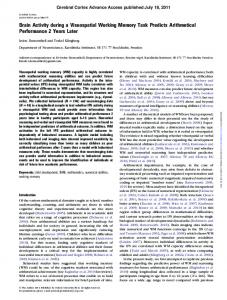
Results Subjects made only few errors in the different tasks. Errors in the visual short-term memory condition (mean 4.0) were significantly more frequent than errors in the phonological (mean 0.87) and updating (mean 2.25) working memory tasks [ANOVA, F(2,14) = 15.8, P = 0.0003]. When the phonological short-term memory task was compared with the visual task (Table 1, Fig. 1), significant activation foci appeared in left premotor frontal gyrus and adjacent Broca's area (BA 6/44), in the left superior temporal gyrus (BA 22/42) and in left and right insula. There was also activation in the inferior part of the left inferior parietal gyrus (BA 40), near the superior temporal gyrus. Increases of rCBF were observed bilaterally in primary sensorimotor areas of mouth and larynx. Moreover, we tentatively report foci of activation in the anterior cingulate gyrus (BA 24), in the medial part of the frontal pole (BA 10), and in the left parahippocampal region. When working memory updating was compared with the phonological short-term memory task (Table 2, Fig. 1), foci of increased rCBF were observed in the right middle frontal gyrus (dorsolateral area, BA 9) and to a lesser extent in the
Downloaded from http://brain.oxfordjournals.org/ by guest on September 2, 2012
Image analysis was performed on a SPARC workstation (Sun Microsystems Inc., Surrey, UK) using statistical parametric mapping (SPM) software 94 (MRC Cyclotron Unit, London, UK; Frackowiak and Friston, 1994) implemented in MATLAB (Mathworks Inc., Sherborn, Mass., USA). Each reconstructed rCBF scan consisting of 31 primary transverse planes was interpolated to 43 planes to render the voxels isotropic. The six acquisitions from each subject were realigned using the first as reference (Woods et al, 1992). The data were then transformed into a standard stereotactic space (Talairach and Tournoux, 1988). A Gaussian filter (16 mm full width at half maximum) was applied to smooth each image to accommodate inter-subject differences in gyral and functional anatomy and to suppress high frequency noise in the images. Such transformation of the data allows for pixel-by-pixel averaging of data across subjects and for direct cross-reference to the anatomical features in the standard stereotactic atlas. Due to variations of initial head positioning, resultant images obtained in all subjects extended from 28 mm below to 44 mm above the intercommissural plane.
Table 1
Downloaded from http://brain.oxfordjournals.org/ by guest on September 2, 2012
VERBAL (versus visual)
UPDATING (versus verbal)
VISUAL (versus verbal)
(P