www.nature.com/scientificreports
OPEN
received: 03 May 2016 accepted: 24 August 2016 Published: 20 September 2016
Fully Automatic System for Accurate Localisation and Analysis of Cephalometric Landmarks in Lateral Cephalograms Claudia Lindner1, Ching-Wei Wang2,3, Cheng-Ta Huang2,3,4, Chung-Hsing Li5,6, Sheng-Wei Chang5,6 & Tim F. Cootes1 Cephalometric tracing is a standard analysis tool for orthodontic diagnosis and treatment planning. The aim of this study was to develop and validate a fully automatic landmark annotation (FALA) system for finding cephalometric landmarks in lateral cephalograms and its application to the classification of skeletal malformations. Digital cephalograms of 400 subjects (age range: 7–76 years) were available. All cephalograms had been manually traced by two experienced orthodontists with 19 cephalometric landmarks, and eight clinical parameters had been calculated for each subject. A FALA system to locate the 19 landmarks in lateral cephalograms was developed. The system was evaluated via comparison to the manual tracings, and the automatically located landmarks were used for classification of the clinical parameters. The system achieved an average point-to-point error of 1.2 mm, and 84.7% of landmarks were located within the clinically accepted precision range of 2.0 mm. The automatic landmark localisation performance was within the inter-observer variability between two clinical experts. The automatic classification achieved an average classification accuracy of 83.4% which was comparable to an experienced orthodontist. The FALA system rapidly and accurately locates and analyses cephalometric landmarks in lateral cephalograms, and has the potential to significantly improve the clinical work flow in orthodontic treatment. Cephalometric radiography is commonly used as a standard tool in orthodontic diagnosis and treatment planning as well as in corrective and plastic surgery planning. Marking of anatomical landmarks of the skull and surrounding soft tissue in lateral cephalograms is an essential part of the diagnosis and planning process. Cephalometric landmarks are used for a number of orthodontic analyses (e.g. Schwarz, Steiner, Ricketts) where several linear and angular measurements are calculated from their positions1. The accuracy with which the landmarks are located has a direct impact on the results of the performed analyses and resulting treatment decisions. Identifying cephalometric landmarks in lateral cephalograms is a challenging problem. The skull is a highly complex 3D object which in a cephalogram is projected onto a single 2D plane, leading to overlapping structures. Furthermore, facial asymmetry, head positioning variations during image acquisition, and radiographic distortion cause the left and right outlines to not be perfectly superposed, leading to duplicated structures. This, combined with individual anatomical variation, in particular in pathological cases, makes it very difficult to reliably position cephalometric landmarks2. Currently in clinical practice, cephalometric landmark positions are identified manually or semi-automatically which is very tedious, time-consuming and prone to inconsistencies within and across orthodontists. Varying levels of orthodontic training and experience may have an impact on inter-observer variations, and time-constraints and other commitments may have an impact on intra-observer consistency3. 1
Centre for Imaging Sciences, The University of Manchester, Oxford Road, M13 9PT Manchester, United Kingdom. Graduate Institute of Biomedical Engineering, National Taiwan University of Science and Technology, Taiwan. 3 NTUST Centre of Computer Vision and Medical Imaging, National Taiwan University of Science and Technology, Taiwan. 4Department of Information Management, Oriental Institute of Technology Taiwan. 5Orthodontics and Dentofacial Orthopedics Division, Dental Department, Tri-Service General Hospital, Taiwan. 6School of Dentistry & Graduated Institute of Dental Science, National Defense Medical Center, Taiwan. Correspondence and requests for materials should be addressed to C.-W.W. (email:
[email protected]) 2
Scientific Reports | 6:33581 | DOI: 10.1038/srep33581
1
www.nature.com/scientificreports/
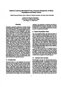
Figure 1. Cephalogram annotation example showing the 19 landmark positions used in this study. A description of all landmarks is given in Supplementary Table S2.
A computerised system for automatic cephalometric landmark identification would help to overcome time-constraints and inconsistencies within and across observers. Furthermore, given the landmark positions, in today’s orthodontic software packages the landmark-based linear and angular measurements are automatically calculated. Therefore, if a computerised system was able to locate cephalometric landmarks with sufficient accuracy then this would have the potential to significantly improve the clinical work flow in orthodontic treatment. Over the last three decades, several attempts had been made to create such a computerised system4–11. However, due to the complexity of this problem, the developed methods were unable to compete with manual landmark identification. In recent years, a number of IEEE International Symposium on Biomedical Imaging (ISBI) Grand Challenges were organised on this topic to encourage the development of better algorithms. The results are summarised in refs 12 and 13 and provide a benchmark for any future development. Preliminary results of our approach were presented at the 2015 ISBI Grand Challenge in Dental X-ray Image Analysis, where our system was awarded the first prize13,14. Supplementary Table S1 gives a quantitative comparison of all seven techniques that were submitted to the 2014 and 2015 ISBI Grand Challenges when tested on the same dataset of 100 images. This shows that our methodology achieved an average point-to-point error of 1.66 mm compared to errors ranging from 1.85 mm to 2.85 mm for all other techniques, demonstrating that our method performed significantly better than any of the other six techniques (p 5.7°
83.2°
>80.5°
0.75
>31.4°
=0 mm
C3
78.7°