atrioventricular pathway which was used as the retrograde limb of two circus movement tachycar- dias. Tachycardia No.1, reflecting anterograde fast pathway ...
Br Heart J 1982; 48: 75-7
Slowing of paroxysmal tachycardia with loss of functional bundle-branch block ROBERT A BAUERNFEIND, BORIS STRASBERG, KENNETH M ROSENt From the Section of Cardiology, Deparme ofMedicine, Abraham Lincoln School ofMedicine, University of Illinois CoUege of Medicine, Chicago, Illinois, USA SUMMARY Electrophysiological studies in a patient with paroxysmal supraventricular tachycardia disclosed anterograde dual atrioventricular nodal pathways, and a concealed left-sided anomalous atrioventricular pathway which was used as the retrograde limb of two circus movement tachycardias. Tachycardia No.1, reflecting anterograde fast pathway conduction, was characterised by functional left bundle-branch block and a stable cycle length of 330 ms. Paroxysmal loss of bundlebranch block resulted in tachycardia No.2, which reflected anterograde slow pathway conduction, and was characterised by narrow QRS and a stable cycle length of 355 ms. Tachycardia No.2 had a longer cycle length than tachycardia No.1 because the increment in AH interval (slow pathway instead of fast pathway conduction) more than compensated for the decrement in ventriculoatrial interval (narrow QRS instead of bundle-branch block).
valve prolapse and recurrent paroxysmal supraventricular tachycardia. After informed written consent was obtained, an electrophysiological study was performed, using standard techniques and definitions.1 Recordings during sinus rhythm (rate 67 beats/min) disclosed a short AH interval (41 ms, normal 54 to 130) and normal PA and HV intervals (36 and 52 ms, respectively). Atrial pacing from multiple sites failed to show evidence of ventricular pre-excitation. Extrastimulus testing from the high right atrium (driven cycle length 500 ms) showed discontinuous Al-A2, A2-H2 and Al-A2, H,-H2 conduction curves, suggesting the presence of anterograde dual (fast and slow) atrioventricular nodal pathways.4 The fast pathway was characterised by A2-H2 intervals of 60 to 100 ms and an effective refractory period of 380 ms, whereas the slow pathway was characterised by A2-H2 intervals of 165 to 260 ms and an effective refractory period of 270 ms. Extrastimuli which conducted over the slow pathway with an A2-H2 interval of 210 ms or longer resulted in induction of tachycardia No.2 (see below). Incremental pacing from the high atrium at rates of 100 to 130 beats/min disclosed AH intervals of 60 to 90 ms. At an atrial paced rate of 140 beats/min, two sets of stable AH intervals (100 to 220 ms) were observed, again suggesting the presence of antero-
The mechanism of paroxysmal supraventricular tachycardia can usually be elucidated by detailed cardiac electrophysiological studies. I An important diagnostic clue is frequently provided by paroxysmal loss of functional bundle-branch block during induced supraventricular tachycardia. When the loss of bundle-branch block results in a decrement in ventriculoatrial interval, a diagnosis of atrioventricular re-entry using an anomalous atrioventricular pathway ipsilateral to the blocked bundle-branch is strongly suggested.2 The decrement in ventriculoatrial interval is usually reflected by a decrement in the cycle length of tachycardia.3 The decrement in cycle length, however, may be relatively small, because an increment in AH interval may partially compensate for the decrement in ventriculoatrial interval.2 We report a patient in whom paroxysmal loss of functional bundle-branch block during induced supraventricular tachycardia resulted in an increment in cycle length of tachycardia. The mechanism of this unusual observation is discussed. Case report The patient was a 33-year-old woman with mitral Supported in part by a NHLBI Institutional Training grant, and research grants. tDr Rosen died after the paper was accepted.
75
Bauernfeind, Strasberg, Rosen
76
grade dual atrioventricular nodal pathways. At atrial tachycardia reflected a circus movement using the paced rates of 150 to 170 beats/min, AH intervals anomalous pathway as the retrograde limb. I The AH were 220 to 270 ms. Termination of atrial pacing dur- interval was relatively long (130 ms), suggesting that ing conduction over the slow pathway resulted in the anterograde limb of the circus movement was the induction of tachycardia No.2 (see below). slow atrioventricular nodal pathway (see Discussion). Ventricular incremental pacing, at rates of 100 to Tachycardia No. 1 (induced only once, by ventricu240 beats/min, showed intact ventriculoatrial conduc- lar pacing) was characterised by functional left tion with a fixed ventriculoatrial conduction time and bundle-branch block and a stable cycle length of 330 an eccentric sequence of retrograde atrial activation ms. The sequence of atrial activation during this (left atrium depolarised first). These findings sug- tachycardia was identical to the sequence observed gested the presence of a concealed left-sided anomal- during tachycardia No.2, suggesting that tachycardia ous atrioventricular pathway.' Termination of ven- No. 1 also reflected a circus movement which used the tricular pacing at a rate of 240 beats/min resulted in anomalous pathway as the retrograde limb. During induction of tachycardia No.1 (see below). this tachycardia, the AH interval was very short (30 ms), suggesting that the anterograde limb of the cirINDUCED SUPRAVENTRICULAR TACHYCARDIAS cus movement was the fast atrioventricular nodal Two distinct tachycardias were induced by program- pathway (see Discussion). med stimulation. Tachycardia No.2 (induced many During tachycardia No. 1 paroxysmal loss of functimes by atrial stimulation) was always characterised tional left bundle-branch block resulted in tachycarby narrow QRS complexes and a stable cycle length of dia No.2 (Fig.). This loss of bundle-branch block 355 ms. During this tachycardia, the sequence of caused shortening of the intervals from onset of QRS atrial activation was eccentric (distal coronary sinus, complexes to each of the atrial electrograms by 75 ms proximal coronary sinus, high right atrial, and low (from 160 to 85 ms for the distal coronary sinus elecseptal right atrial electrograms followed the onset of trogram). The cycle length of tachycardia, however, QRS complexes by 85, 105, 150, and 175 ms, respec- did not shorten but lengthened by 25 ms. The lengtively), and the atria could be reset by ventricular thening of the tachycardia cycle length reflected an extrastimuli introduced when the His bundle was increment in AH interval of 100 ms (from 30 to 130 being depolarised anterogradely, suggesting that this ms) which more than compensated for the decrement 3
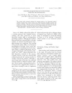
CL'35
ms
I
VI
I
I I I I I I I I I I I I
A
AJ
I I I I
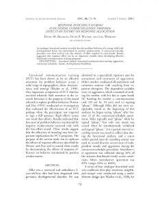
Fig. Paroxysmal loss offunctional left bwmdle-branch block during atnoventricular re-entrant tachycardia. Ekrocardiographic leads I and VI, as wel as high right atrial (HRA), His bundle (HBE), and distal coronary sinus (DCS) electrograms, are shown. AH intervals are shown below HBE. Time lines (bottom) are at 200 ms interals. Thefirstfour beats of re-entrant tachycardia are conducted with left bundle-branch block and have cycle lngth (CL) 330 ms. Note that the sequence ofretrograde atrial activation is eccentic, proceeding fnom DCS to HRA and then to the low septal right atrion (HBE), and dtat the AH iterval is 30 ms (fast atrioventiuar nodal pathway conduction). At thefifth beat there is sudden loss ofleft bundle-branch block, resulting in shortenig of the vencuatrial internal. The tachardia continues, with the last five beats ofparoxysmal supravtricular tachycardia being conducted normaly and having CL 355 me. Note that the sequence of retograde atrial activaium is unchanged, but that the AH inral is 130 ms (slow atioventrcular nodal pathway conduction).
Slowing ofparoxysmal tachycardia in ventriculoatrial interval. The HV interval ms during each of the tachycardias.
was
50
Discussion
Occasional patients have dual atrioventricular pathcoexisting with anomalous atrioventricular pathways.56 These patients have the potential for sevways
eral distinct circus
movement
tachycardias.56 Our
77 not given. We suspect that their patient had anterograde dual atrioventricular nodal pathways, which were not shown during atrial extrastimulus testing because the fast pathway effective refractory period was shorter than the atrial functional refractory period at the cycle length employed. Our observations provide an additional surface electrocardiographic clue to the diagnosis of a specific mechanism of paroxysmal supraventricular tachycardia. Slowing of tachycardia with loss of functional bundle-branch block (or speeding of tachycardia with the occurrence of functional bundle-branch block) suggests the possibility of anterograde dual atrioventricular nodal pathways coexisting with an anomalous atrioventricular pathway ipsilateral to the site of functional bundle-branch block.
patient had anterograde dual atrioventricular nodal pathways, and a concealed left-sided anomalous atrioventricular pathway which was used as the retrograde limb of two circus movements. One circus movement used the slow atrioventricular nodal pathway (AH intervals of 130 ms) as the anterograde limb, and was conducted with normal QRS complexes. The other circus movement used the fast atrioventricular nodal pathway (AH intervals of 30 ms) as the anterograde limb, and was conducted with left bundle- References branch block. Fast and slow pathway AH intervals 1 Wu D, Denes P, Amat-y-Leon F, et al. Clinical, elecwere shorter during these circus movements than durtrocardiographic and electrophysiologic observations in ing high right atrial pacing. This probably reflected patients with supraventricular tachycardia. Am J Cardiol the fact that during these circus movements input to 1978; 41: 1045-51. the atrioventricular node was from the left atrium, as 2 Pritchett ELC, Tonkin AM, Dugan FA, Wallace AG, Gallagher JJ. Ventriculoatrial conduction time during AH intervals are significantly shorter during coronary reciprocating tachycardia with intermittent l?undlesinus pacing than during high right pacing at equivalbranch block in Wolff-Parkinson-White syndrome. Br ent rates.7 HeartJ 1976; 38: 1058-64. Our patient presented an almost unique finding. 3 Spurrell RAJ, Krikler DM, Sowton E. Retrograde invaDuring induced atrioventricular re-entrant tachycarsion of the bundle branches producing aberration of the dia, paroxysmal loss of functional bundle-branch QRS complex during supraventricular tachycardia block resulted in sudden lengthening of the cycle studied by programmed electrical stimulation. Circulalength of tachycardia. The explanation for this finding ion 1974; 50: 487-95. is as follows. When the functional bundle-branch 4 Denes P, Wu D, Dhingra RC, Chuquimia R, Rosen block was lost, the ventriculoatrial interval shortened, KM. Demonstration of dual A-V nodal pathways in resulting in early atrial depolarisation and early patients with paroxysmal supraventricular tachycardia. Circulation 1973; 48: 549-55. anterograde input into the atrioventricular node. This S Spurrell RAJ, Krikler D, Sowton E. Two or more intra early input found the fast atrioventricular nodal A-V nodal pathways in association with either a James or pathway refractory, and conducted anterogradely Kent extranodal bypass in 3 patients with paroxysmal over the slow atrioventricular nodal pathway. Subsesupraventricular tachycardia. Br Heart J 1973; 35: 113quent beats of the narrow QRS tachycardia also con22. ducted over the slow pathway, probably because of 6 Amat-y-Leon F, Wyndham C, Wu D, Denes P, Dhingra retrograde concealment into the fast pathway. The RC, Rosen KM. Participation of fast and slow A-V nodal cycle length of tachycardia increased because the pathways in tachycardia complicating the WolffParkinson-White syndrome. Circulation 1977; 55: 663-8. increment in AH interval (slow pathway instead of 7 Amat-y-Leon F, Denes P, Wu D, Pietras RJ, Rosen fast pathway conduction) more than compensated for KM. Effects of atrial pacing site on atrial and atrioventhe decrement in ventriculoatrial interval (narrow tricular nodal function. Br Heart J 1975; 37: 576-82. QRS instead of left bundle-branch block). 8 Akhtar M, Damato AN, Ruskin JN, et. al. Antegrade We are aware of only one patient similar to ours. and retrograde conduction characteristics in three patAkhtar and co-workers8 reported a patient with terns of paroxysmal atrioventricular junctional reentrant induced atrioventricular re-entrant tachycardia, durtachycardia. Am Heart J 1978; 95: 22-42. ing which loss of functional right bundle-branch block resulted in a 20 ms increase in cycle length of tachycardia, because the AH interval increased by 65 Requests for reprints to Dr Robert A Bauernfeind, Cardiology Section, University of Illonois Hospital, ms whereas the ventriculoatrial interval decreased by only 45 ms. An explanation for this observation was PO Box 6998, Chicago, Illinois 60680, USA.