we show that the kinase domain of Fyn is sufficient for association with MomT ... We further demonstrate that a Fyn mutant lacking the SH2 domain is able to bind ...
JOURNAL OF VIROLOGY, Jan. 1997, p. 199–206 0022-538X/97/$04.0010 Copyright q 1997, American Society for Microbiology
Vol. 71, No. 1
Functional Interaction between the SH2 Domain of Fyn and Tyrosine 324 of Hamster Polyomavirus Middle-T Antigen NICOLAS M. DUNANT, ANJA S. MESSERSCHMITT,
AND
KURT BALLMER-HOFER*
Friedrich Miescher Institute, CH-4002 Basel, Switzerland Received 4 June 1996/Accepted 1 October 1996
Middle-T antigen of mouse polyomavirus (MomT) associates with the cellular tyrosine kinases c-Src, c-Yes, and Fyn, while middle-T antigen of hamster polyomavirus (HamT) exclusively binds Fyn. This interaction is essential for polyomavirus-mediated transformation of cells in culture and tumor formation in animals. Here we show that the kinase domain of Fyn is sufficient for association with MomT but not for binding of HamT. We further demonstrate that a Fyn mutant lacking the SH2 domain is able to bind MomT but fails to associate with HamT, indicating that the SH2 domain of Fyn is essential for stable association with HamT. HamT, but not MomT, contains a tyrosine residue, Tyr-324, in the sequence context YEEI. Mutation of Tyr-324 to phenylalanine led to a drastic reduction of associated Fyn and abolished the oncogenicity of HamT. This suggests that Tyr-324 is the major phosphotyrosine residue mediating the binding of HamT to the SH2 domain of Fyn. These findings show that mouse and hamster polyomaviruses use different strategies to target Srcrelated tyrosine kinases. carboxy-terminal 30 amino acids, which contain a putative transmembrane sequence. Middle-T proteins of both polyomaviruses have been shown to associate with and thereby change the activities of several cellular proteins that play a key role in the growth regulation of cells, including phosphatase 2A (8, 31, 47), Src-related tyrosine kinases (reviewed in reference 12), PI 3-kinase (8, 48), phospholipase C-g1 (2, 42), and 14-3-3 proteins (30). MomT was also shown to bind the adapter protein SHC (3, 13), thus activating the Ras pathway (46). It is not clear whether HamT also binds SHC, since it lacks the corresponding binding site present in MomT (NPTY motif). The transforming potential of MomT was shown to strictly depend on its associated tyrosine kinase activity (reviewed in reference 12). The tyrosine kinases binding to MomT are c-Src (9) and c-Yes (23), and, to a lesser extent, Fyn (6, 24). While c-Src and c-Yes are dramatically activated upon association with MomT, Fyn activity is not increased. In contrast to MomT, HamT has been shown to bind exclusively Fyn, which is activated about twofold (8). It has been proposed that the tumor profile of hamster polyomavirus could, at least in part, be dictated by the specific activation of Fyn, since this kinase is expressed at high levels in lymphocytes and is involved in signal transduction from the T-cell receptor (8). Src-related tyrosine kinases carry acylation signals close to the amino terminus (reviewed in reference 35) followed by a unique domain which is not conserved among Src family kinases and SH3 and SH2 domains, regions of homology for all Src-related tyrosine kinases (Fig. 1). The carboxy-terminal half comprises the catalytic or kinase (SH1) domain, which is highly conserved within the family, as well as a carboxy-terminal regulatory sequence, the kinase tail, which contains a conserved tyrosine residue, Tyr-527 in c-Src, that represses kinase activity (7) upon phosphorylation by a regulatory tyrosine kinase, Csk (29). Negative regulation of kinase activity by the tail is thought to result from intramolecular association of phosphorylated Tyr-527 with the SH2 domain (25, 37). The SH3 domain seems to cooperate with the SH2 domain in repressing kinase activity (43). SH2 and SH3 domains were also reported to mediate intermolecular interactions of tyrosine kinases with various proteins such as growth factor receptors or effectors
Polyomaviruses are oncogenic DNA viruses of the papovavirus family. Two polyomaviruses, one from the mouse and another from the hamster, are known. While mouse polyomavirus causes a wide variety of tumors (carcinomas, epitheliomas, and hemangiomas but never lymphomas and leukemias) in newborn mice (22), hamster polyomavirus was originally isolated from hair follicle tumors of hamsters and then shown to induce lymphomas and leukemias upon infection of newborn hamsters (17). Polyomaviruses encode three proteins in the early region of their genome, the so-called tumor antigens (T antigens), large-, middle-, and small-T antigens, which are translated from alternatively spliced forms of the primary transcript. Mouse and hamster polyomaviruses are the only two papovaviruses known to encode a middle-T protein. The T antigens are responsible for the oncogenic properties of these viruses (11, 44). While large-T antigen is able to immortalize primary rodent fibroblasts, both middle-T and large-T antigens are necessary for induction of a fully transformed phenotype and small-T antigen potentiates the efficiency of transformation (reviewed in reference 10). Middle-T antigen of mouse polyomavirus (MomT) is sufficient for transformation of established cells (45), while middle-T antigen of hamster polyomavirus (HamT) (8) was shown to transform established F111 rat cells only in the presence of small-T antigen (16). The middle-T proteins of mouse and hamster polyomaviruses have an overall sequence identity of 41% (8). Regions of higher homology are clustered along the sequence. The first 79 amino acids of middle-T antigen are shared by all three T antigens. This region shows 48% identity between MomT and HamT. Amino acids 80 to 192 are common to middle-T and small-T antigens and show 59% identity between the two species, while the rest of the sequence, which is unique to middle-T antigen, shows only 22% identity. The homologies in the unique region are located in two sequence stretches: (i) the sequence surrounding tyrosine 315 (MomT numbering), the phosphatidylinositol (PI) 3-kinase binding site; and (ii) the
* Corresponding author. Mailing address: Friedrich Miescher Institute, P.O. Box 2543, CH-4002 Basel, Switzerland. Phone: 61 697 6689. Fax: 61 697 3976. E-mail: ballmer#@fmi.ch. 199
200
DUNANT ET AL.
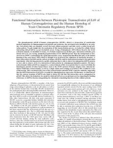
FIG. 1. Domain structures of Fyn constructs and middle-T antigens. (A) Fyn deletion mutants. The approximate epitopes for the antibodies (anti-FYN3, MAb 12CA5, and anti-SRC2) are indicated. 1, association; 2, no association; wt, wild type. (B) Middle-T antigens of mouse and hamster polyomaviruses. Conserved regions (amino-terminal half and membrane anchor) are shaded. The amino acid sequence around tyrosine 324 is indicated in one-letter code.
involved in downstream signalling (reviewed in reference 32). While SH2 domains recognize specific peptide sequences containing a phosphorylated tyrosine residue (40), SH3 domains bind to proline-rich sequences (34). We have shown previously that the kinase domain of Srcrelated tyrosine kinases was sufficient for both association with and phosphorylation of MomT (15). Here we show that the SH2 domain of Fyn is required for association with HamT. This protein carries a binding motif specific for Src family SH2 domains that is not present in MomT. This demonstrates that MomT and HamT target Src-related tyrosine kinases differently. MATERIALS AND METHODS Plasmid constructs. Construction of the various plasmids was carried out by using standard molecular cloning techniques. The fyn cDNA (38) was subcloned as an EcoRI fragment into a pSP72 vector (Promega). The pSP72fyn plasmid was used as template for PCRs. The constructs encoding DSH2Fyn, DSH3Fyn, and DUFyn were generated by the method of splicing by overlap extension-PCR (18, 19). The sequence encoding K-Fyn (starting at codon 262 of fyn cDNA) was amplified from pSP72fyn by PCR. The primer used introduced a HindIII site and a novel start codon and fused a nucleotide sequence specifying the 9-amino-acid peptide YPYDVPDYA derived from the influenza virus hemagglutinin (HA) protein (HA tag) together with a sequence encoding an 11-amino-acid linker peptide KLMGLVVMNIT in frame to the 59 end of the truncated fyn coding sequence. PCR products were ligated as HindIII/ClaI fragments into a pSV expression vector (28), yielding the plasmids pSVDSH2fyn, pSVDSH3fyn,
J. VIROL. pSVDUfyn, and pSVk-fyn. The vector pSVfyn carries the wild-type fyn cDNA. pSVsT is an expression vector for mouse polyomavirus small-T antigen under the control of the simian virus 40 early promoter. pcDNAmT is an expression vector derived from pcDNA1Neo (Invitrogen) carrying the cDNA sequence of MomT. pGHamT (obtained from J. Feunteun) is an expression vector for HamT under the control of the simian virus 40 early promoter (16). The mutant pGHamTY324F was constructed by splicing by overlap extension-PCR using primers changing the codon for Tyr-324 to a phenylalanine codon. All mutations were confirmed by restriction enzyme analysis and DNA sequencing. Antibodies. Wild-type Fyn, DSH2Fyn, and DSH3Fyn were immunoprecipitated with anti-FYN3, a rabbit polyclonal antiserum raised against a peptide corresponding to residues 29 to 48 in the unique domain of human Fyn (Santa Cruz Biotechnology). DUFyn was immunoprecipitated with anti-SRC2 (Santa Cruz Biotechnology), a rabbit polyclonal antibody raised against a peptide corresponding to residues 509 to 533, i.e., the carboxy terminus, of c-Src. Anti-SRC2 also recognizes the carboxy terminus of Fyn. The HA-tagged K-Fyn protein was detected with monoclonal antibody (MAb) 12CA5 (Boehringer). MomT was immunoprecipitated with monoclonal papovavirus protein antibody (PAb) 762 (27). HamT was immunoprecipitated with TBH serum, obtained from tumor-bearing hamsters infected with hamster polyomavirus (provided by J. Feunteun) (8), or with anti-GST/HamT, a rabbit serum raised against the fusion protein glutathione S-transferase (GST)-HamT (2), a gift from S. Courtneidge. Cell lines and DNA transfection. NIH 3T3 cells were maintained in Dulbecco’s modified Eagle’s medium (DMEM) supplemented with 10% calf serum at 378C in a 10% CO2 incubator. Src fibroblasts (provided by P. Soriano), a cell line derived from transgenic mice lacking both fyn alleles and expressing only an inactive fragment of c-Yes (41), were maintained in DMEM with 10% fetal calf serum. Transient transfections were performed by the Lipofectamine method. Twenty micrograms of plasmid DNA together with 60 ml of Lipofectamine (GIBCO) in 4 ml of Optimem 1 medium (GIBCO) was added to 106 NIH 3T3 cells or Src-fibroblasts plated on 10-cm-diameter dishes and incubated for 5 h. Four milliliters of DMEM containing 20% serum was added to the transfection mixture. Eighteen hours after transfection, cells were lysed for immunoprecipitation or used for metabolic labeling. Focus formation on F111 rat fibroblasts was performed as previously described (33). Metabolic labeling with Tran35S-Label. In vivo labeling with Tran35S-Label ([35S]methionine-[35S]cysteine; ICN Pharmaceuticals) was performed as follows. Eighteen hours after transfection, cells were washed with DMEM without methionine and cysteine followed by incubation for 6 h with 250 mCi of Tran35SLabel in 2.5 ml of DMEM lacking methionine and cysteine and supplemented with 1% serum. Immunoprecipitations and in vitro kinase assays. Cell lysis with buffer containing Nonidet P-40, immunoprecipitations using protein A-Sepharose CL-4B (Pharmacia Biotech), and in vitro kinase assays with [g-32P]ATP were performed as described previously (20). Immunoprecipitates were analyzed by sodium dodecyl sulfate (SDS)-polyacrylamide gel electrophoresis. Gels from kinase assays were treated with 1 M NaOH at 658C for 30 min prior to autoradiography. For reprecipitation, immunoprecipitates were boiled for 10 min in 2% SDS–5% 2-mercaptoethanol to disrupt the antibodies and centrifuged briefly. The supernatant was vacuum concentrated, dissolved in 20 times the original volume with Nonidet P-40 lysis buffer, and incubated overnight with the appropriate antisera. Immune complexes were harvested with protein A-Sepharose CL-4B and washed as described previously (20).
RESULTS Fyn associates with MomT and HamT. We tested the ability of wild-type Fyn to bind and phosphorylate MomT and HamT after transient transfection of expression vectors coding for Fyn, MomT, or HamT into Src fibroblasts, cells derived from a knockout mouse lacking Fyn and expressing only a truncated, inactive form of c-Yes (36). Lysates of cells expressing Fyn without or with MomT or HamT were used for immunoprecipitations with anti-FYN3 serum. Immunoprecipitates were phosphorylated in vitro and analyzed by SDS-polyacrylamide gel electrophoresis followed by autoradiography. Anti-FYN3 immunoprecipitates from cells expressing Fyn gave rise to a phosphorylated protein with a molecular mass of 60 kDa (Fig. 2, lanes 2), while anti-FYN3 immunoprecipitates from cells expressing Fyn together with MomT gave rise to labeled bands corresponding to molecular masses of 56, 60, and 85 kDa (Fig. 2A, lane 3). The 56-, 60-, and 85-kDa bands were analyzed by V8 protease mapping and were identified as MomT, Fyn, and the phosphorylated form of the p85 subunit of PI 3-kinase,
VOL. 71, 1997
HAMSTER POLYOMAVIRUS MIDDLE-T ASSOCIATION WITH Fyn
201
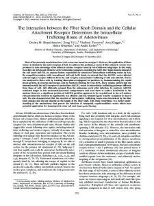
FIG. 2. The SH2 domain of Fyn is required for coprecipitation of HamT but not of MomT. Immunoprecipitations were performed with lysates from control (2), MomT-, and HamT-expressing Src fibroblasts transfected with expression vectors for Fyn, DSH2Fyn, and DSH3Fyn. (A) Complex formation of Fyn mutants with MomT. Immunoprecipitates prepared with anti-FYN3 from control cells (lane 1) or cells expressing Fyn (lane 2), DSH2-Fyn (lane 4), or DSH3-Fyn (lane 6) alone or together with MomT (lanes 3, 5, and 7) were phosphorylated in vitro with [g-32P]ATP and analyzed on SDS–10% polyacrylamide gels. Phosphorylated proteins were detected by autoradiography. Anti-FYN3 immunoprecipitates were also used for reprecipitation of MomT with PAb 762 (lanes 8 to 13). (B) Complex formation of Fyn mutants with HamT. Immunoprecipitates prepared with anti-FYN3 from control cells (lane 1) or cells expressing Fyn (lane 2), DSH2-Fyn (lane 4), or DSH3-Fyn (lane 6) alone or together with HamT (lanes 3, 5, and 7) were analyzed by the same procedure used for panel A. HamT was reprecipitated with anti-GST/HamT (lanes 8 to 13). Bars, bands corresponding to the respective kinase variants; arrows, bands corresponding to MomT or HamT; asterisks, the position of the p85 subunit of PI 3-kinase.
respectively, by comparison with published maps (4, 15, 21) (data not shown). MomT was reprecipitated with PAb 762 from anti-FYN3 immunoprecipitates prepared from cells expressing both Fyn and MomT (Fig. 2A, lane 9) but not from cells expressing only Fyn (Fig. 2A, lane 8). This finding is in agreement with the published observation that MomT is able to form a complex with Fyn (6, 24). Anti-FYN3 immunoprecipitates from cells expressing both Fyn and HamT gave rise to labeled bands corresponding to apparent molecular masses of about 60 and 85 kDa and to a weaker band corresponding to an apparent molecular mass of about 55 kDa (Fig. 2B, lane 3). The 85-kDa band was identified by V8 mapping as the p85 subunit of PI 3-kinase (data not shown). Coprecipitation of p85 with Fyn in cells expressing HamT has been shown previously to be indicative of complex formation between Fyn and HamT (8). The band corresponding to a molecular mass of about 55 kDa was identified as HamT by reprecipitation with anti-GST/HamT (Fig. 2B, lane 9). HamT was also directly immunoprecipitated with TBH serum, a serum obtained from tumor-bearing hamsters infected with hamster polyomavirus (8), or with anti-GST/HamT serum (2). Neither serum cross-reacts with Fyn, therefore al-
lowing the monitoring of complex formation of Fyn with HamT. Immunoprecipitates prepared with TBH or anti-GST/ HamT serum were used for kinase assays followed by autoradiography. Immunoprecipitates from cells expressing both HamT and Fyn gave rise to a prominent band of about 60 kDa (Fig. 3, lane 2) and to less intense bands of about 55 and 85 kDa which were detected upon longer exposure (Fig. 3, lane 8). The prominent 60-kDa band was shown to be Fyn by reprecipitation with anti-FYN3 serum (Fig. 3, lane 10) and by V8 protease mapping (data not shown). The intensity of the band corresponding to HamT was always significantly lower than that of the band representing Fyn. The kinase domain of Fyn is sufficient for binding MomT but not HamT. We have shown earlier that the kinase domain of c-Src is sufficient for binding and phosphorylation of MomT (15). This raised the question of whether the kinase domain of Fyn is also sufficient for binding and phosphorylation of HamT. We therefore constructed a mutant Fyn protein consisting of the kinase domain (residues 262 to 537) with an HA tag fused to the amino terminus. The ability of this mutant protein, K-Fyn (Fig. 1), to associate with MomT and HamT was tested by expression of K-Fyn alone or together with MomT or HamT
202
DUNANT ET AL.
J. VIROL.
FIG. 3. Coprecipitation of Fyn and DSH3Fyn but not DSH2Fyn with HamT. Immunoprecipitates prepared with TBH serum from cells expressing Fyn, DSH2Fyn, or DSH3Fyn alone (2; lanes 1, 3, and 5, respectively) or together with HamT (lanes 2, 4, and 6) were phosphorylated in vitro with [g-32P]ATP and analyzed on SDS–10% polyacrylamide gels. Phosphorylated proteins were detected by autoradiography. Lanes 7 and 8 correspond to lanes 1 and 2, but the exposure time was four times longer. The immunoprecipitates analyzed in lanes 1 to 6 were also used for reprecipitation of Fyn variants with anti-FYN3 (lanes 9 to 14). Bars, bands corresponding to the respective Fyn variants; asterisks, the position of the p85 subunit of PI 3-kinase.
in NIH 3T3 cells followed by immunoprecipitation with MAb 12CA5, a monoclonal antibody specific for the HA tag. Immunoprecipitates were phosphorylated in vitro with [g-32P]ATP and analyzed by SDS-polyacrylamide gel electrophoresis and autoradiography. Immunoprecipitates from cells expressing KFyn gave rise to a phosphoprotein of approximately 32 kDa, as
FIG. 4. MomT but not HamT is coprecipitated with the kinase domain of Fyn. Immunoprecipitates prepared with MAb 12CA5 by using lysates from NIH 3T3 cells expressing K-Fyn alone (2; lanes 1 and 3) or together with MomT (lane 2) or HamT (lane 4) were phosphorylated in vitro with [g-32P]ATP and analyzed on SDS–10% polyacrylamide gels. Phosphorylated proteins were detected by autoradiography. As a control for HamT expression, the lysate from cells expressing K-Fyn together with HamT was also used for immunoprecipitation with anti-FYN3 serum and analyzed as described above (lane 5). Bars, bands corresponding to the expected phosphoprotein; arrows, bands corresponding to MomT or HamT; asterisk, the position of the p85 subunit of PI 3-kinase.
expected from the calculated molecular mass (Fig. 4, lanes 1 and 3). MomT was coprecipitated with K-Fyn and phosphorylated (Fig. 4, lane 2). In contrast, no coprecipitation of HamT with K-Fyn was observed in NIH 3T3 cells expressing both K-Fyn and HamT (Fig. 4, lane 4). Expression of HamT in these NIH 3T3 cells was confirmed by the fact that HamT and p85 were coprecipitated with endogenous wild-type Fyn by using anti-FYN3 serum (Fig. 4, lane 5). The SH2 domain of Fyn is required for association with HamT. The fact that K-Fyn did not bind HamT suggests that sequences located in the amino-terminal half of Fyn are required for association with HamT. HamT contains a tyrosine residue (Tyr-324) in the sequence context YEEI that corresponds to a phosphopeptide shown to specifically bind the SH2 domain of Src-related tyrosine kinases in vitro (40). This observation led us to ask whether HamT binds to the SH2 domain of Fyn. To test this hypothesis, we constructed a Fyn deletion mutant lacking the SH2 domain (residues 144 to 248), DSH2Fyn (Fig. 1). The ability of DSH2Fyn to bind MomT or HamT was evaluated by coexpression of DSH2Fyn with MomT or HamT in Src fibroblasts lacking endogenous Fyn. Immunoprecipitates prepared with anti-FYN3 serum showed that DSH2Fyn migrated as a band with an apparent molecular mass of 49 kDa, as expected (Fig. 2, lanes 4). While MomT and p85 were coprecipitated efficiently with DSH2Fyn by using anti-FYN3 (Fig. 2A, lane 5), no phosphorylated HamT or p85 was precipitated together with DSH2Fyn from cells expressing this mutant and HamT (Fig. 2B, lane 5). No HamT protein could be reprecipitated with anti-GST/HamT serum from anti-FYN3 immunoprecipitates, indicating that HamT did not bind DSH2Fyn (Fig. 2B, lane 11). These findings were confirmed by immunoprecipitation with TBH serum. TBH serum immunoprecipitates from Src fibroblasts expressing both Fyn and HamT gave rise to a prominent band of 60 kDa corresponding to Fyn (Fig. 3, lane 2). In contrast, TBH serum immunoprecipitates from cells expressing DSH2Fyn together with HamT did not contain DSH2Fyn (Fig. 3, lane 4). While wild-type Fyn was reprecipitated with anti-FYN3 serum from immunoprecipitates prepared with
VOL. 71, 1997
HAMSTER POLYOMAVIRUS MIDDLE-T ASSOCIATION WITH Fyn
FIG. 5. Reduced association of Fyn with Y324F HamT. Immunoprecipitates prepared from equal amounts of Src fibroblasts expressing HamT and Y324F HamT alone (2; lanes 2 and 4, respectively) or together with Fyn (lanes 3 and 5, respectively) or from control cells (lane 1) with anti-GST/HamT serum were phosphorylated in vitro with [g-32P]ATP and analyzed on SDS–10% polyacrylamide gels. Equal amounts of labeled immunoprecipitates were used for reprecipitation of Fyn with anti-FYN3 (lanes 6 to 10). Src fibroblasts expressing HamT and Y324F HamT alone (2; lanes 12 and 14, respectively) or together with Fyn (lanes 13 and 15, respectively) or control cells (lane 11) were metabolically labeled with Tran35S-Label (35S-Met/-Cys) and used for immunoprecipitation with anti-GST/HamT serum to determine the relative expression levels of HamT and Y324F HamT. The same lysates from metabolically labeled cells were used for immunoprecipitation with anti-FYN3 to determine the level of Fyn expression (lanes 16 to 20). Asterisk, the position of the p85 subunit of PI 3-kinase.
TBH serum (Fig. 3, lane 10), no DSH2Fyn was reprecipitated (Fig. 3, lane 12), indicating that this mutant failed to associate stably with HamT. HamT expression was assayed by in vivo labeling with Tran35S-Label followed by immunoprecipitation with anti-GST/HamT, showing that the expression levels of HamT protein in cells expressing wild-type Fyn and DSH2Fyn were similar (data not shown). Mutation of Tyr-324 of HamT to phenylalanine leads to reduced association of Fyn and abolishes oncogenic transformation by HamT. To test whether phosphorylated Tyr-324 of HamT is necessary for association of HamT with Fyn in vivo, we mutated this residue to phenylalanine. Src fibroblasts were transfected with pGHamT or pGHamTY324F, an expression vector carrying the Y324F HamT mutant, alone or together with pSVfyn. HamT and Y324F HamT were expressed at comparable levels, as shown by immunoprecipitation from metabolically labeled cells (Fig. 5, lanes 12 to 15). In vitro phosphorylation of Y324F HamT coexpressed with Fyn (Fig. 5, lane 5) was reduced when compared with that of wild-type HamT (lane 3), although the levels of Fyn expression in cells expressing wild-type and mutant HamT were similar (lanes 18 and 20). The amount of Fyn coprecipitated with Y324F HamT (Fig. 5,
203
lane 5) was about 30% of that coprecipitated with wild-type HamT (lane 3). This result was confirmed by reprecipitation of Fyn with anti-FYN3 serum from in vitro-phosphorylated antiGST/HamT immunoprecipitates prepared from cells expressing HamT and Y324F HamT (Fig. 5, lanes 8 and 10, respectively). Our findings show that Tyr-324 is a major determinant for association of HamT with Fyn. However, the fact that association of Fyn with Y324F HamT was not completely abolished indicates that other sequence elements may also be involved in mediating specific association with Fyn. The ability of Y324F HamT to induce cell transformation was tested in focus formation assays with F111 rat fibroblasts. Expression vectors for Y324F HamT and, as controls, HamT and MomT were transfected alone or together with pSVsT, a vector encoding small-T antigen, into F111 rat fibroblasts, and focus formation on F111 cell monolayers was assayed under low-serum conditions (Fig. 6). As described before, the presence of small-T antigen was necessary for efficient focus formation by HamT (16). This is in contrast to MomT, which also formed foci in the absence of small-T antigen. Foci formed by HamT in the presence of small-T antigen were smaller than those induced by MomT. Y324F HamT failed to induce focus formation even in the presence of small-T antigen, showing that mutation of Tyr-324 abolished the oncogenicity of HamT. The SH3 domain and the unique domain of Fyn are dispensable for association with HamT. HamT contains several proline-rich motifs which are putative binding sites for SH3 domains (34). We therefore tested the ability of a Fyn mutant lacking the SH3 domain (residues 83 to 142), DSH3Fyn (Fig. 1), to bind MomT or HamT in Src fibroblasts by the approach described above. Moreover, the unique domain (residues 21 to 82) of Fyn was deleted from DUFyn (Fig. 1) and the ability of this mutant to bind HamT was evaluated. Both DSH3Fyn (Fig. 2B) and DUFyn (Fig. 7) were fully competent to associate with HamT, indicating that the SH3 domain and the unique domain are dispensable for association of Fyn with HamT. DISCUSSION MomT has been shown to associate with the tyrosine kinases c-Src, c-Yes, and Fyn (reviewed in reference 12). In contrast, HamT exclusively binds Fyn in fibroblasts (8). We have demonstrated earlier that the kinase domain of c-Src, including the regulatory tail, is sufficient for both binding and phosphorylation of MomT (15). Here we report that the kinase domain of the highly homologous tyrosine kinase Fyn is also sufficient for binding MomT but does not associate with HamT. This suggests that sequences located in the amino-terminal half of Fyn are essential for stable association with HamT. We therefore constructed a series of Fyn mutants lacking either the SH2, SH3, or unique domain, three domains known to mediate the association of Src family tyrosine kinases with various cellular proteins (32, 39). While association of DSH3Fyn or DUFyn with HamT was not reduced, DSH2Fyn failed to bind HamT, indicating that the SH2 domain of Fyn is necessary for association with HamT. In an earlier work, peptides containing a phosphotyrosine residue in the context YEEI were identified as ligands of Src family SH2 domains (40). HamT contains a tyrosine residue, Tyr-324, in the sequence context YEEI. We therefore argued that HamT might associate with the SH2 domain of Fyn via phosphorylated Tyr-324. In order to test this hypothesis, we mutated Tyr-324 of HamT to a nonphosphorylatable phenylalanine residue. This mutant, Y324F HamT, showed a threefold reduction of coprecipitated Fyn when analyzed in kinase assays. This allows two possible explanations.
204
DUNANT ET AL.
J. VIROL.
FIG. 6. Y324F HamT is transformation defective. F111 rat fibroblasts were transfected with vectors encoding HamT, Y324F HamT, and MomT alone or together with (1) a vector encoding small-T antigen (sT). Foci formed on the cell monolayer 3 weeks after transfection are shown. Each plate is representative of at least two independent experiments. 2, mock transfection.
(i) The amount of Fyn protein coprecipitated with mutant HamT was reduced as a consequence of a reduction in the binding affinity. (ii) Fyn might become less activated by Y324F HamT, resulting in a reduction in Fyn autophosphorylation. We cannot discriminate between these two possibilities, since we were not able to detect Fyn by coprecipitation with HamT in lysates from metabolically labeled cells or by immunoblotting, probably because the amount of Fyn protein complexed with HamT is very small. The residual association of Fyn with Y324F HamT indicates that other sequence elements besides phosphorylated Tyr-324 may contribute to specific binding to Fyn. HamT contains only one additional tyrosine residue in a sequence context with homology to YEEI, namely, Tyr-330 in the context YLEL. We constructed a double mutant of HamT with both Tyr-324 and Tyr-330 mutated to phenylalanine. The association of Fyn with this mutant was not further decreased (data not shown), indicating that Tyr-330 is not a binding site for Fyn. Therefore, it is unlikely that other phosphotyrosine residues take over the function of Tyr-324 in the Y324F HamT mutant. We consider it more probable that the residual binding of Fyn to Y324F HamT is phosphotyrosine independent. The fact that Y324F HamT is transformation defective shows that Tyr-324 is essential for the oncogenicity of HamT and that the residual binding of Fyn to Y324F HamT is not functional. Our data establish that MomT and HamT bind and activate Src-related tyrosine kinases through different mechanisms. Kinase-inactive c-Src is able to bind MomT, indicating that association with c-Src does not require tyrosine phosphorylation of MomT (5). In contrast, the observation that mutation of HamT at Tyr-324 to a nonphosphorylatable phenylalanine residue reduced association with Fyn suggests that this residue binds the SH2 domain of Fyn upon phosphorylation. In vivo, phosphorylation of HamT at Tyr-324 by Fyn followed by stable association with the SH2 domain might result in activation of the kinase through stabilization of the open conformation.
This concept is in agreement with the finding that cytosolic tyrosine kinases preferentially phosphorylate peptides capable of binding their own SH2 domains (49). One might predict that catalytically inactive Fyn fails to associate with HamT. However, we could not test this hypothesis, since complex formation between Fyn and HamT could be detected only by in vitro kinase assays. Due to the very low stoichiometry of phosphorylation of HamT, it was also not possible to show that Tyr-324 is phosphorylated in vivo. The fact that mouse and hamster polyomaviruses use different strategies to target Src-related tyrosine kinases with their respective middle-T proteins is surprising when one considers the close relatedness of the two viruses. Mouse and hamster polyomaviruses are the only papovaviruses known to encode a middle-T antigen. Their genome organization is highly conserved, and the viral proteins show an average homology of 50%. However, the unique regions of the two middle-T proteins, which are essential for downstream signalling, are strongly divergent. It has been shown that sequences in the amino-terminal half of MomT as well as residues 203 to 218 in the unique region of MomT are essential for association with c-Src. These conclusions were based on mutational analysis (1, 12, 26) and on the finding that MAbs recognizing the peptide sequence from residues 203 to 218 fail to coprecipitate tyrosine kinase activity (14). Residues 203 to 218 are not conserved in HamT. On the other hand, the YEEI motif of HamT, which we have shown to contain a tyrosine residue required for formation of a functional complex with Fyn, is not present in MomT. Taken together, these findings indicate that the two middle-T antigens may have evolved separately. What are the structural determinants responsible for the fact that HamT selectively associates with Fyn and not with highly homologous tyrosine kinases like c-Src or c-Yes? It is unlikely that the YEEI motif alone is sufficient for determining binding specificity, since in vitro this sequence binds the SH2 domains
VOL. 71, 1997
HAMSTER POLYOMAVIRUS MIDDLE-T ASSOCIATION WITH Fyn
205
FIG. 7. The unique domain of Fyn is dispensable for association with HamT. Immunoprecipitates were prepared with anti-SRC2 serum from lysates of cells expressing Fyn alone (2; lane 1) or together with HamT (lane 2) or DUFyn alone (lane 9) or together with HamT (lane 10). Alternatively, immunoprecipitates were prepared from the same lysates with TBH serum (lanes 5 and 6 for Fyn alone and together with HamT, respectively; lanes 13 and 14 for DUFyn alone and together with HamT, respectively). Immunoprecipitates were phosphorylated in vitro with [g-32P]ATP and analyzed on SDS–10% polyacrylamide gels. Phosphorylated proteins were detected by autoradiography. The immunoprecipitates analyzed in lanes 1, 2, 9, and 10 were also used for reprecipitation of HamT with anti-GST/HamT serum (lanes 3, 4, 11, and 12), and the immunoprecipitates analyzed in lanes 5, 6, 13, and 14 were used for reprecipitation of Fyn and DUFyn with anti-SRC2 (lanes 7, 8, 15, and 16). Arrows, the position of HamT; bars, bands corresponding to the respective Fyn variants; asterisks, the position of the p85 subunit of PI 3-kinase.
of different Src family members, such as c-Src, Fyn, Lck, and c-Fgr, with similar affinities (40). Therefore, other sequences in HamT might contribute to the specific binding of this protein to Fyn. Future experiments will be aimed at the identification of determinants of specificity in both HamT and Fyn.
3.
4.
ACKNOWLEDGMENTS N.M.D. and A.S.M. contributed equally to this work. We thank Sara Courtneidge, Redwood City, Calif., for anti-GST/ HamT; Stephen Dilworth, London, England, for PAb 762; Jean Feunteun, Villejuif, France, for TBH serum; and P. Soriano, Seattle, Wash., for Src fibroblasts. We are grateful to our colleagues Stefano Fumagalli, Brian Hemmings, and David Stover for critically reading the manuscript. N.M.D. was supported by a fellowship from Stipendienfonds der Basler Chemischen Industrie.
5.
6.
7.
REFERENCES
8.
1. Brizuela, L., L. M. Olcese, and S. A. Courtneidge. 1994. Transformation by middle T antigens. Semin. Virol. 5:381–389. 2. Brizuela, L., E. T. Ulug, M. A. Jones, and S. A. Courtneidge. 1995. Induction
9.
of interleukin-2 transcription by the hamster polyomavirus middle T antigen: a role for Fyn in T cell signal transduction. Eur. J. Immunol. 25:385–393. Campbell, K. S., E. Ogris, B. Burke, W. Su, K. R. Auger, B. J. Druker, B. S. Schaffhausen, T. M. Roberts, and D. C. Pallas. 1994. Polyoma middle tumor antigen interacts with SHC protein via the NPTY (Asn-Pro-Thr-Tyr) motif in middle tumor antigen. Proc. Natl. Acad. Sci. USA 91:6344–6348. Cheng, S. H., P. C. Espino, J. Marshall, R. Harvey, J. Merrill, and A. E. Smith. 1991. Structural elements that regulate pp59c-fyn catalytic activity, transforming potential, and ability to associate with polyomavirus middle-T antigen. J. Virol. 65:170–179. Cheng, S. H., P. C. Espino, J. Marshall, R. Harvey, and A. E. Smith. 1990. Stoichiometry of cellular and viral components in the polyomavirus middle-T antigen-tyrosine kinase complex. Mol. Cell. Biol. 10:5569–5574. Cheng, S. H., R. Harvey, P. C. Espino, K. Semba, T. Yamamoto, K. Toyoshima, and A. E. Smith. 1988. Peptide antibodies to the human c-fyn gene product demonstrate pp59c-fyn is capable of complex formation with the middle-T antigen of polyomavirus. EMBO J. 7:3845–3855. Cooper, J. A., K. L. Gould, C. A. Cartwright, and T. Hunter. 1986. Tyr527 is phosphorylated in pp60c-src: implications for regulation. Science 231:1431– 1434. Courtneidge, S. A., L. Goutebroze, A. Cartwright, A. Heber, S. Scherneck, and J. Feunteun. 1991. Identification and characterization of the hamster polyomavirus middle-T antigen. J. Virol. 65:3301–3308. Courtneidge, S. A., and A. E. Smith. 1983. Polyoma virus transforming
206
10. 11. 12. 13. 14. 15. 16. 17.
18. 19. 20. 21.
22. 23. 24. 25. 26. 27. 28. 29. 30.
DUNANT ET AL. protein associates with the product of the c-src cellular gene. Nature 303: 435–439. Cuzin, F. 1984. The polyoma virus oncogenes. Biochim. Biophys. Acta 781: 193–204. Delmas, V., C. Bastien, S. Scherneck, and J. Feunteun. 1985. A new member of the polyomavirus family: the hamster papovavirus complete nucleotide sequence and transformation properties. EMBO J. 4:1279–1286. Dilworth, S. M. 1995. Polyoma virus middle T antigen: meddler or mimic? Trends Microbiol. 3:31–35. Dilworth, S. M., C. E. Brewster, M. D. Jones, L. Lanfrancone, G. Pelicci, and P. G. Pelicci. 1994. Transformation by polyoma virus middle T-antigen involves the binding and tyrosine phosphorylation of Shc. Nature 367:87–90. Dilworth, S. M., and V. P. Horner. 1993. Novel monoclonal antibodies that differentiate between the binding of pp60c-src or protein phosphatase 2A by polyomavirus middle-T antigen. J. Virol. 67:2235–2244. Dunant, N. M., M. Senften, and K. Ballmer-Hofer. 1996. Polyomavirus middle-T antigen associates with the kinase domain of Src-related tyrosine kinases. J. Virol. 70:1323–1330. Goutebroze, L., and J. Feunteun. 1992. Transformation by hamster polyomavirus: identification and functional analysis of the early genes. J. Virol. 66:2495–2504. Graffi, A., E. Bender, T. Schramm, W. Kuhn, and F. Schneiders. 1969. Induction of transmissible lymphomas in Syrian hamsters by application of DNA from viral hamster papovavirus-induced tumors and by cell-free filtrates from human tumors. Med. Sci. 64:1172–1175. Ho, S. N., H. D. Hunt, R. M. Horton, J. K. Pullen, and L. R. Pease. 1989. Site-directed mutagenesis by overlap extension using the polymerase chain reaction. Gene 77:51–59. Horton, R. M., H. D. Hunt, S. N. Ho, J. K. Pullen, and L. R. Pease. 1989. Engineering hybrid genes without the use of restriction enzymes: gene splicing by overlap extension. Gene 77:61–68. Kaech, S., L. Covic, A. Wyss, and K. Ballmer-Hofer. 1991. Association of p60c-src with polyoma virus middle-T antigen abrogating mitosis-specific activation. Nature 350:431–433. Kaplan, D. R., M. Whitman, B. S. Schaffhausen, D. C. Pallas, M. White, L. Cantley, and T. M. Roberts. 1987. Common elements in growth factor stimulation and oncogenic transformation: 85 kd phosphoprotein and phosphatidylinositol kinase activity. Cell 50:1021–1029. Kiefer, F., S. A. Courtneidge, and E. F. Wagner. 1994. Oncogenic properties of the middle T antigens of polyomaviruses. Adv. Cancer Res. 64:125–157. Kornbluth, S., M. Sudol, and H. Hanafusa. 1987. Association of the polyomavirus middle-T antigen with c-yes protein. Nature 325:171–173. Kypta, R. M., A. Hemming, and S. A. Courtneidge. 1988. Identification and characterization of p59fyn (a src-like protein tyrosine kinase) in normal and polyoma virus transformed cells. EMBO J. 7:3837–3844. Liu, X., S. R. Brodeur, G. Gish, S. Zhou, L. C. Cantley, A. P. Laudano, and T. Pawson. 1993. Regulation of c-Src tyrosine kinase activity by the Src SH2 domain. Oncogene 8:1119–1126. Markland, W., and A. E. Smith. 1987. Mapping of the amino-terminal half of polyomavirus middle-T antigen indicates that this region is the binding domain for pp60c-src. J. Virol. 61:285–292. Messerschmitt, A., C. Disela, S. Dilworth, A. G. Marti, and K. BallmerHofer. 1996. Polyomavirus middle-T antigen lacking a membrane anchor sequence accumulates in the nucleus. J. Gen. Virol. 77:17–26. Muser, J., S. Kaech, C. Moroni, and K. Ballmer-Hofer. 1989. Stimulation of pp60c-src kinase activity in FDC-P1 cells by polyoma middle-T antigen and hematopoietic growth factors. Oncogene 4:1433–1439. Okada, M., S. Nada, Y. Yamanashi, T. Yamamoto, and H. Nakagawa. 1991. CSK: a protein-tyrosine kinase involved in regulation of src family kinases. J. Biol. Chem. 266:24249–24252. Pallas, D. C., H. Fu, L. C. Haehnel, W. Weller, R. J. Collier, and T. M. Roberts. 1994. Association of polyomavirus middle tumor antigen with 14-
J. VIROL. 3-3 proteins. Science 265:535–537. 31. Pallas, D. C., L. K. Shahrik, B. L. Martin, S. Jaspers, T. B. Miller, D. L. Brautigan, and T. M. Roberts. 1990. Polyoma small and middle T antigens and SV40 small t antigen form stable complexes with protein phosphatase 2A. Cell 60:167–176. 32. Pawson, T. 1994. SH2 and SH3 domains in signal transduction. Adv. Cancer Res. 64:87–110. 33. Pe´rez, L., A. Paasinen, B. Schnierle, S. Kaech, M. Senften, and K. BallmerHofer. 1993. Mitosis-specific phosphorylation of polyomavirus middle-sized tumor antigen and its role during cell transformation. Proc. Natl. Acad. Sci. USA 90:8113–8117. 34. Ren, R., B. J. Mayer, P. Cicchetti, and D. Baltimore. 1993. Identification of a ten-amino acid proline-rich SH3 binding site. Science 259:1157–1161. 35. Resh, M. D. 1994. Myristylation and palmitylation of Src family members: the fats of the matter. Cell 76:411–413. 36. Roche, S., S. Fumagalli, and S. A. Courtneidge. 1995. Requirement for Src family protein tyrosine kinases in G2 for fibroblast cell division. Science 269:1567–1569. 37. Roussel, R. R., S. R. Brodeur, D. Shalloway, and A. P. Laudano. 1991. Selective binding of activated pp60c-src by an immobilized synthetic phosphopeptide modeled on the carboxyl terminus of pp60c-src. Proc. Natl. Acad. Sci. USA 88:10696–10700. 38. Semba, K., M. Nishizawa, N. Miyajima, M. C. Yoshida, J. Sukegawa, M. Sasaki, T. Yamamoto, and K. Toyoshima. 1986. yes-related protooncogene, syn, belongs to the protein-tyrosine kinase family. Proc. Natl. Acad. Sci. USA 83:5459–5463. 39. Shaw, A. S., K. E. Amrein, C. Hammond, D. F. Stern, B. M. Sefton, and J. K. Rose. 1989. The lck tyrosine protein kinase interacts with the cytoplasmic tail of the CD4 glycoprotein through its unique amino-terminal domain. Cell 59:627–636. 40. Songyang, Z., S. E. Sheolson, M. Chaudhuri, G. Gish, T. Pawson, W. G. Haser, F. King, T. Roberts, S. Ratnofsky, R. J. Lechleider, B. G. Neel, R. B. Birge, J. E. Fajardo, M. M. Chou, H. Hanafusa, B. Schaffhausen, and L. C. Cantley. 1993. SH2 domains recognize specific phosphopeptide sequences. Cell 72:767–778. 41. Stein, P. L., H. Vogel, and P. Soriano. 1994. Combined deficiencies of Src, Fyn, and Yes tyrosine kinases in mutant mice. Genes Dev. 8:1999–2007. 42. Su, W., W. Liu, B. S. Schaffhausen, and T. M. Roberts. 1995. Association of polyomavirus middle tumor antigen with phospholipase C-gamma 1. J. Biol. Chem. 270:12331–12334. 43. Superti Furga, G., S. Fumagalli, M. Koegl, S. A. Courtneidge, and G. Draetta. 1993. Csk inhibition of c-Src activity requires both the SH2 and SH3 domains of Src. EMBO J. 12:2625–2634. 44. Tooze, J. 1980. DNA tumor viruses. Cold Spring Harbor Laboratory Press, Cold Spring Harbor, N.Y. 45. Treisman, R., U. Novak, J. Favaloro, and R. Kamen. 1981. Transformation of rat cells by an altered polyoma virus genome expressing only the middle-T protein. Nature 292:595–600. 46. Urich, M., M. Y. El Shemerly, D. Besser, Y. Nagamine, and K. BallmerHofer. 1995. Activation and nuclear translocation of mitogen-activated protein kinases by polyomavirus middle-T or serum depend on phosphatidylinositol 3-kinase. J. Biol. Chem. 270:29286–29292. 47. Walter, G., R. Ruediger, C. Slaughter, and M. Mumby. 1990. Association of protein phosphatase 2A with polyoma virus medium tumor antigen. Proc. Natl. Acad. Sci. USA 87:2521–2525. 48. Whitman, M., D. R. Kaplan, B. S. Schaffhausen, L. Cantley, and T. M. Roberts. 1985. Association of phosphatidylinositol kinase activity with polyoma middle-T competent for transformation. Nature 315:239–242. 49. Zhou, S., K. L. Carraway, M. J. Eck, S. C. Harrison, R. A. Feldman, M. Mohammadi, J. Schlessinger, S. R. Hubbard, D. P. Smith, and C. Eng. 1995. Catalytic specificity of protein-tyrosine kinases is critical for selective signalling. Nature 373:536–539.