Biochimica et Biophysica Acta 1818 (2012) 551–558
Contents lists available at SciVerse ScienceDirect
Biochimica et Biophysica Acta journal homepage: www.elsevier.com/locate/bbamem
Functional interactions between voltage-gated Ca 2 + channels and Rab3-interacting molecules (RIMs): New insights into stimulus–secretion coupling María A. Gandini, Ricardo Felix ⁎ Department of Cell Biology, Center for Research and Advanced Studies of the National Polytechnic Institute (Cinvestav-IPN), Mexico City, Mexico
a r t i c l e
i n f o
Article history: Received 4 October 2011 Received in revised form 8 December 2011 Accepted 9 December 2011 Available online 16 December 2011 Keywords: CaV channels RIM Exocytosis Stimulus–secretion coupling Hormone secretion Neurotransmitter release
a b s t r a c t Stimulus–secretion coupling is a complex set of intracellular reactions initiated by an external stimulus that result in the release of hormones and neurotransmitters. Under physiological conditions this signaling process takes a few milliseconds, and to minimize delays cells have developed a formidable integrated network, in which the relevant molecules are tightly packed on the nanometer scale. Active zones, the sites of release, are composed of several different proteins including voltage-gated Ca2 + (CaV) channels. It is well acknowledged that hormone and neurotransmitter release is initiated by the activation of these channels located close to docked vesicles, though the mechanisms that enrich channels at release sites are largely unknown. Interestingly, Rab3 binding proteins (RIMs), a diverse multidomain family of proteins that operate as effectors of the small G protein Rab3 involved in secretory vesicle trafficking, have recently identified as binding partners of CaV channels, placing both proteins in the center of an interaction network in the molecular anatomy of the active zones that influence different aspects of secretion. Here, we review recent evidences providing support for the notion that RIMs directly bind to the pore-forming and auxiliary β subunits of CaV channels and with RIM-binding protein, another interactor of the channels. Through these interactions, RIMs regulate the biophysical properties of the channels and their anchoring relative to active zones, significantly influencing hormone and neurotransmitter release. © 2011 Elsevier B.V. All rights reserved.
1. Introduction There is hardly any cellular function that is not influenced directly or indirectly by transient changes in the intracellular concentration of Ca 2 +. Stimulus–secretion coupling, a process that connects the receipt of a stimulus with the release of molecules from membrane bound vesicles by exocytosis, illustrates nicely the role of Ca 2 + as a triggering and controlling event in cell behavior. A classical example of this process is the link between membrane depolarization at the presynaptic terminal and the release of neurotransmitter into the synaptic cleft. However, stimulus–secretion coupling is an essential process occurring not only in neurons but in all secretory cells including, neuroendocrine, endocrine, and exocrine cells. Secretion of hormones and neurotransmitters involves an elaborate molecular dialogue between voltage-gated Ca 2 + (CaV) channels and the exocytotic machinery. The temporal precision of exocytosis requires a tight spatial coupling between docked vesicles and plasma membrane Ca 2 + channels. The opening and closing of these channels by depolarizing stimuli, such as action potentials, allows Ca 2 + ions to
enter cells down a steep electrochemical gradient, producing the transient intracellular Ca 2 + signals that triggers exocytosis. As a consequence, regulation of Ca 2 + channel activity through activation of second messenger cascades or by protein–protein interactions modulates hormone release and synaptic transmission. Experimental evidence suggests that Ca 2 + channels are indeed physically coupled to various proteins in the vesicle release machinery such as syntaxin 1, SNAP-25, and synaptotagmin [1] which enables a short diffusional distance between the channels and the Ca 2 + sensor for vesicle fusion. However, the list of proteins and molecular interactions that enable an enrichment of Ca 2 + channels at the active zones is far from complete. Indeed, the presynaptic active zone of synapses consists of a dense accumulation of cytomatrix proteins. Among them, the scaffolding Rab3-interacting molecules (RIMs), has received increasing attention because they appear to play a crucial role in CaV channel localization and regulation. Hence, the purpose of the present paper is to review recent advances in our understanding of the interaction between CaV channels and RIMs and its possible physiological significance. 2. Rab3-interacting molecules (RIMs)
⁎ Corresponding author at: Departamento de Biología Celular, Cinvestav-IPN, Avenida IPN #2508, Col. Zacatenco, México D.F., CP 07300, México. Tel.:+ 52 55 57 47 39 88; fax: + 52 55 57 47 33 93. E-mail address:
[email protected] (R. Felix). 0005-2736/$ – see front matter © 2011 Elsevier B.V. All rights reserved. doi:10.1016/j.bbamem.2011.12.011
RIM proteins were originally identified as putative effectors for the synaptic vesicle protein Rab3 [2]. Though a single gene has been detected in invertebrates, experimental data show that in vertebrates
552
M.A. Gandini, R. Felix / Biochimica et Biophysica Acta 1818 (2012) 551–558
RIMs are encoded by a complex gene family of four members (RIM1– 4). Of these, the RIM2 gene includes three independent promoters that specify the three isoforms of the RIM2 protein, whereas the other three genes appear to have only a single promoter that directs transcription of either RIM1 or RIM3 and RIM4 [3,4]. However, the multiplicity of RIMs in vertebrates is also related to extensive alternative splicing. Three canonical sites of alternative splicing referred to as sites A, B and C has been identified in the sequence analyses of the RIM genes. In addition to any intrinsic activity of the alternatively spliced sequences, alternative splicing may alter the way Rab3 and RIM-binding proteins (RBPs) bind to distinct RIM domains [3,4]. All known RIM proteins share a common structural and functional domain architecture (Fig. 1A), with a C2B module at their C termini [3] which serves as an interaction site with multiple binding partners. RIM1α and RIM2α (hereafter referred to as RIM1 and RIM2) are highly homologous proteins expressed in distinct patterns in the brain and other tissues (Table 1), and are the only members of the protein family that bind Rab3 [5,6]. They are formed by an N-terminal Zn 2 + finger, a central PDZ domain and two “degenerated” C2-domains which fail to bind Ca 2 +. These functional domains confer RIM1 and RIM2 a central role as scaffold proteins able to interact with a variety of proteins involved in exocytosis (Fig. 1A) including SNAP25, synaptotagmin, 14-3-3, ELSK/CAST, RBPs and CaV channels [7-11]. These molecular interactions regulate vesicle fusion, and therefore contribute to determine the process of hormone and neurotransmitter release triggered by depolarization-induced Ca 2 + influx. Furthermore, at least one of these two RIM variants is required for effective synaptic transmission [5].
The RIM protein family also includes RIM1β and RIM2β members, which differ from the α-RIMs by the lack of the zinc finger, and RIM2γ–4γ proteins comprising only the C-terminal C2 domain and a flanking N-terminal region [4]. These proteins have been also shown to regulate exocytosis [3]. Excellent reviews have been written on RIM proteins with different emphases, mostly on their role in synaptic plasticity and neurotransmitter release, which are the most studied molecular functions of these proteins [12-14]. 3. Voltage-gated Ca 2 + (CaV) channels CaV channels are transmembrane proteins that mediate Ca 2 + ions to enter to the cell from the extracellular space in response to membrane depolarization, coupling the electrical signals in the cell surface to intracellular processes such as muscle contraction, hormone secretion or neurotransmitter release, among many others [15-18]. According to their electrophysiological properties, CaV channels have been classified into low voltage-activated (LVA or T-type) and high voltage-activated (HVA) channels, a class that includes the L-, N-, P/Q- and R-types, which can be distinguished using pharmacological approaches [16,17,19]. Ca 2 + channels are also named using the chemical symbol of the permeating ion (Ca) with the principal physiological regulator (voltage) indicated as a subscript (CaV). The numerical identifier corresponds to the CaV channel transmembrane pore-forming α1-subunit gene subfamily (1 to 3 at present) and the order of discovery of the α1-subunit within that subfamily. Hence, the CaV1 subfamily (CaV1.1 to CaV1.4) includes channels containing α1S, α1C, α1D, and α1F which mediate L-type currents. The CaV2 family (CaV2.1 to CaV2.3)
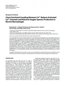
Fig. 1. Molecular structure of RIM proteins, CaVβ subunits, and the HVA CaV channel complex. A) RIM 1/2α is formed by one Zn2 +-finger-like domain (Zn2 +), one PDZ domain (PDZ), two C2 domains (C2A and C2B) and a proline-rich region (PXXP). RIMs 1/2β lack the Zn2 +-finger-like domain and RIMs 2γ–4γ have comprised only the C2 domains. CaVβ subunits are formed by three conserved domains: PDZ, Scr homology 3 (SH3) and guanylate kinase (GK). The β-interaction domain (BID) is one of the regions involved in the interaction of the protein with the CaVα1 pore-forming subunit. Both protein structures are depicted in N- to C-terminal direction. B) HVA CaV channels are oligomeric proteins of a pore-forming α1 subunit, and auxiliary subunits β and α2δ. The α1 subunit is formed by four homologous membrane-spanning repeated domains (I–IV), each with six transmembrane-helixes (S1–S6) and a pore-forming reentrant loop between S5 and S6. The voltage sensor is located in S4 and is indicated in yellow.
M.A. Gandini, R. Felix / Biochimica et Biophysica Acta 1818 (2012) 551–558
553
Table 1 Summary of RIM isoforms expressed in different cell types and tissues. Tissue/cell line
Specie
Isoform
Reference
Brain
Rat Mouse Rat Mouse Rat Mouse Rat Mouse Mouse Mouse Rat Rat Mouse Human Chicken Rat Rat Mouse Bovine Rat Rat Rat Mouse Rat Rat Mouse Hamster
RIM1α/RIM2α/RIM2β/RIM2γ/RIM3γ/RIM4γ RIM1α/RIM2α/RIM2β/RIM3γ/RIM4γ RIM1α RIM1α/RIM1β/RIM2α/RIM2β/RIM2γ RIM1α/RIM2α/RIM3γ/RIM4γ RIM1α RIM1α/RIM2α RIM1α/RIM2α/RIM1β/RIM2β RIM2α RIM1α/RIM2α/RIM2β/RIM3γ RIM1α/RIM2α/RIM3γ/RIM4γ RIM1α RIM1α/RIM2α RIM1α RIM1α RIM1α/RIM2α/RIM3γ RIM1α RIM1α/RIM2α RIM1α/RIM2α RIM1α/RIM2α/RIM3γ/RIM4γ RIM2α/RIM4γ RIM1α RIM2α RIM1α RIM2α RIM2α RIM1α
[2-5] [5,43,48] [9] [6,57,61,77-80] [11,43] [79,81-83] [84] [60] [48] [48] [43] [2] [48,49] [58] [45,85] [43] [2] [5] [8] [11,43] [43] [86] [56,87] [47] [7,51] [50,56] [86]
Hippocampus Cerebellum Calyx of Held Inner hair cells (immature) Organ of Corti (immature) Eye Photoreceptor cells
Ciliary ganglion calyx Dorsal root ganglion Spinal cord Adrenal chromaffin cells PC12 Pancreas β Pancreatic cells RIN-m5f INS-1 MIN-6 HIT-T15
includes channels containing α1A, α1B, and α1E, which originate the N-, P/Q- and R-type currents, respectively. Last, the T-type channels are named CaV3.1 through CaV3.3, corresponding to α1G, α1H, and α1I [16,17,19]. The expression of the CaV1.1 channels is restricted to skeletal muscle where they play key roles in excitation–contraction coupling and regulation of gene transcription [19,20]. Hypokalemic periodic paralysis and malignant hyperthermia sensitivity are human diseases associated to mutations in the gene encoding the CaV1.1α1 subunit [20-22]. The CaV1.2 isoform is preferentially expressed in cardiac muscle where it is responsible of excitation–contraction coupling, enzyme activity regulation and gene transcription. The CaV1.3 isoform displays differential expression patterns in many cell types and tissues including neurons, smooth muscle and inner ear cells as well as in pancreatic islets [20,22,23]. The Timothy and Brugada syndromes have been shown to be associated with mutations in the CACNA1C gene which encodes the CaV1.2α1 subunit. Both diseases are characterized by abnormal electrocardiogram findings and an increased risk of sudden cardiac death. The CaV1.4 channels are expressed in the retina and their function is related to visual transduction. Different mutations in the gene encoding the CaV1.4α1 subunit have been associated to an X-linked form of congenital stationary night blindness [20-22]. The CaV2.1 and CaV2.2 channels are primarily localized in nerve terminals and are responsible for neurotransmitter release [19,20]. Mutations in the gene coding the CaV2.1α1 subunit cause several neurologic disorders including familial hemiplegic migraine type 1, episodic ataxia type 2 and spinocerebellar ataxia type 6 [21,24]. Likewise, CaV2.3 channels are also expressed in neurons and are involved in repetitive firing, dendritic Ca 2 + transients and neurotransmission. Last, CaV3 (LVA; T-type) channels are mainly expressed in neurons and cardiac myocytes and its role has been related to pacemaking and repetitive firing. Interestingly, a mutation in one of the three T-type channel isoforms (CaV3.2) has been associated with childhood absence epilepsy [19,20]. In addition to the CaVα1 subunit, CaV1 and CaV2 channels arise from the multimerization of other proteins including a mostly extracellular CaVα2δ subunit and a cytoplasmic CaVβ subunit (Fig. 1B). The
CaVα1 subunit is the principal component of the channels and is responsible for their unique biophysical and pharmacological properties. However, proper trafficking and functioning of the channels require the auxiliary subunits. The CaVα2δ auxiliary subunit is a glycosylated protein encoded by a single gene that is proteolytically processed to generate two peptides (α2 and δ) linked by a single disulfide bridge [25]. The principal effect of this subunit is to increase the functional expression of the channels [26-29], as a consequence of increased trafficking [28]. Structurally the mature CaVα2δ subunit has been viewed as a type I transmembrane (TM) spanning protein (Fig. 1B), and from the functional point of view there is now compelling evidence for a major involvement of this protein in targeting the Ca 2 + channel complex to lipid rafts [30-33]. Although a recent study has challenged this model and offered a new mechanism for CaV channel raft localization by suggesting the CaVα2δ subunit associates with the plasma membrane via a glycosylphosphatidylinositol (GPI) anchor in the δ peptide [34], raft localization of CaVα2δ seems to be preserved even after replacement of the reported GPI anchoring motif with the TM domain of a functionally inert protein PIN-G. Conversely, the GPI-anchoring motif is not sufficient to target PIN-G to lipid rafts [33]. These data are most consistent with a model where CaVα2δ retains its type I TM topology and its targeting to lipid rafts is governed by sequences upstream of the putative GPI anchor that promote protein–protein rather than lipid–lipid interactions [33]. The CaVβ auxiliary subunits also regulate the functional expression and the biophysical properties of the CaV1 and CaV2 channels [17,35-37]. Two mechanisms have been proposed for this, an enhancement of expression and a direct effect on gating. With respect to the effect on expression, the CaVβ subunits have been suggested to enhance channel trafficking to the membrane by masking an endoplasmic reticulum retention signal in the CaVα1 subunit [35,38] as well as by protecting the channel complex from proteasomal degradation [39,40]. The CaVβ subunits have also been found to hyperpolarize the voltage dependence of activation and increase the channel open probability, which increase current through individual channels and therefore result in augmented macroscopic current density [36,41,42]. The CaVβ auxiliary subunits have a modular structure
554
M.A. Gandini, R. Felix / Biochimica et Biophysica Acta 1818 (2012) 551–558
consisting of five distinct regions (Fig. 1A). The first, third, and fifth regions are variable in length and amino acid sequence, whereas the second and fourth regions are highly conserved and are homologous to the Src homology 3 (SH3) and guanylate kinase (GK) domains, respectively [36]. These domains are interaction modules engaged in protein–protein interactions. 4. Interactions between CaV channels and the RIM family members Although it is well established that Ca 2 + entering cells through CaV channels in the vicinity of docked vesicles is primarily responsible for initiating hormone release and synaptic transmission, the mechanisms that maintain CaV channels at release sites are virtually unknown. Initial studies by Mori and colleagues (2007) have identified a direct interaction between the CaVβ auxiliary subunit and RIM1 by performing yeast two-hybrid screening, pull down and coimmunoprecipitation assays [11]. Using different constructs with several structural motifs of CaVβ4 as bait, they determined that the SH3 and GK domains (Fig. 1A) were necessary for the interaction with RIM1. They also found an interaction with the CaVβ2 subunit, indicating that the association of RIM1 with the channel complex was not dependent on the CaVβ isoform [11]. More recently, the same group extended these studies and showed that members of the four subfamilies of CaVβ subunits bind RIM1, RIM2, RIM3γ and RIM4γ, using GST fusion RIM proteins incubated with cell lysates obtained from transfected HEK-293 cells. Though the affinity of CaVβ for RIMγs seemed to be lower than that for RIMαs [43], its interaction has also important functional consequences as we shall discuss later. 4.1. RIMs may interact with CaV channels via its β auxiliary subunits In their seminal paper, Kiyonaka and co-workers (2007) identified the RIM1 C-terminus as a major CaVβ binding domain by using in vitro pull down assays. In the brain, the association of RIM1 with native CaV channels (of the CaV2.1 class) was determined by coimmunoprecipitation assays. Interestingly this association was disrupted when the channels were incubated with a dominant negative construct of CaVβ4 (BADN), which is capable of binding RIM1 but not the CaVα1 subunit, providing evidence for a physical interaction between the channel and the synaptic protein in a more physiological context. Likewise, in cultured hippocampal neurons, RIM1 and CaVβ4b both accumulated near presynaptic termini in parallel with CaV2.1 clustering, suggesting that the RIM1–CaVβ interaction may regulate the localization of channels at the presynaptic membrane [11]. It is worth mentioning also that the Stanley group has argued against the idea of the physical association between RIMs and CaV channels based mainly on co-immunoprecipitation experiments using different antibodies against RIM and the CaV2.2 channel [4446]. Though the reason for this discrepancy is presently unknown, according to these authors the differing results may be reconciled, in a model where CaV channels are interconnected by a scaffold and together form the backbone of the active zone release apparatus; a separate scaffold projects from the channel to the docked secretory vesicles. The model accommodates attachment of new vesicles and their release and, hence, the link between the channels and the vesicles could be cycling between high- and low-affinity states [44-46]. In this scenario, RIMs could be components of the complex, tethering the docking site/vesicle to the channel and responsible for the highto low-affinity switch. Recently, our group found that the coupling between RIM1 and the CaVβ auxiliary subunits is also operational in L-type channels (CaV1.2 and CaV1.3). By using co-immunoprecipitation experiments, a RIM1–CaV channel complex formed by direct interaction of the CaVβ2 and CaVβ3 subunits was identified in a heterologous system and also in RIN-m5F cells, an insulin-secreting cell line [47]. Consistent with this, Striessnig and his colleagues demonstrated that the
presence of the auxiliary subunit of the CaV channels is necessary for the interaction of RIM1 with the CaV1.3 channel complex heterologously expressed in tsA-201 cells, and showed also that RIM proteins are expressed in cochlear inner hair cell, where they substantially modify the biophysical properties of the channels [48]. The possible interaction of L-type channels and RIMs is also supported by the observation that the CaV1.3α1 subunit is localized at the active zone of retinal photoreceptor ribbon synapses forming tight clusters with different proteins including RIM2 [49]. 4.2. RIMs may also bind directly to the CaV channel ion-conducting α1 subunit A direct interaction between the pore-forming CaVα1 subunit and members of the RIM protein family has been also documented [7,50,51]. The first evidence of this interaction was obtained using GST fusion proteins containing the C2 domains of RIMs immobilized on glutathione-agarose beads and incubated with in vitro translated proteins representing the cytoplasmic loop connecting domain II and III of CaV1.2α1 and CaV2.2α1 subunits [7]. These experiments showed that two binding sequences in the C2 domains, KRRT and KKKT, appear to be important for the interaction of RIMs and the II-III loop of the CaV2.2α1 subunit. Likewise, the KKKT sequence in the C2 domain is allegedly responsible for binding the exocytosis-regulatory protein synaptotagmin I, through the synaptic protein interaction (synprint) site of neuronal CaV channels (N- and P/Q-type) located in the cytoplasmic loop connecting domains II and III of the pore-forming subunit [52,53]. Encouraged by these findings, Shibasaki and colleagues examined whether RIM2 and another scaffolding protein of the active zone called Piccolo, both of which have C2 domains, also interact with the CaVα1 subunit. Their results indicated that the C2 domains of RIM2 and Piccolo bind directly to the cytoplasmic loop of the CaV1.2α1 subunit of the L-type channels [50]. It is worth mentioning that these two proteins have been proposed as downstream effectors of EPAC2 (exchange protein activated directly by cyclic AMP type 2) a cAMP-regulated guanine nucleotide exchange factor (GEF) that mediate protein kinase A (PKA)-independent signal transduction properties of the second messenger cAMP [54,55]. Piccolo dimerizes with RIM2 in a Ca 2 +-dependent manner [56], and both are implicated in potentiation of glucose-stimulated insulin secretion via EPAC2 in pancreatic β-cells. These studies also suggest that CaV channels and the secretory granules are targeted to the same region in the pancreatic β cell and that the molecular organization is critical in regulated exocytosis within a zone of voltage-dependent Ca 2 + entry [50]. In addition, Jacobo and her colleagues using peptides heterologously expressed in insulinoma INS-1 cells confirmed the interaction of the CaV1.2α1/II-III loop with RIM2 and Piccolo, and also demonstrated that the II-III loops of CaV1.2α1 and CaV1.3α1 target their respective channels to membrane microdomains (lipid rafts) possibly through physical interactions with other lipid raft-resident proteins including RIM2. This association appears to be critical for efficient potentiation of glucose-dependent insulin secretion by the hormone glucagon-like peptide-1 in pancreatic β-cells, given that RIM2, acting as an anchor that positions the CaV1.2 channels within a lipid raft domain, may serve as an assembly point for effectors of cAMP signaling via EPAC2 [51]. Recent studies by the Südhof group have addressed questions about how the N- and P/Q-type CaV channels are selectively recruited to presynaptic active zones. By using C-terminal sequences of these channels as baits and distinct fragments of RIM1 as preys they localized the interaction domain to the PDZ region of RIM1. Then, to test whether RIMs act to localize CaV channel to active zones, the authors generated conditional double knockout (KO) mice of all RIM isoforms that contain PDZ-domains and using electrophysiology and Ca 2 +-imaging, as well as quantitative immunostaining of the channels, they
M.A. Gandini, R. Felix / Biochimica et Biophysica Acta 1818 (2012) 551–558
showed that the RIM PDZ-domains are required for localizing the channels to the release sites. They also found that the RIM prolinerich sequences which interact with RBPs also play a key role in tethering the channels to the presynaptic active zones [57]. A possible role for RBPs in synaptic transmission by creating a functional link between the synaptic-vesicle tethering apparatus and the CaV channels had been previously suggested by Hibino and colleagues (2002). Thus, it has been proposed that RIMs tether CaV channels to active zones via distinct parallel interactions, i) by directly binding of the CaVα1 subunit with the RIM PDZ-domains (interaction that is specific for N- and P/Q-type channels), ii) by indirectly interacting through RBPs, and iii) by association via the CaVβ auxiliary subunit. The latter two types of interaction are shared by different types of channels [10,11,43,57]. As we shall discuss in the next section, the formation of RIM–CaV channel complexes has relevant physiological implications. 5. Physiological relevance of the interaction between CaV channels and RIMs The kinetic analysis of the ion channel macroscopic currents has revealed some interesting aspects related to the functional significance of the RIM–CaV channel coupling. The most prominent effects of RIMs on CaV channel currents occur in the inactivation parameters (Fig. 2). RIM1 drastically slows current inactivation through neuronal CaV2 and L-type CaV1.2 and CaV1.3 channels [11,47]. Similar effects have been reported for RIM2 on CaV2.1 and CaV1.3 channel currents [43,48]. Interestingly, CaV2.1 current inactivation may be also markedly decelerated by RIM3γ and RIM4γ [43] suggesting that changing the time course of inactivation is a common feature of RIM regulation on CaV channel currents. As mentioned earlier, the studies of Kiyonaka and colleagues (2007) revealed a physical association between RIM and the CaVβ auxiliary subunits that results in a marked suppression of voltagedependent inactivation of different neuronal CaV channels. Consistent with this, docking of secretory vesicles and acetylcholine (ACh) release is significantly enhanced by transfection of RIM1 constructs in neuroendocrine PC12 cells. Conversely, transfection of a dominant
555
negative β construct that disrupts the RIM1–CaVβ association, accelerated inactivation of native channels, suppressed vesicle docking as well as ACh release in PC12 cells, and produced a significant inhibition of glutamate release in cultured cerebellar neurons. It should be mentioned that RIMγ knockdown attenuates glutamate release in a less pronounced manner than RIMα in cerebellar neurons and its over-expression enhances exocytosis in PC12 cells [3,43]. Interestingly, RIMγs also block localization of the vesicles near the cell membrane [43], confirming that changes in channel function may be a general feature of RIM-mediated regulation, whereas anchoring the vesicles to the channels may depend on the type RIM involved in the interaction [43]. Taking advantage of genetic analyses showing an association between the gene encoding RIM1 and the autosomal dominant conerod dystrophy CORD7 [58], the Mori group tested whether the RIM1 mutation associated to CORD7 affected CaV regulation by RIM1 [59]. Their data raised the interesting possibility that CORD7 phenotypes including retinal deficits and enhanced cognition are at least partly due to altered regulation of presynaptic CaV channels. Hence, a nucleotide (G/A) substitution, that replaces Arg-655 with His in the middle C2A domain, which binds to CaV channels, was introduced in the RIM1 cDNA to yield a construct which carried a mutation corresponding to the human CORD7 mutation. Functional analysis showed that in P/Q-type CaV2.1 channels, the CORD7 mutation enhanced the RIM1-mediated suppression of inactivation and augmented current density, likely leading to enhanced neurotransmitter release and synaptic transmission. In contrast, for L-type CaV1.4 channels, the CORD7 mutation abolished the RIM1-mediated hyperpolarization of current activation, likely resulting in impaired synaptic transmission at ribbon synapses of the visual system [59]. Heterologous expression of L-type CaV1.3 channels together with RIM constructs have revealed also that RIMs slow both Ca2 +-dependent inactivation (CDI) and voltage-dependent inactivation (VDI) through binding to the CaVβ auxiliary subunits [48]. By slowing inactivation over a large voltage range and by substantially increasing the noninactivating component of the macroscopic current, RIM-associated channel complexes may carry larger CaV1.3 window currents during
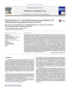
Fig. 2. A hypothetical scheme of the regulation of CaV channel inactivation by RIM proteins. Although the crystallographic structure of CaV channels is not available, the model is compatible with many experimentally derived data [87]. A) At rest, the channels are closed. B) When an action potential arrives, the channels open allowing the flow of Ca2 + ions down their electrochemical gradient. C) During sustained membrane depolarization the channels undergo a conformational change in which the inactivation gate occludes the ion-conducting pore. D) The presence of the RIM proteins results in noticeable changes in channel function, in particular a significant slowdown in inactivation kinetics, as illustrated in the scheme showing the time course of hypothetical macroscopic current traces through CaV channels in absence (E) or presence (F) of RIMs.
556
M.A. Gandini, R. Felix / Biochimica et Biophysica Acta 1818 (2012) 551–558
prolonged depolarization. Interestingly, neurotransmitter release and spontaneous action potentials during cochlear inner hair cell (IHC) development depend on the activity of CaV1.3 channels which voltageand Ca2 +-dependent inactivation kinetics are slower than in other tissues. It has been reported that RIM2 mRNA is expressed in immature cochlear IHCs and the protein co-localizes with CaV1.3 channels in the same presynaptic compartment of IHCs [48]. Therefore, association with RIMs may represent a possible molecular mechanism to inhibit CDI and VDI of Cav1.3 channels in IHCs which may be important for the specific signaling functions of these channels during different developmental stages with immature IHCs generating spontaneous, regenerative Ca2 + action potentials important for normal development. Likewise, Gandini and her colleagues (2011) studied the effect of RIM1 on recombinant L-type CaV1.3 as well as of the CaV1.2 channels expressed in HEK-293 cells and their functional impact on insulin secretion. Their results show that RIM1 does not alter Ba2 + current density through these channels but drastically slows down their inactivation kinetics. As a consequence, the amount of charge mobilized is increased during depolarization, which means that a larger amount of ions passed through the channels when RIM1 is present. Interestingly, the effect of RIM1 on channel inactivation was prevented when CaVβ was absent, confirming that the interaction between the channels and RIM1 may occur via the auxiliary subunit. The relevance of this association was assessed by electrophysiological recordings and insulin secretion measurements in the insulinoma RIN-m5f cells after RIM1 gene silencing. Knock down of endogenous RIM1 increased inactivation of whole cell native currents and decreased insulin release triggered by Ca2 + influx in response to K +-induced membrane depolarization [47]. Using a combined experimental strategy based on protein/protein interaction studies, generation of conditional RIM1/2 knockout mice and electrophysiology, recent studies have shed new light on the physiology of RIMs–CaV channel interaction [57,60,61]. Kaeser and colleagues (2011) proposed that RIMs have two alternative forms of interaction with CaV channels: a direct one via their PDZ-domains that is specific for N- and P/Q-type channels, and an indirect (nonspecific) interaction via RBPs. Using rescue experiments in RIMdeficient hippocampal neurons, these authors found that the RIM PDZ-domain is required to reverse the impairment in presynaptic Ca 2 + influx in RIM-deficient cells and for localizing the channels to the presynaptic boutons [57]. Similar to that in hippocampal synapses, several lines of evidence suggested that genetic elimination of RIMs interfered with the coupling between CaV channels and transmitter release at the calyx of Held, a glutamatergic synapse in the auditory brainstem accessible to quantitative analysis of transmitter release [60]. These studies also show that at both the hippocampal and the calyx of Held synapses, the size of the releasable pool of vesicles is reduced in the RIM1/2 double knockout mouse [57,60,61], corroborating that RIMs are central organizers of active zones. Interestingly, the Zn 2 + finger domain of RIMs seems to be necessary and sufficient for the vesicle priming function, and given this site interacts with Munc13, it was suggested that the RIMs effects on priming are mediated by Munc13 specifically by preventing its homodimerization [61]. It is well established that G protein coupled receptors (GPCRs) orchestrate precise regulation neurotransmitter and hormone release through inhibition of CaV2 channels [62,63]. A unifying property of this GPCR-mediated inhibition of CaV is its sensitivity to pertussis toxin, thus implicating Gαi and/or Gαo proteins. This inhibition exhibits three major biophysical characteristics: i) it slows down the activation kinetics of the inhibited channels, ii) shifts the activation voltage to a more depolarized potential and, iii) can be relieved by a strong conditioning depolarizing potential, resulting in the so-called prepulse facilitation [62,63]. These actions are mediated by the interaction of the G-protein βγ dimer (Gβγ) [64,65] with the alpha interacting domain (AID) located in the cytoplasmic loop (I-II loop)
connecting the first two homologous repeats of the CaVα1 subunit (Fig. 2B) which also constitutes the high-affinity CaVβ-binding site [66,67]. For this reason, and given that some functional effects of the Gβγ dimer are the opposites of those of CaVβ subunits, it had raised the question whether Gβγ and CaVβ compete with each other. However, recent studies suggest that the voltage dependence of Gβγ inhibition of HVA channels arises from the movement of IS6, and that CaVβ and a rigid IS6-AID linker play a pivotal role in translating this movement to Gβγ dissociation [68]. As mentioned earlier, the CaV2α1 subunit contains a synprint region that allows synaptic proteins to regulate channel activity [7,69,70], .i.e. binding of syntaxin 1 and SNAP-25 results in a hyperpolarizing shift in the half-inactivation potential, thus reducing channel availability [69,71,72]. However, a second action of syntaxin 1 on CaV channel regulation has been reported by Zamponi and his colleagues. Co-expression of this protein with N-type channels results in a tonic G protein-mediated channel inhibition that does not involve receptor activation [73]. This appears to be due to a syntaxin 1 mediated colocalization of the channel and the Gβγ dimer that ultimately culminates in tonic channel inhibition. Likewise, it is worth mentioning that the existence of a syntaxin-Cav3.2 (T-type) channel complex has been recently revealed in central neurons. Though this interaction
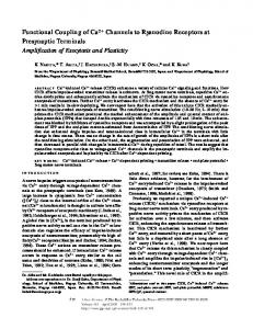
Fig. 3. Functional coupling between CaV channels and RIMs. A) RIMs anchor synaptic vesicles next to channels through its interaction with zone-specific proteins (Rab3, Munc13) and the CaVβ auxiliary subunit. B) After depolarization, RIMs regulate the time course of channel inactivation resulting in a sustained Ca2 + influx. D) These molecular interactions favor hormone and neurotransmitter release (C).
M.A. Gandini, R. Felix / Biochimica et Biophysica Acta 1818 (2012) 551–558
may involve different molecular determinants from those found in HVA channels, it modulates channel gating and appears essential for T-type channel-triggered low-threshold exocytosis [74]. Likewise, interesting actions of RIM1 on G-protein regulation of CaV2.2 channels heterologously expressed in HEK-293 have been recently reported [75]. As expected, RIM1 potently decreases the extent of CaV2.2 channel inactivation and the application of DAMGO, an agonist of the μ-opioid receptor, induced direct G-protein regulation. Interestingly, RIM1 did not alter the association of the Gβγ dimer onto the channel, but the kinetic and extent of recovery from G-protein inhibition were significantly affected in RIM1-expressing cells. These findings provided the first evidence for an efficient RIM1-dependent modulation of direct G-protein regulation of neuronal CaV2.2 channels, and emphasize the importance of the constitutive proteins from the presynaptic vesicle machinery, particularly RIMs, for both neurotransmitter secretion and fine-tuning of CaV channel activity [75]. In summary, neurotransmitter release is initiated by the activation of CaV channels close to docked vesicles. Although the mechanisms that enrich channels at release sites are largely unknown, recent studies suggest that RIM proteins may affect many aspects of neurotransmitter release [8–13, 76]. Combined, these studies support a model predicting a dual function for the RIM/CaV channel interaction in hormone and neurotransmitter release by coordinating the molecular constituents and Ca 2 + signaling in the active zones (Fig. 3). Acknowledgements Work in the laboratory of R.F. is supported by Conacyt, Mexico (grant 128707). M.A.G. is the recipient of a doctoral fellowship from Conacyt. The authors thank Andrés S. Gandini for art work, and three anonymous reviewers for helpful comments that significantly improved the manuscript. References [1] J.D. Spafford, G.W. Zamponi, Functional interactions between presynaptic calcium channels and the neurotransmitter release machinery, Curr. Opin. Neurobiol. 13 (2003) 308–314. [2] Y. Wang, M. Okamoto, F. Schmitz, K. Hofmann, T.C. Südhof, Rim is a putative Rab3 effector in regulating synaptic-vesicle fusion, Nature 388 (1997) 593–598. [3] Y. Wang, S. Sugita, T.C. Südhof, The RIM/NIM family of neuronal C2 domain proteins. Interactions with Rab3 and a new class of Src homology 3 domain proteins, J. Biol. Chem. 275 (2000) 20033–20044. [4] Y. Wang, T.C. Südhof, Genomic definition of RIM proteins: evolutionary amplification of a family of synaptic regulatory proteins (small star, filled), Genomics 81 (2003) 126–137. [5] S. Schoch, T. Mittelstaedt, P.S. Kaeser, D. Padgett, N. Feldmann, V. Chevaleyre, P.E. Castillo, R.E. Hammer, W. Han, F. Schmitz, W. Lin, T.C. Südhof, Redundant functions of RIM1α and RIM2α in Ca2 +-triggered neurotransmitter release, EMBO J. 25 (2006) 5852–5863. [6] P.S. Kaeser, H.B. Kwon, C.Q. Chiu, L. Deng, P.E. Castillo, T.C. Südhof, RIM1α and RIM1β are synthesized from distinct promoters of the RIM1 gene to mediate differential but overlapping synaptic functions, J. Neurosci. 28 (2008) 13435–13447. [7] T. Coppola, S. Magnin-Luthi, V. Perret-Menoud, S. Gattesco, G. Schiavo, R. Regazzi, Direct interaction of the Rab3 effector RIM with Ca2 + channels, SNAP-25, and synaptotagmin, J. Biol. Chem. 276 (2001) 32756–32762. [8] L. Sun, M.A. Bittner, R.W. Holz, Rim, a component of the presynaptic active zone and modulator of exocytosis, binds 14-3-3 through its N terminus, J. Biol. Chem. 278 (2003) 38301–38309. [9] E. Takao-Rikitsu, S. Mochida, E. Inoue, M. Deguchi-Tawarada, M. Inoue, T. Ohtsuka, Y. Takai, Physical and functional interaction of the active zone proteins, CAST, RIM1, and Bassoon, in neurotransmitter release, J. Cell Biol. 164 (2004) 301–311. [10] H. Hibino, R. Pironkova, O. Onwumere, M. Vologodskaia, A.J. Hudspeth, F. Lesage, RIM binding proteins (RBPs) couple Rab3-interacting molecules (RIMs) to voltage-gated Ca2 + channels, Neuron 34 (2002) 411–423. [11] S. Kiyonaka, M. Wakamori, T. Miki, Y. Uriu, M. Nonaka, H. Bito, A.M. Beedle, E. Mori, Y. Hara, M. De Waard, M. Kanagawa, M. Itakura, M. Takahashi, K.P. Campbell, Y. Mori, RIM1 confers sustained activity and neurotransmitter vesicle anchoring to presynaptic Ca2 + channels, Nat. Neurosci. 10 (2007) 691–701. [12] T. Mittelstaedt, E. Alvarez-Baron, S. Schoch, RIM proteins and their role in synapse function, Biol. Chem. 391 (2010) 599–606. [13] P.S. Kaeser, T.C. Südhof, RIM function in short- and long-term synaptic plasticity, Biochem. Soc. Trans. 33 (2005) 1345–1349. [14] A. Pernia-Andrade, P. Jonas, The multiple faces of RIM, Neuron 69 (2011) 185–187.
557
[15] W.A. Catterall, Structure and regulation of voltage-gated Ca2 + channels, Annu. Rev. Cell Dev. Biol. 16 (2000) 521–555. [16] R. Felix, Molecular regulation of voltage-gated Ca2 + channels, J. Recept. Signal. Transduction Res. 25 (2005) 57–71. [17] L. Lacinova, Voltage-dependent calcium channels, Gen. Physiol. Biophys. 24 (Suppl 1) (2005) 1–78. [18] S.N. Yang, P.O. Berggren, The role of voltage-gated calcium channels in pancreatic β-cell physiology and pathophysiology, Endocr. Rev. 27 (2006) 621–676. [19] W.A. Catterall, E. Perez-Reyes, T.P. Snutch, J. Striessnig, International Union of Pharmacology. XLVIII. Nomenclature and structure-function relationships of voltage-gated calcium channels, Pharmacol. Rev. 57 (2005) 411–425. [20] W.A. Catterall, Voltage-gated calcium channels, Cold Spring Harb. Perspect. Biol. 3 (2011) a003947. [21] R. Felix, Calcium channelopathies, Neuromolecular Med. 8 (2006) 307–318. [22] J. Striessnig, H.J. Bolz, A. Koschak, Channelopathies in CaV1.1, CaV1.3, and CaV1.4 voltage-gated L-type Ca2 + channels, Pflugers Arch. 460 (2010) 361–374. [23] A. Zuccotti, S. Clementi, T. Reinbothe, A. Torrente, D.H. Vandael, A. Pirone, Structural and functional differences between L-type calcium channels: crucial issues for future selective targeting, Trends Pharmacol. Sci. 32 (2011) 366–375. [24] D. Pietrobon, Insights into migraine mechanisms and CaV2.1 calcium channel function from mouse models of familial hemiplegic migraine, J. Physiol. 588 (2010) 1871–1878. [25] R. Felix, Voltage-dependent Ca2 + channel α2δ auxiliary subunit: structure, function and regulation, Receptors Channels 6 (1999) 351–362. [26] R. Felix, C.A. Gurnett, M. De Waard, K.P. Campbell, Dissection of functional domains of the voltage-dependent Ca2 + channel α2δ subunit, J. Neurosci. 17 (1997) 6884–6891. [27] N. Klugbauer, E. Marais, F. Hofmann, Calcium channel α2δ subunits: differential expression, function, and drug binding, J. Bioenerg. Biomembr. 35 (2003) 639–647. [28] C. Cantí, M. Nieto-Rostro, I. Foucault, F. Heblich, J. Wratten, M.W. Richards, J. Hendrich, L. Douglas, K.M. Page, A. Davies, A.C. Dolphin, The metal-iondependent adhesion site in the Von Willebrand factor A domain of α2δ subunits is key to trafficking voltage-gated Ca2 + channels, Proc. Natl. Acad. Sci. U. S. A. 102 (2005) 11230–11235. [29] A. Andrade, A. Sandoval, R. Gonzalez-Ramirez, D. Lipscombe, K.P. Campbell, R. Felix, The α2δ subunit augments functional expression and modifies the pharmacology of CaV1.3 L-type channels, Cell Calcium 46 (2009) 282–292. [30] C.A. Gurnett, M. De Waard, K.P. Campbell, Dual function of the voltage-dependent Ca2 + channel alpha 2 delta subunit in current stimulation and subunit interaction, Neuron 16 (1996) 431–440. [31] A. Davies, L. Douglas, J. Hendrich, J. Wratten, A. Tran Van Minh, I. Foucault, D. Koch, W.S. Pratt, H.R. Saibil, A.C. Dolphin, The calcium channel α2δ-2 subunit partitions with CaV2.1 into lipid rafts in cerebellum: implications for localization and function, J. Neurosci. 26 (2006) 8748–8757. [32] P. Robinson, S. Etheridge, L. Song, P. Armenise, O.T. Jones, E.M. Fitzgerald, Formation of N-type (Cav2.2) voltage-gated calcium channel membrane microdomains: lipid raft association and clustering, Cell Calcium 48 (2010) 183–194. [33] P. Robinson, S. Etheridge, L. Song, R. Shah, E.M. Fitzgerald, O.T. Jones, Targeting of voltage-gated calcium channel α2δ-1 subunit to lipid rafts is independent from a GPI-anchoring motif, PLoS One 6 (2011) e19802. [34] A. Davies, I. Kadurin, A. Alvarez-Laviada, L. Douglas, M. Nieto-Rostro, C.S. Bauer, W.S. Pratt, A.C. Dolphin, The α2δ subunits of voltage-gated calcium channels form GPI-anchored proteins, a posttranslational modification essential for function, Proc. Natl. Acad. Sci. U. S. A. 107 (2010) 1654–1659. [35] D. Bichet, V. Cornet, S. Geib, E. Carlier, S. Volsen, T. Hoshi, Y. Mori, M. De Waard, The I-II loop of the Ca2 + channel α1 subunit contains an endoplasmic reticulum retention signal antagonized by the beta subunit, Neuron 25 (2000) 177–190. [36] Z. Buraei, J. Yang, The β subunit of voltage-gated Ca2 + channels, Physiol. Rev. 90 (2010) 1461–1506. [37] D. Walker, D. Bichet, K.P. Campbell, M. De Waard, A b4 isoform-specific interaction site in the carboxyl-terminal region of the voltage-dependent Ca2 + channel α1A subunit, J. Biol. Chem. 273 (1998) 2361–2367. [38] K. Fang, H.M. Colecraft, Mechanism of auxiliary beta-subunit-mediated membrane targeting of L-type (Ca(V)1.2) channels, J. Physiol. 589 (2011) 4437–4455. [39] C. Altier, A. Garcia-Caballero, B. Simms, H. You, L. Chen, J. Walcher, H.W. Tedford, T. Hermosilla, G.W. Zamponi, The Cavbeta subunit prevents RFP2-mediated ubiquitination and proteasomal degradation of L-type channels, Nat. Neurosci. 14 (2011) 173–180. [40] D. Waithe, L. Ferron, K.M. Page, K. Chaggar, A.C. Dolphin, β-Subunits promote the expression of CaV2.2 channels by reducing their proteasomal degradation, J. Biol. Chem. 286 (2011) 9598–9611. [41] D. Walker, M. De Waard, Subunit interaction sites in voltage-dependent Ca2 + channels: role in channel function, Trends Neurosci. 21 (1998) 148–154. [42] P. Hidalgo, A. Neely, Multiplicity of protein interactions and functions of the voltage-gated calcium channel β-subunit, Cell Calcium 42 (2007) 389–396. [43] Y. Uriu, S. Kiyonaka, T. Miki, M. Yagi, S. Akiyama, E. Mori, A. Nakao, A.M. Beedle, K.P. Campbell, M. Wakamori, Y. Mori, Rab3-interacting molecule gamma isoforms lacking the Rab3-binding domain induce long lasting currents but block neurotransmitter vesicle anchoring in voltage-dependent P/Q-type Ca2 + channels, J. Biol. Chem. 285 (2010) 21750–21767. [44] E.F. Stanley, T.S. Reese, G.Z. Wang, Molecular scaffold reorganization at the transmitter release site with vesicle exocytosis or botulinum toxin C1, Eur. J. Neurosci. 18 (2003) 2403–2407. [45] R. Khanna, Q. Li, J. Bewersdorf, E.F. Stanley, The presynaptic CaV2.2 channeltransmitter release site core complex, Eur. J. Neurosci. 26 (2007) 547–559.
558
M.A. Gandini, R. Felix / Biochimica et Biophysica Acta 1818 (2012) 551–558
[46] F.K. Wong, E.F. Stanley, Rab3a interacting molecule (RIM) and the tethering of presynaptic transmitter release site-associated CaV2.2 calcium channels, J. Neurochem. 112 (2010) 463–473. [47] M.A. Gandini, A. Sandoval, R. Gonzalez-Ramirez, Y. Mori, M. de Waard, R. Felix, Functional coupling of Rab3-interacting molecule 1 (RIM1) and L-type Ca2 + channels in insulin release, J. Biol. Chem. 286 (2011) 15757–15765. [48] M. Gebhart, G. Juhasz-Vedres, A. Zuccotti, N. Brandt, J. Engel, A. Trockenbacher, G. Kaur, G.J. Obermair, M. Knipper, A. Koschak, J. Striessnig, Modulation of Cav1.3 Ca2 + channel gating by Rab3 interacting molecule, Mol. Cell. Neurosci. 44 (2010) 246–259. [49] S. tom Dieck, W.D. Altrock, M.M. Kessels, B. Qualmann, H. Regus, D. Brauner, A. Fejtova, O. Bracko, E.D. Gundelfinger, J.H. Brandstatter, Molecular dissection of the photoreceptor ribbon synapse: physical interaction of Bassoon and RIBEYE is essential for the assembly of the ribbon complex, J. Cell Biol. 168 (2005) 825–836. [50] T. Shibasaki, Y. Sunaga, K. Fujimoto, Y. Kashima, S. Seino, Interaction of ATP sensor, cAMP sensor, Ca2 + sensor, and voltage-dependent Ca2 + channel in insulin granule exocytosis, J. Biol. Chem. 279 (2004) 7956–7961. [51] S.M. Jacobo, M.L. Guerra, R.E. Jarrard, J.A. Przybyla, G. Liu, V.J. Watts, G.H. Hockerman, The intracellular II-III loops of Cav1.2 and Cav1.3 uncouple L-type voltage-gated Ca2 + channels from glucagon-like peptide-1 potentiation of insulin secretion in INS-1 cells via displacement from lipid rafts, J. Pharmacol. Exp. Ther. 330 (2009) 283–293. [52] Z.H. Sheng, C.T. Yokoyama, W.A. Catterall, Interaction of the synprint site of N-type Ca2 + channels with the C2B domain of synaptotagmin I, Proc. Natl. Acad. Sci. U. S. A. 94 (1997) 5405–5410. [53] N. Charvin, C. L'Eveque, D. Walker, F. Berton, C. Raymond, M. Kataoka, Y. ShojiKasai, M. Takahashi, M. De Waard, M.J. Seagar, Direct interaction of the calcium sensor protein synaptotagmin I with a cytoplasmic domain of the alpha1A subunit of the P/Q-type calcium channel, EMBO J. 16 (1997) 4591–4596. [54] J.L. Bos, Epac: a new cAMP target and new avenues in cAMP research, Nat. Rev. Mol. Cell Biol. 4 (2003) 733–738. [55] M. Gloerich, J.L. Bos, Epac: defining a new mechanism for cAMP action, Annu. Rev. Pharmacol. Toxicol. 50 (2010) 355–375. [56] K. Fujimoto, T. Shibasaki, N. Yokoi, Y. Kashima, M. Matsumoto, T. Sasaki, N. Tajima, T. Iwanaga, S. Seino, Piccolo, a Ca2 + sensor in pancreatic beta-cells. Involvement of cAMP-GEFII. Rim2. Piccolo complex in cAMP-dependent exocytosis, J. Biol. Chem. 277 (2002) 50497–50502. [57] P.S. Kaeser, L. Deng, Y. Wang, I. Dulubova, X. Liu, J. Rizo, T.C. Südhof, RIM proteins tether Ca2 + channels to presynaptic active zones via a direct PDZ-domain interaction, Cell 144 (2011) 282–295. [58] M. Michaelides, G.E. Holder, D.M. Hunt, F.W. Fitzke, A.C. Bird, A.T. Moore, A detailed study of the phenotype of an autosomal dominant cone-rod dystrophy (CORD7) associated with mutation in the gene for RIM1, Br. J. Ophthalmol. 89 (2005) 198–206. [59] T. Miki, S. Kiyonaka, Y. Uriu, M. De Waard, M. Wakamori, A.M. Beedle, K.P. Campbell, Y. Mori, Mutation associated with an autosomal dominant cone-rod dystrophy CORD7 modifies RIM1-mediated modulation of voltage-dependent Ca 2 + channels, Channels (Austin) 1 (2007) 144–147. [60] Y. Han, P.S. Kaeser, T.C. Südhof, R. Schneggenburger, RIM determines Ca2 + channel density and vesicle docking at the presynaptic active zone, Neuron 69 (2011) 304–316. [61] L. Deng, P.S. Kaeser, W. Xu, T.C. Südhof, RIM proteins activate vesicle priming by reversing autoinhibitory homodimerization of Munc13, Neuron 69 (2011) 317–331. [62] H.W. Tedford, G.W. Zamponi, Direct G protein modulation of Cav2 calcium channels, Pharmacol. Rev. 58 (2006) 837–862. [63] K.P. Currie, G protein modulation of CaV2 voltage-gated calcium channels, Channels (Austin) 4 (2010) 497–509. [64] S.R. Ikeda, Voltage-dependent modulation of N-type calcium channels by G-protein beta gamma subunits, Nature 380 (1996) 255–258. [65] S. Herlitze, D.E. Garcia, K. Mackie, B. Hille, T. Scheuer, W.A. Catterall, Modulation of Ca2 + channels by G-protein beta gamma subunits, Nature 380 (1996) 258–262. [66] M. Pragnell, M. De Waard, Y. Mori, T. Tanabe, T.P. Snutch, K.P. Campbell, Calcium channel beta-subunit binds to a conserved motif in the I-II cytoplasmic linker of the alpha 1-subunit, Nature 368 (1994) 67–70.
[67] Y. Opatowsky, C.C. Chen, K.P. Campbell, J.A. Hirsch, Structural analysis of the voltage-dependent calcium channel beta subunit functional core and its complex with the alpha 1 interaction domain, Neuron 42 (2004) 387–399. [68] Y. Zhang, Y.H. Chen, S.D. Bangaru, L. He, K. Abele, S. Tanabe, T. Kozasa, J. Yang, Origin of the voltage dependence of G-protein regulation of P/Q-type Ca2 + channels, J. Neurosci. 28 (2008) 14176–14188. [69] S.E. Jarvis, G.W. Zamponi, Distinct molecular determinants govern syntaxin 1Amediated inactivation and G-protein inhibition of N-type calcium channels, J. Neurosci. 21 (2001) 2939–2948. [70] A.E. Kisilevsky, G.W. Zamponi, Presynaptic calcium channels: structure, regulators, and blockers, Handb. Exp. Pharmacol. (2008) 45–75. [71] I. Bezprozvanny, R.H. Scheller, R.W. Tsien, Functional impact of syntaxin on gating of N-type and Q-type calcium channels, Nature 378 (1995) 623–626. [72] E.F. Stanley, Syntaxin I modulation of presynaptic calcium channel inactivation revealed by botulinum toxin C1, Eur. J. Neurosci. 17 (2003) 1303–1305. [73] S.E. Jarvis, J.M. Magga, A.M. Beedle, J.E. Braun, G.W. Zamponi, G protein modulation of N-type calcium channels is facilitated by physical interactions between syntaxin 1A and Gbetagamma, J. Biol. Chem. 275 (2000) 6388–6394. [74] N. Weiss, S. Hameed, J.M. Fernandez-Fernandez, K. Fablet, M. Karmazinova, C. Poillot, J. Proft, L. Chen, I. Bidaud, A. Monteil, S. Huc-Brandt, L. Lacinova, P. Lory, G.W. Zamponi, M. De Waard, A Cav3.2/syntaxin-1A signaling complex controls T-type channel activity and low-threshold exocytosis, J. Biol. Chem. (2011). [75] N. Weiss, A. Sandoval, S. Kyonaka, R. Felix, Y. Mori, M. De Waard, Rim1 modulates direct G-protein regulation of CaV2.2 channels, Pflugers Arch. 461 (2011) 447–459. [76] N. Calakos, S. Schoch, T.C. Sudhof, R.C. Malenka, Multiple roles for the active zone protein RIM1alpha in late stages of neurotransmitter release, Neuron 42 (2004) 889–896. [77] S. Schoch, P.E. Castillo, T. Jo, K. Mukherjee, M. Geppert, Y. Wang, F. Schmitz, R.C. Malenka, T.C. Sudhof, RIM1alpha forms a protein scaffold for regulating neurotransmitter release at the active zone, Nature 415 (2002) 321–326. [78] P.E. Castillo, S. Schoch, F. Schmitz, T.C. Sudhof, R.C. Malenka, RIM1alpha is required for presynaptic long-term potentiation, Nature 415 (2002) 327–330. [79] V. Chevaleyre, B.D. Heifets, P.S. Kaeser, T.C. Sudhof, P.E. Castillo, Endocannabinoidmediated long-term plasticity requires cAMP/PKA signaling and RIM1alpha, Neuron 54 (2007) 801–812. [80] G. Lonart, S. Schoch, P.S. Kaeser, C.J. Larkin, T.C. Sudhof, D.J. Linden, Phosphorylation of RIM1alpha by PKA triggers presynaptic long-term potentiation at cerebellar parallel fiber synapses, Cell 115 (2003) 49–60. [81] F. Simsek-Duran, D.J. Linden, G. Lonart, Adapter protein 14-3-3 is required for a presynaptic form of LTP in the cerebellum, Nat. Neurosci. 7 (2004) 1296–1298. [82] P.M. Lachamp, Y. Liu, S.J. Liu, Glutamatergic modulation of cerebellar interneuron activity is mediated by an enhancement of GABA release and requires protein kinase A/RIM1alpha signaling, J. Neurosci. 29 (2009) 381–392. [83] I. Dulubova, X. Lou, J. Lu, I. Huryeva, A. Alam, R. Schneggenburger, T.C. Sudhof, J. Rizo, A Munc13/RIM/Rab3 tripartite complex: from priming to plasticity? EMBO J. 24 (2005) 2839–2850. [84] R. Khanna, Q. Li, L. Sun, T.J. Collins, E.F. Stanley, N type Ca2 + channels and RIM scaffold protein covary at the presynaptic transmitter release face but are components of independent protein complexes, Neuroscience 140 (2006) 1201–1208. [85] M. Iezzi, R. Regazzi, C.B. Wollheim, The Rab3-interacting molecule RIM is expressed in pancreatic beta-cells and is implicated in insulin exocytosis, FEBS Lett. 474 (2000) 66–70. [86] T. Yasuda, T. Shibasaki, K. Minami, H. Takahashi, A. Mizoguchi, Y. Uriu, T. Numata, Y. Mori, J. Miyazaki, T. Miki, S. Seino, Rim2α determines docking and priming states in insulin granule exocytosis, Cell Metab. 12 (2010) 117–129. [87] S.C. Stotz, S.E. Jarvis, G.W. Zamponi, Functional roles of cytoplasmic loops and pore lining transmembrane helices in the voltage-dependent inactivation of HVA calcium channels, J. Physiol. 554 (2004) 263–273.