YALE JOURNAL OF BIOLOGY AND MEDICINE 90 (2017), pp.543-551.
Review
Gene Therapy for Color Blindness Mark M. Hassalla,b,*, Alun R. Barnarda,b, and Robert E. MacLarena,b Nuffield Laboratory of Ophthalmology, Department of Clinical Neurosciences, University of Oxford, Oxford, UK; bOxford Eye Hospital, Oxford University Hospitals NHS Foundation Trust, Oxford, UK a
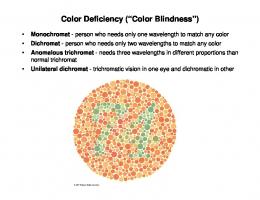
Achromatopsia is a rare congenital cause of vision loss due to isolated cone photoreceptor dysfunction. The most common underlying genetic mutations are autosomal recessive changes in CNGA3, CNGB3, GNAT2, PDE6H, PDE6C, or ATF6. Animal models of Cnga3, Cngb3, and Gnat2 have been rescued using AAV gene therapy; showing partial restoration of cone electrophysiology and integration of this new photopic vision in reflexive and behavioral visual tests. Three gene therapy phase I/II trials are currently being conducted in human patients in the USA, the UK, and Germany. This review details the AAV gene therapy treatments of achromatopsia to date. We also present novel data showing rescue of a Cnga3-/mouse model using an rAAV.CBA.CNGA3 vector. We conclude by synthesizing the implications of this animal work for ongoing human trials, particularly, the challenge of restoring integrated cone retinofugal pathways in an adult visual system. The evidence to date suggests that gene therapy for achromatopsia will need to be applied early in childhood to be effective. INTRODUCTION Achromatopsia is a rare (1 in 30,000 to 80,000) congenital visual condition characterized by diminished or absent cone photoreceptor function [1]. Patients have severely reduced visual acuity (~20/200), nystagmus and photophobia [2]. Causative autosomal recessive mutations affect genes of the cone phototransduction cascade, CNGA3, CNGB3, GNAT2, PDE6H, PDE6C, (Table 1) [3–8] or the transcription factor ATF6 [9]. A subset of patients have unidentified causative mutations [10]. Patients typically show complete cone function loss, as shown by absent cone-isolating electroretinogram (ERG†) recordings [2]. An incomplete phenotype occurs more rarely, but is not clearly correlated to the underlying mutation [1]. Achromatopsia affects all three classes of photoreceptor and hence, by definition, relates to genes
that are expressed in all cone subtypes, as listed above. Conversely, blue monochromatism, tritanopia, and redgreen color blindness (deuteranopia and protanopia) are caused by mutations in genes specific to certain subtypes of cone photoreceptors; an important difference to achromatopsia. The blue cone opsin gene is found on chromosome 7 and the red and green opsins are both on the X-chromosome. For instance, Bornholm disease can affect the green or red opsin gene only, or blue cone monochromatism can affect both [11]. Tritanopia (loss of the S-cone opsin) is rare because unlike the X-linked diseases, a recessive variant requires a mutation on both alleles. More commonly tritanopia results from a dominantly inherited S-opsin mutation that leads to progressive loss of the S-cones, similar to the rod loss caused by rhodopsin gene mutations in retinitis pigmentosa.
*To whom all correspondence should be addressed: Dr. Mark M. Hassall, Nuffield Laboratory of Ophthalmology, Level 6, West Wing, John Radcliffe Hospital, Headley Way, Oxford, UK, OX3 9DU, Tel: +44 1865 234768, Email:
[email protected]. †Abbreviations: ERG, electroretinogram; bp, base pairs; AAV, adeno-associated Virus; gp, genome particle; OMR, optomotor response; CBA, chicken beta actin; IHC, immunohistochemistry; TKO, triple knock out; fMRI, functional magnetic resonance imaging; CNTF, Ciliary Neurotrophic Factor. Keywords: Gene therapy, Achromatopsia, Gene editing, Cone photoreceptors Copyright © 2017
543
544
Hassall et al.: Gene therapy for color blindness
Table 1. Summary of the genes known to cause achromatopsia. Gene
Full name
Location
Gene size (kB)
Exons
CDS length (bp)
First report
Percentage of international cases (Kohl, 2004)
Percent of Netherlands cases (Thiadins, 2009)
CNGB3
cyclic nucleotide gated channel beta 3
8q21.3
170
19
2430
(Kohl, 2000)
47
87
CNGA3
cyclic nucleotide gated channel alpha 3
2q11.2
53
9
2085
(Kohl, 1998)
23
5
GNAT2
G protein subunit alpha transducin 2
1p13.3
16.6
9
1065
(Kohl 2002; Aligianis, 2002)
2
0
PDE6C
phosphodiesterase 10q23.33 6C
69
22
2577
(Chang, 2009)
Undiscovered
Not tested
PDE6H
phosphodiesterase 12p12.3 6H
9.6
4
252
(Kohl, 2012)
Undiscovered
Undiscovered
ATF6
Activating Transcription Factor 6
197.8
17
2013
(Kohl, 2015)
Undiscovered
Undiscovered
1q23.3
A well reported achromatopsia population lived on the South Pacific island of Pingelap [12]. A genetic bottleneck following Typhoon Lengkeki in 1775 created a founder effect and over 10 percent of the island population had complete achromatopsia [13]. The story is notably recorded in The Island of the Colorblind by the neurologist and author, Oliver Sacks. The causative cyclic nucleotide gated channel beta 3 subunit (CNGB3) gene mutation was subsequently genotyped [13]. CNGA3 was the first achromatopsia gene to be identified [3] and since then five other autosomal (non-X-linked) genes have been discovered [4–9]. The coding sequences of all the genes causing achromatopsia are under 2,600 base pairs (bp), making them all small enough to package in an adeno-associated Virus (AAV) gene therapy vector [14]. Indeed, phase I clinical trials to treat achromatopsia using an AAV vector to deliver CNGA3 have already started in Germany (NCT02610582), CNGA3 in the USA and Israel (NCT02935517), CNGB3 in the USA (NCT02599922), and CNGB3 in the UK (NCT03001310) (Table 2). This review will examine the examples of successful AAV rescue in animal models of achromatopsia, the registered human gene therapy trials and the potential barriers still to be overcome; particularly the reactivation of the dormant retinofugal pathway. PRE-CLINICAL RESCUE OF ANIMAL MODELS Eight different animal models of achromatopsia have been treated using AAV vectors utilizing either species-
specific or cross-species transgenes. There are also two instances of AAV transgene restoring normal spectral sensitivity in dichromatic or monochromatic animal models. After summarizing these preclinical experiments, we also present original data from our own experience with gene therapy for Cnga3 achromatopsia in mice. The first successful restoration of color vision using gene therapy in an animal model of achromatopsia delivered Gnat2 cDNA to the Gnatcpfl3 mouse. The transgene was packaged in AAV5 under control of a fragment of human red cone opsin gene promoter that expresses in L/Mcones but not S-cones [15]. The vector was delivered by subretinal injection at a 1010 genome particle (gp) dose. Restoration of cone function was demonstrated on light adapted (photopic) electroretinography (ERG) and in a optomotor response (OMR) test. This work showed proof of principle for gene therapy to restore cone function in achromatopsia caused by a naturally occurring missense mutation in Gnat2. Two canine models of Cngb3 achromatopsia (missense in exon 6 or full deletion of Cngb3) were subsequently rescued using a AAV5 vector expressing human CNGB3 [16]. The transgene was expressed with the same fragment of human red-green opsin promoter (PR2.1). The vector was delivered by transvitreal subretinal injection at a 1012 gp dose. Treatment improved both photopic electrophysiological response and functional vision in a photopic maze task. This study was the first to rescue a larger, cone-enriched model of achromatopsia and confirmed that human coding sequence could also rescue an animal model with the orthologous
Hassall et al.: Gene therapy for color blindness
gene defect. Dogs receiving treatment after a year of age however gained little benefit, which was discussed by the researchers as possibly relating to retinal cone pathway remodeling during the period of visual development. A Cnga3 knock-out mouse model had been previously developed by targeted deletion of exon 7 [17]. The first gene therapy rescue of this Cnga3 mouse model also led to an improvement in cone-isolating ERG test steps and showed improved visually-mediated water maze behavior [18]. This study showed that restored cone signaling was integrated into higher cortical function, not just tectally-mediated optomotor responses. The Cnga3 knockout mouse model was rescued using a mouse Cnga3 transgene under control of a 0.5 kB mouse short wavelength opsin promoter. This was packaged in a mutant AAV5Y719F capsid with 6x10⁹ gp injected into the subretinal space of 2-week-old mice [18]. A modest cone ERG rescue was observed, approximately 40 to 50 percent. The magnitude of the ERG rescue is likely to have been diminished because the mouse blue opsin promoter only expresses in S-cones and a subset of M-cones [19]. To explore this promoter question further, the same research group subsequently rescued a different Cnga3 mouse model (cpfl5) using the ubiquitous chicken beta actin (CBA) promoter [20]. This cpfl5 mouse model of achromatopsia results from a missense mutation in exon 5 of the Cnga3 gene. Modified CBA.Cnga3 vector restored cone ERG responses to 80 percent of the wild type levels three weeks after treatment, more than was gained with the first S-cone promoter [18]. This difference in promoter efficacy in restoring normal electrophysiology to all subpopulations of cones may be a significant factor in the human trials currently underway. The cpfl5 model of Cnga3 achromatopsia also had a slow cone degeneration; a phenotype that is also seen in human patients. AAV rescue largely prevented cone degeneration and the transgene rescued disrupted cone opsin trafficking [20]. Work by Pang et al. (2012) provides independent confirmation that the CBA promoter drives therapeutic levels of transgene expression in cones. This may be relevant to the RPE65 gene therapy clinical trials which also use the CBA promoter, because RPE65 is expressed in cones in some species [21]. Carvalho et al. (2011) explored the therapeutic age window for gene therapy in achromatopsia. A mouse model caused by a premature stop codon was rescued with human CNGB3 transgene packaged in an AAV8 capsid. The use of a cone arrestin promoter fragment resulted in universal expression in all cone subpopulations, an alternative solution to the use of the ubiquitous CBA promoter. The subretinal vector rescued retinal function, as tested by ERG, at a range of ages, but again there were poor visual responses in mice treated at later ages. This
545
confirmed that there is an optimal therapeutic window at a younger age [22]. The limited benefit at later ages may be a result of slow retinal degeneration in this model or amblyopia. Cone outer segment abnormalities have also been noted in achromatopsia animal models and it has been shown that regenerating the outer segments with intravitreal ciliary neurotrophic factor just before achromatopsia gene therapy may improve the responses significantly in older Cngb3 deficient mice [23]. Interestingly, a clinical trial using CNTF delivered by an encapsulated cell-based device aimed to see if achromatopsia in humans could be improved by a similar mechanism on outer segments found no evidence for improved cone function [24]. This trial showed a decline in rod function, which is a critical issue for an achromatopsia patient fully dependent on rod function. These cells do not generally decline in achromatopsia, although there are rare exceptions [25]. The importance of maintaining baseline rod function following achromatopsia gene therapy creates additional challenges compared to typical cone-rod dystrophies in which there is no residual normal population of cells to maintain. Two serendipitous discoveries of day blindness in unrelated Awassi sheep flocks in Israel yielded two new large-animal models of Cnga3 achromatopsia. Both models—a premature stop codon [26] and a missense mutation [27]—were rescued using the same human PR2.1-CNGA3 vector described by Komaromy et al. (2010). As expected, photopic ERG was restored. Treated animals were also able to use the restored cone function to navigate a maze in photopic conditions. A mouse Cnga3 transgene also provided a similar rescue effect [26]. All the above studies delivered the vector by subretinal injection. The outer retina is difficult to target efficiently from intravitreal injections and the risk of off-target transduction of inner retinal cells is increased in an intravitreal approach [28]. Regarding other retinal conditions and the comparison of intravitreal vs subretinal delivery; the optimal details of different celltype promoters, optimized viral capsid serotypes and target cells-types are beyond the scope of this review. These animal studies have largely explored if the developing visual pathways of young adults benefit from gene therapy and the evidence suggests that the response to treatment gets less as the visual system matures. There is however evidence that plasticity of the mammalian cone visual pathways exists elsewhere. Two related papers explore cone opsin gene therapy in naive visual systems. Human L-opsin gene therapy produces trichromatic vision in Old World dichromatic adult primates. Multifocal-ERG and modified Cambridge color test show new spectral sensitivity in treated monkeys [29]. Secondly, a vector containing rat M-opsin restored M-cone ERG function in a rat model of congenital S-cone
546
Hassall et al.: Gene therapy for color blindness
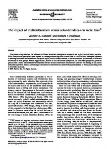
Figure 1. (A) Representative photopic ERG traces recorded from CNGA3 (red) treated eye and untreated eye (black trace) of a TKO mouse. The response of an untreated wildtype control is also shown (grey trace). Scale bar is 50µV by 50 ms. (B) Immunohistochemical staining for CNGA3 in retinas of TKO mice following injection with rAAV2/5.CBA. CNGA3 but no labelling in an area of distant to the injection in the same eye (C). Scale bar 50µM.
monochromatism [30]. These studies in animal models do, however, differ from complete achromatopsia in that both have residual pathways of cone subpopulation function. They therefore presumably have parvo-cellular retinofugal pathways and development of cortical areas such as the blobs in layers 2 and 3 of V1 and the V4 area that mediate color vision [31]. This perhaps proves indirectly that the failure to restore full color vision in the adult achromatopsia models has more to do with re-modelling of the cone visual pathway more than any intrinsic deficiencies in the cone photoreceptors themselves. The implications for human achromatopsia gene therapy are that the treatment will most likely need to be applied in early childhood to be maximally effective. ORIGINAL DATA ON CNGA3 RESCUE IN MICE Mouse models of achromatopsia remain heavily dependent upon rod function and the contribution from the non-image forming melanopsin photoreceptor pathway can also be significant [32]. To isolate any functional vision improvement mediated by cone rescue, we treated a triple knock out (TKO) mouse line that has almost no intrinsic light sensitivity [32]. The mouse is homozygous for targeted mutations in three genes: Gnat1 (rodspecific alpha transducin), Opn4 (ganglion cell specific melanopsin) and Cnga3. We demonstrated effective cone
transduction and rescue of cone ERG following gene therapy with an AAV vector expressing Cnga3. Five to 6-week-old TKO mice received 1µl subretinal injections containing 1 x 10⁹ genome particles of rAAV2/5.CBA.CNGA3. The mice received no injection in the fellow eye. Two to 3 months after injection, ERGs were recorded to assess restoration of visual function of transduced retinae in vivo. Eyes were removed postmortem, fixed in 4 percent paraformaldehyde, embedded and sectioned (18 µm) for immunohistochemistry (IHC) to detect and localize CNGA3 protein. In vivo, treated eyes showed an approximately 20 percent improvement in photopic ERG b-wave amplitude (Figure 1). The restoration of a proportion of visual function in TKO mice shows that CNGA3 transgene expression acts solely through cone photoreceptors. This is a finding that was not empirically evident from AAV rescue of other achromatopsia models that had other remaining photosensitivity pathways. The functional rescue supported findings by earlier groups that human transgene can successfully rescue mouse models of achromatopsia. On IHC, the CNGA3 signal localized to outer segments, with no CNGA3 signal in the uninjected eyes (Figure 1). This demonstrated effective vector transduction of the cone photoreceptors and correct transport and localization of the transgene. It was interesting to note that Cnga3 staining was not seen in rod photoreceptors, despite use of the ubiquitous CBA promoter. A reason
Hassall et al.: Gene therapy for color blindness
may be that CNGA3 needs to form a 3:1 tetrameric protein with CNGB3 to produce a functional cyclic GMP channel [33]. THE HUMAN RETINA IN ACHROMATOPSIA Achromatopsia fundi are grossly normal. Yet, cone structure and density, as measured with high resolution adaptive-optics SLO, may show subtle changes. Studies of CNGB3 and CNGA3 patients show variable foveal appearance on OCT and cone density [34,35]. Notably, patients with Gnat2 mutations may have almost normal cone density [34,36]. Thus, the causative mutation and cone count may limit the magnitude of any treatment effect and illustrates the overlap between achromatopsia and a slow cone dystrophy. Over half of adult CNGB3 patients have some degree of foveal hypoplasia. Foveal cone density is less than normal adults, yet cone inner segments remain intact; the cone mosaic pattern is often irregular. Cones also have abnormal reflectance patterns, however, cone density has not been shown to decline with age and is likely a static feature throughout life [35]. This metric is likely not an important influence or barrier to selecting patient age in a trial. The use of intravitreal CNTF injections to regenerate cone outer segments as in the animal models may have a role in these patients [23]. One caution is that all these studies on cone mosaics rely on manual quantification by an unreported number of observers. It is unclear if these studies accommodated the inter-observer variability of manual cone mosaic analysis in achromatopsia [37]. Animal models of achromatopsia with cone degeneration show late-stage reduction in rod ERG [20]. However, the scotopic retinal electrophysiology of affected patients is generally normal [38]. Rod photoreceptor electrophysiology is not usually affected in achromatopsia, although there are rare exceptions of CNGA3 missense changes leading to rod ERG reduction. This is presumed to be due to synaptic changes, since CNGA3 is not expressed in rods. The canine models of achromatopsia however show normal rod function [16]. In one clinical study, adult patients with congenital achromatopsia viewed photopic and scotopic visual stimuli; functional magnetic resonance imaging (fMRI) recorded the cortical retinotopic representations of foveal function [39]. The study controlled for the effects of achromatopsia nystagmus on vision. Normal controls demonstrated a region of V1 occipital cortex dedicated to the cone-specific fovea. In achromatopsia patients, this same rod-free foveolar representation in the visual cortex was activated by rod-isolating visual tests. Rod visual pathways had monopolized the unused cortical space normally dedicated to cones [39]. Thus, absence of cone visual input during childhood development remodels
547
and re-allocates retinofugal pathways and this fits with psychophysics studies which suggest that achromatopsia patients have faster rates of dark adaptation than normal [40]. ACHROMATOPSIA AND CIRCADIAN RHYTHM The human circadian rhythm is a roughly 24hour cycle; the internal body clock. It is endogenously generated, but modulated by external cues, such as light levels. Cones initiate the circadian response to light and melanopsin provides sustained input for ongoing response [41]. Cone photoreceptor electrophysiological response to light also varies by an intrinsic 24-hour cycle [42]. What effect does achromatopsia have on photopic entrainment and the sleep-wake cycle? No evidence is available for the effect of achromatopsia on circadian rhythm in humans. A small study of patients with red-green color deficiency showed no effect on serum melatonin suppression by light. Melatonin is a signaling molecule secreted in a 24-hr circadian pattern by the pineal gland. It correlates with other markers of circadian rhythm [43]. Yet, mice lacking medium wavelength cones exhibit impaired photopic photo-entrainment and phase shifting [44]. More clinical data from achromatopsia patients is needed to explore circadian disruption. GENE THERAPY CLINICAL TRIALS FOR ACHROMATOPSIA As of June 2017, there are four registered clinical trials for gene therapy in achromatopsia, of which three have commenced. All trials use the human transgene driven by cone-specific promoter fragments and packaged in an AAV capsid; details of the vectors and study design are listed in Table 2. All vectors have been previously shown to rescue animal models of achromatopsia. Patients receive delivery of the vector by subretinal injection after vitrectomy. None of the constructs include regulatory elements that increase transgene expression such as WPRE [45] or intron/exon splice sites. All trials are phase I/II and the primary outcome measure is safety. It is unclear whether the CNG-naive immune system of null mutation achromatopsia patients may react to transgene proteins expressed in the cell membrane. Mutations in CNGA3 and CNGB3 account for 70 percent of mutations in human patients; both are channel proteins that insert into the cell membrane. The modest pro-inflammatory stimulus of retinal surgery and viral capsid proteins may potentiate any pro-inflammatory effects of these transmembrane proteins. None of the clinical trials have publicly recorded plans for routine
Gene
CNGB3
CNGA3
CNGA3
CNGB3
ClinicalTrials Identifier
NCT03001310
NCT02610582
NCT02935517
NCT02599922
rAAV2tYF-PR1.7hCNGB3
rAAV2tYF-PR1.7hCNGA3 (‘AGTC402’)
rAAV.hCNGA3
AAV2/8-hCARp. hCNGB3
Vector
AAV2tYF
AAV2tYF
rAAV8
AAV5
Capsid
PR1.7 (Fragment L-Opsin pr)
PR1.7 (Fragment L-Opsin pr)
Potentially, full length red-cone opsin promoter
Human cone arrestin fragment
Promoter
Three doses, unspecified
Three doses, unspecified
low dose: ≤ 1x10e10 gp (n=3) intermediate dose: ≤ 5x10e10 vgp (n=3) high dose: ≤ 1x10e11 gp (n=3)
Unreported
Dose
Vit + subretinal injection
Unreported
Unreported
Unreported
Vit + subretinal injection
Vit + subretinal injection
Unreported
Routine immunosuppression
Vitrectomy + subretinal injection
Delivery route
Table 2. Summary of the publicly registered clinical trials for gene therapy in Achromatopsia.
USA
USA, Israel
Tubingham, Germany
United Kingdom
Locations
548 Hassall et al.: Gene therapy for color blindness
Hassall et al.: Gene therapy for color blindness
immunosuppression in treated patients. Secondary outcomes for the trials include visual acuity, electrophysiology, and color vision function. It is possible that AAV transgene expression may rescue electrophysiology without providing integrated central visual functions like color or acuity. The trials are designed to initially test the safety profile in adult patients, but three of the trials plan to include patients as young as 6 years in later groups. By comparing older and younger patients, the trials will examine differences in integrated functional cone rescue between age groups. If the pattern observed in the animal trials holds true, there may be a difference in benefit due to patient age at time of treatment. Such a difference may arise from reorganization of cortical pathways [39] or foveal hypoplasia [35]. In the one other therapeutic trial registered for achromatopsia (ClinicalTrials.gov number, NCT01648452), six CNGB3 achromatopsia patients received intraocular implants that released Ciliary Neurotrophic Factor (CNTF) [24]. CNTF provides neuroprotection to rod and cone photoreceptors in degenerative retinal disease [46]. Concurrent treatment with intravitreal CNTF in a canine model of CNGB3 achromatopsia potentiated gene therapy rescue. CNTF improved ERG rescue in older animals, overcoming a notable inability of gene therapy alone to benefit most dogs over 1 year of age [23]. Limited evidence suggested that CNTF pre-treatment de-differentiated the cone photoreceptors by elongating the outer segments to facilitate CNGA3/B3 tetrameric protein assembly. As mentioned above, in the CNTF only trial, no functional improvement in cone function occurred. There are no current plans to include CNTF pre-treatment in any of the registered human gene therapy trials. This discussion may be reignited if the early trials show difficulty in rescuing cone function in older patients. CONCLUSION AND OUTLOOK Animal trials have demonstrated that gene therapy can restore the electrophysiological function of cones. The three most common causative mutations (Cnga3, Cngb3, and Gnat2) have all been rescued, often by multiple groups using different vectors. There is some evidence that cone density declines with age. This metric is unlikely to affect the therapeutic window. Nonetheless, in a mouse model showing atypical cone degeneration, Cnga3 re-expression halted cell loss and corrected opsin mislocalization. The electrophysiological treatment effect is longlasting; over 180 days in mice and over 3 years in sheep. The effect is maximal in younger animals and more modest in older mice and dogs; making age an important consideration in study design. CNTF potentiates cone
549
rescue in animals and is an example of the adjuvant therapies that may be needed to successfully treat older patients. Optimally, treatment should occur in childhood (under 6 years). Thus, allowing functional cones to direct retinofugal pathway development. In such young patients there would be additional technical challenges to the injection surgery, but a greater potential for integrated functional rescue. Acknowledgments: Grant support: Rhodes Trust, Fight for Sight, NIHR Oxford Biomedical Research Centre, Royal College of Surgeons of Edinburgh. Disclosures: The authors have no relevant disclosures relating to achromatopsia. REFERENCES 1. Michaelides M, Hunt DM, Moore AT. The cone dysfunction syndromes. Br J Ophthalmol. 2004 Feb 1;88(2):291–7. 2. Simunovic MP, Moore AT. The cone dystrophies. Eye . 1998;12 ( Pt 3b):553–65. 3. Kohl S, Marx T, Giddings I, Jägle H, Jacobson SG, Apfelstedt-Sylla E, et al. Total colourblindness is caused by mutations in the gene encoding the alpha-subunit of the cone photoreceptor cGMP-gated cation channel. Nat Genet. 1998 Jul 1;19(3):257–9. 4. Kohl S, Baumann B, Broghammer M, Jägle H, Sieving P, Kellner U, et al. Mutations in the CNGB3 gene encoding the beta-subunit of the cone photoreceptor cGMP-gated channel are responsible for achromatopsia (ACHM3) linked to chromosome 8q21. Hum Mol Genet. 2000 Sep 1;9(14):2107–16. 5. Kohl S, Baumann B, Rosenberg T, Kellner U, Lorenz B, Vadalà M, et al. Mutations in the cone photoreceptor G-protein alpha-subunit gene GNAT2 in patients with achromatopsia. Am J Hum Genet. 2002 Aug 1;71(2):422–5. 6. Aligianis IA, Forshew T, Johnson S, Michaelides M, Johnson CA, Trembath RC, et al. Mapping of a novel locus for achromatopsia (ACHM4) to 1p and identification of a germline mutation in the alpha subunit of cone transducin (GNAT2). J Med Genet. 2002 Sep 1;39(9):656–60. 7. Chang B, Grau T, Dangel S, Hurd R, Jurklies B, Sener EC, et al. A homologous genetic basis of the murine cpfl1 mutant and human achromatopsia linked to mutations in the PDE6C gene. Proc Natl Acad Sci U S A. 2009 Nov 17;106(46):19581–6. 8. Kohl S, Coppieters F, Meire F, Schaich S, Roosing S, Brennenstuhl C, et al. A nonsense mutation in PDE6H causes autosomal-recessive incomplete achromatopsia. Am J Hum Genet. 2012 Sep 7;91(3):527–32. 9. Kohl S, Zobor D, Chiang W-C, Weisschuh N, Staller J, Menendez IG, et al. Mutations in the unfolded protein response regulator ATF6 cause the cone dysfunction disorder achromatopsia. Nat Genet. 2015 Jul 1;47(7):757 – +. 10. Mayer AK, Van Cauwenbergh C, Rother C, Baumann B, Reuter P, ACHM Study Group, et al. CNGB3 mutation spectrum including copy number variations in 485 achro-
550
Hassall et al.: Gene therapy for color blindness
matopsia patients. Hum Mutat. 2017;38(11):1579-1591. 11. McClements M, Davies WIL, Michaelides M, Young T, Neitz M, MacLaren RE, et al. Variations in Opsin Coding Sequences Cause X-Linked Cone Dysfunction Syndrome with Myopia and Dichromacy. Invest Ophthalmol Vis Sci. 2013 Feb 1;54(2):1361–9. 12. Hussels IE, Morton NE. Pingelap and Mokil Atolls: achromatopsia. Am J Hum Genet. 1972 May;24(3):304–9. 13. Sheffield VC. The vision of Typhoon Lengkieki. Nat Med. 2000 Jul 1;6(7):746–7. 14. Kotterman MA, Schaffer DV. Engineering adeno-associated viruses for clinical gene therapy. Nat Rev Genet. 2014 Jul;15(7):445–51. 15. Alexander JJ, Umino Y, Everhart D, Chang B, Min SH, Li Q, et al. Restoration of cone vision in a mouse model of achromatopsia. Nat Med. 2007 Jun;13(6):685–7. 16. Komáromy AM, Alexander JJ, Rowlan JS, Garcia MM, Chiodo VA, Kaya A, et al. Gene therapy rescues cone function in congenital achromatopsia. Hum Mol Genet. 2010 Apr 8;19(13):2581–93. 17. Biel M, Seeliger M, Pfeifer A, Kohler K, Gerstner A, Ludwig A, et al. Selective loss of cone function in mice lacking the cyclic nucleotide-gated channel CNG3. Proceedings of the National Academy of Sciences. 1999 Jun 22;96(13):7553–7. 18. Michalakis S, Mühlfriedel R, Tanimoto N, Krishnamoorthy V, Koch S, Fischer MD, et al. Restoration of cone vision in the CNGA3-/- mouse model of congenital complete lack of cone photoreceptor function. Mol Ther. 2010 Dec;18(12):2057–63. 19. Akimoto M, Filippova E, Gage PJ, Zhu X, Craft CM, Swaroop A. Transgenic mice expressing Cre-recombinase specifically in M- or S-cone photoreceptors. Invest Ophthalmol Vis Sci. 2004 Jan 1;45(1):42–7. 20. Pang J-J, Deng W-T, Dai X, Lei B, Everhart D, Umino Y, et al. AAV-mediated cone rescue in a naturally occurring mouse model of CNGA3-achromatopsia. PLoS One. 2012 Apr 16;7(4):e35250. 21. MacLaren RE. An analysis of retinal gene therapy clinical trials. Curr Opin Mol Ther. 2009 Oct 1;11(5):540–6. 22. Carvalho LS, Xu J, Pearson RA, Smith AJ, Bainbridge JW, Morris LM, et al. Long-term and age-dependent restoration of visual function in a mouse model of CNGB3-associated achromatopsia following gene therapy. Hum Mol Genet. 2011 Aug 15;20(16):3161–75. 23. Komáromy AM, Rowlan JS, Corr ATP, Reinstein SL, Boye SL, Cooper AE, et al. Transient photoreceptor deconstruction by CNTF enhances rAAV-mediated cone functional rescue in late stage CNGB3-achromatopsia. Mol Ther. 2013 Jun;21(6):1131–41. 24. Zein WM, Jeffrey BG, Wiley HE, Turriff AE, Tumminia SJ, Tao W, et al. CNGB3-Achromatopsia Clinical Trial With CNTF: Diminished Rod Pathway Responses With No Evidence of Improvement in Cone Function. Invest Ophthalmol Vis Sci. 2014 Oct 1;55(10):6301–8. 25. Shaikh RS, Reuter P, Sisk RA, Kausar T, Shahzad M, Maqsood MI, et al. Homozygous missense variant in the human CNGA3 channel causes cone-rod dystrophy. Eur J Hum Genet. 2015 Apr;23(4):473–80. 26. Banin E, Gootwine E, Obolensky A, Ezra-Elia R, Ejzen-
berg A, Zelinger L, et al. Gene Augmentation Therapy Restores Retinal Function and Visual Behavior in a Sheep Model of CNGA3 Achromatopsia. Mol Ther. 2015 Sep;23(9):1423–33. 27. Gootwine E, Abu-Siam M, Obolensky A, Rosov A, Honig H, Nitzan T, et al. Gene Augmentation Therapy for a Missense Substitution in the cGMP-Binding Domain of Ovine CNGA3 Gene Restores Vision in Day-Blind Sheep. Invest Ophthalmol Vis Sci. 2017 Mar 1;58(3):1577–84. 28. MacLaren RE, Bennett J, Schwartz SD. Gene Therapy and Stem Cell Transplantation in Retinal Disease: The New Frontier. Ophthalmology. 2016 Oct 1;123(10S):S98–106. 29. Mancuso K, Hauswirth WW, Li Q, Connor TB, Kuchenbecker JA, Mauck MC, et al. Gene therapy for red-green colour blindness in adult primates. Nature. 2009 Jan 1;461(7265):784–U34. 30. Zhang Z, Pang J, Xia F, Guo Q, Li L, An J, et al. AAV-mediated Gene Therapy Restores Cone Function In A Rat With An M-cone Opsin Deficiency, A Model For Blue Cone Monochromacy. Invest Ophthalmol Vis Sci. 2011 Apr 22;52(14):1403–1403. 31. Zeki S, Bartels A. The clinical and functional measurement of cortical (in)activity in the visual brain, with special reference to the two subdivisions (V4 and V4 alpha) of the human colour centre. Philos Trans R Soc Lond B Biol Sci. 1999 Jul 29;354(1387):1371–82. 32. Hattar S, Lucas RJ, Mrosovsky N, Thompson S, Douglas RH, Hankins MW, et al. Melanopsin and rod-cone photoreceptive systems account for all major accessory visual functions in mice. Nature. 2003 Jan 1;424(6944):76–81. 33. Peng C, Rich ED, Varnum MD. Subunit configuration of heteromeric cone cyclic nucleotide-gated channels. Neuron. 2004 May 13;42(3):401–10. 34. Dubis AM, Cooper RF, Aboshiha J, Langlo CS, Sundaram V, Liu B, et al. Genotype-dependent variability in residual cone structure in achromatopsia: toward developing metrics for assessing cone health. Invest Ophthalmol Vis Sci. 2014 Oct 2;55(11):7303–11. 35. Langlo CS, Patterson EJ, Higgins BP, Summerfelt P, Razeen MM, Erker LR, et al. Residual Foveal Cone Structure in CNGB3-associated Achromatopsia. Invest Ophthalmol Vis Sci. 2016 Aug 1;57(10):3984–95. 36. Ueno S, Nakanishi A, Kominami T, Ito Y, Hayashi T, Yoshitake K, et al. In vivo imaging of a cone mosaic in a patient with achromatopsia associated with a GNAT2 variant. Jpn J Ophthalmol. 2017 Jan;61(1):92–8. 37. Abozaid MA, Langlo CS, Dubis AM, Michaelides M, Tarima S, Carroll J. Reliability and Repeatability of Cone Density Measurements in Patients with Congenital Achromatopsia. Adv Exp Med Biol. 2016;854(Chapter 37):277–83. 38. Zobor D, Werner A, Stanzial F, Benedicenti F, Rudolph G, Kellner U, et al. The clinical phenotype of CNGA3-related achromatopsia: Pretreatment characterization in preparation of a gene replacement therapy trial. Invest Ophthalmol Vis Sci. 2017 Feb 1;58(2):821–32. 39. Baseler HA, Brewer AA, Sharpe LT, Morland AB, Jägle H, Wandell BA. Reorganization of human cortical maps caused by inherited photoreceptor abnormalities. Nat Neurosci. 2002 Apr 1;5(4):364–70.
Hassall et al.: Gene therapy for color blindness 40. Aboshiha J, Luong V, Cowing J, Dubis AM, Bainbridge JW, Ali RR, et al. Dark-Adaptation Functions in Molecularly Confirmed Achromatopsia and the Implications for Assessment in Retinal Therapy Trials. Invest Ophthalmol Vis Sci. 2014 Oct 1;55(10):6340–9. 41. Dollet A, Albrecht U, Cooper HM, Dkhissi-Benyahya O. Cones are required for normal temporal responses to light of phase shifts and clock gene expression. Chronobiol Int. 2010 Jun;27(4):768–81. 42. Cameron MA, Barnard AR, Hut RA, Bonnefont X, van der Horst GTJ, Hankins MW, et al. Electroretinography of wild-type and Cry mutant mice reveals circadian tuning of photopic and mesopic retinal responses. J Biol Rhythms. 2008 Dec;23(6):489–501. 43. Ruberg FL, Skene DJ, Hanifin JP, Rollag MD, English J, Arendt J, et al. Melatonin regulation in humans with color vision deficiencies. J Clin Endocrinol Metab. 1996 Aug;81(8):2980–5. 44. Dkhissi-Benyahya O, Gronfier C, De Vanssay W, Flamant F, Cooper HM. Modeling the role of mid-wavelength cones in circadian responses to light. Neuron. 2007 Mar 1;53(5):677–87. 45. Patrício MI, Barnard AR, Orlans HO, McClements ME, MacLaren RE. Inclusion of the Woodchuck Hepatitis Virus Posttranscriptional Regulatory Element Enhances AAV2-Driven Transduction of Mouse and Human Retina. Mol Ther Nucleic Acids. 2017 Mar 17;6:198–208. 46. Lipinski DM, Barnard AR, Singh MS, Martin C, Lee EJ, Davies WIL, et al. CNTF Gene Therapy Confers Lifelong Neuroprotection in a Mouse Model of Human Retinitis Pigmentosa. Mol Ther. 2015 Aug 1;23(8):1308–19.
551