Received: 6 March 2017
|
Revised: 26 April 2017
|
Accepted: 2 May 2017
DOI: 10.1002/dvg.23038
TECHNOLOGY REPORT
Generation and characterization of a conditional allele of Interferon Regulatory Factor 6 Arianna L. Smith1*
|
Youssef A. Kousa2,3*
|
Akira Kinoshita4* | Kate Fodor5 |
Baoli Yang6 | Brian C. Schutte1,7,8 1
Genetics PhD Program, Michigan State University, East Lansing, Michigan
2
Department of Biochemistry and Molecular Biology, Michigan State University, East Lansing, Michigan
3
College of Osteopathic Medicine, Michigan State University, East Lansing, Michigan
4
Department of Pediatrics, Nagasaki University, Nagasaki, Japan
5
College of Veterinary Medicine, Michigan State University, East Lansing, Michigan
6
Department of Obstetrics and Gynecology, The University of Iowa
7
Department of Microbiology and Molecular Genetics, Michigan State University, East Lansing, Michigan
8
Department of Pediatrics and Human Development, Michigan State University, East Lansing, Michigan
Correspondence Brian C. Schutte, 5162 Biomedical and Physical Science Building, Microbiology and Molecular Genetics, Michigan State University, East Lansing, MI 48823, USA. Email:
[email protected] Present addresses Youssef A. Kousa, Pediatric Residency Program, Children's National Health System, 111 Michigan Avenue, NW, Washington, DC, 20010 Baoli Yang, Immortagen, BioVentures Center, 2500 Crosspark Rd e156b, Coralville, IA 52241
Summary Interferon Regulatory Factor 6 (IRF6) is a critical regulator of differentiation, proliferation, and migration of keratinocytes. Mutations in IRF6 cause two autosomal dominant disorders characterized by cleft lip with or without cleft palate. In addition, DNA variation in IRF6 confers significant risk for non-syndromic cleft lip and palate. IRF6 is also implicated in adult onset development and disease processes, including mammary gland development and squamous cell carcinoma. Mice homozygous for a null allele of Irf6 die shortly after birth due to severe skin, limb, and craniofacial defects, thus impeding the study of gene function after birth. To circumvent this, a conditional allele of Irf6 was generated. To validate the functionality of the conditional allele, we used three “deleter” Cre strains: Gdf9-Cre, CAG-Cre, and Ella-Cre. When Cre expression was driven by the Gdf9-Cre or CAG-Cre transgenes, 100% recombination was observed as indicated by DNA genotyping and phenotyping. In contrast, use of the Ella-Cre transgenic line resulted in incomplete recombination, despite expression at the one-cell stage. In sum, we generated a novel tool to delete Irf6 in a tissue specific fashion, allowing for study of gene function past perinatal stages. However, recombination efficiency of this allele was dictated by the Cre-driver used. KEYWORDS
cleft lip and palate, conditional allele, Cre/lox, epithelium, IRF6, Van der Woude Syndrome
Interferon Regulatory Factor 6 (IRF6) is a member of the Interferon
mutations in IRF6 cause two autosomal dominant orofacial clefting dis-
Regulatory Factor family of transcription factors. This nine-member
orders, Van der Woude syndrome (VWS) and Popliteal Pterygium syn-
family contains a highly conserved DNA binding domain and a less
drome (Kondo et al., 2002). In addition, a DNA variant in IRF6, present
conserved protein association domain. With the exception of IRF6,
in 30% of the population worldwide, confers significant risk for non-
other members of the IRF family have been implicated in immune
syndromic cleft lip and palate, one of the most common congenital
response (Tamura, Yanai, Savitsky, & Taniguchi, 2008). Alternatively,
defects (Rahimov et al., 2008; Zucchero et al., 2004). Irf6 regulates the development of multiples tissues during embryo-
*These authors contributed equally.
genesis. 2017;55:e23038. https://doi.org/10.1002/dvg.23038
genesis. Specifically, Irf6 is expressed in embryonic skin and other
wileyonlinelibrary.com/journal/dvg
C 2017 Wiley Periodicals, Inc. V
|
1 of 8
2 of 8
|
SMITH
ET AL.
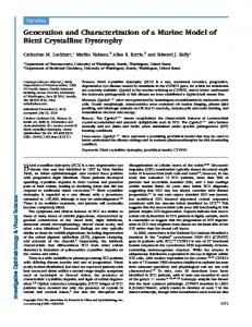
Construction of a conditional allele of Irf6. (a) A targeting vector was generated to contain three LoxP sites (triangles). The targeting vector contained a LoxP flanked PGK-neo cassette inserted 30 of exon four. A third LoxP site was incorporated 50 of exon three. (b) Cells carrying the recombinant allele were detected via PCR. Regions amplified are indicated by green tracks. (c) Southern blot analysis of ES cells. A 50 probe (indicated by blue tracks) and 30 probe (indicated by red tracks) were used. Irf61: WT allele, Irf6nl: null allele
FIGURE 1
ectodermally derived tissues (Ingraham et al., 2006; Kondo et al., 2002;
down-regulation of IRF6 was associated with squamous cell carcinoma
Richardson et al., 2006). More recent reports also indicated Irf6 expres-
(Botti et al., 2011; Restivo et al., 2011; Stransky et al., 2011). In squa-
sion and function in mesoderm derived tissues, including the tongue
mous cell carcinoma biopsies, down-regulation of IRF6 was strongly
(Goudy et al., 2013). Knockout murine models using conventional gene
associated with hypermethylation of CpG islands near the IRF6 pro-
targeting strategies were generated to study Irf6 function during
moter (Botti et al., 2011). Children with VWS have increased risk of
embryogenesis. Mice homozygous for a null allele of Irf6 (Irf6gt/gt) dis-
wound complications following surgical repair of orofacial clefts, sug-
played severe skin, limb, and craniofacial abnormalities (Ingraham et al.,
gesting a role for IRF6 in wound healing (Jones et al., 2010). Studies of
embryos at embryonic
wound closure in Irf6gt/gt embryos indicate a role for Irf6 in the regula-
day 17.5 (E17.5) had increased proliferation of the epidermis. In addi-
tion of cell migration (Biggs, Rhea, Schutte, & Dunnwald, 2012). In sum,
tion, these embryos failed to develop a stratified epidermis, lacking
Irf6 has been implicated in both congenital and adult disease processes
gt/gt
2006; Richardson et al., 2006). Skin from Irf6
both the granular and cornified layers. Thus, Irf6 is an essential regula-
of significant clinical impact. However, studies of Irf6 function post
tor of proliferation and differentiation in keratinocytes (Ingraham et al.,
embryogenesis are precluded by current mouse models because the
2006; Richardson et al., 2006).
loss of Irf6 in the mouse results in perinatal lethality. The aim of this
Other studies indicate a role for Irf6 in adult development and dis-
work was to develop a conditional allele of Irf6 to allow investigation
ease. In the mouse, Irf6 is expressed throughout development of the
of gene function post-embryogenesis in a spatial and temporal fashion.
mammary gland, reaching maximal expression during lactation (Bailey et al., 2009). Interestingly, IRF6 expression is reduced or absent in
1 | RESULTS AND DISCUSSION
breast carcinomas (Bailey et al., 2005). Re-introduction of Irf6, along with its binding partner Maspin, into breast cancer cells resulted in cell
To generate a conditional allele of Irf6 (Irf6fl), we used a three LoxP site
cycle arrest (Bailey and Hendrix, 2008; Bailey et al., 2005). In addition,
strategy (Figure 1a). Two LoxP sites flanked a Pgk-Neo selectable
SMITH
|
ET AL.
Gdf9-Cre mediated recombination of Irf6fl
T A B LE 1
tg
Gdf9-Cre/1;
Irf6
fl/1
1/1
X tg
; Irf6
gt/fl
3 of 8
Table 1). These data were consistent with the perinatal lethality observed in Irf6gt/gt. Additionally, we note the presence of Irf6fl/nl and
#
Irf6fl/1 animals and the absence of Irf6nl/nl or Irf6nl/1 animals, suggesting
Genotype
Expected
Observed
tgGdf9-Cre/1; Irf6gt/nl
5.125
0
tgGdf9-Cre/1; Irf6fl/nl
5.125
7
due to peri-natal lethality of mutant pups, we performed timed matings
tgGdf9-Cre/1; Irf6gt/1
5.125
3
between tgGdf9-Cre/1;Irf6fl/1 females and Irf6gt/1 males. Pregnant
tgGdf9-Cre/1; Irf6fl/1
5.125
6
females were sacrificed at E17.5 and three litters were collected
tg1/1; Irf6gt/nl
5.125
0
tg1/1; Irf6fl/nl
5.125
8
(cKO). In addition to severe skin, limb, and craniofacial abnormalities,
tg1/1; Irf6gt/1
5.125
5
cKO embryos had impaired barrier function (Figure 2c,d) and severe
5.125
12
oral adhesions (Figure 2e,f). These embryos lacked the Irf6fl allele and
1/1
tg
; Irf6
that the conditional allele from the sire did not recombine.
fl/1
CNG
To confirm if the observed underrepresentation of genotypes was
(N 5 34 embryos). We observed nine embryos that phenocopied Irf6gt/ gt
2 (not included)
embryos (Figure 2a,b) and were referred to as conditional knockouts
were positive for the Irf6nl and Irf6gt alleles (Figure 2e). Notably, the mutant phenotype was independent of the presence of the Gdf9-Cre
TOTAL
41
41
transgene, indicating that the Gdf9-Cre mediated recombination of the Irf6fl allele occurred prior to completion of meiosis I. Furthermore, these data demonstrate that the Irf6fl allele is capable of efficient recombina-
marker cassette in intron four, and a third LoxP site was located in intron two (Irf6ne8). Recombination between LoxP sites flanking the selectable marker produced a floxed allele with exons three and four being flanked by LoxP sites. Recombination between the remaining LoxP sites would delete exons three and four (Irf6nl). This allele is predicted to be a null allele, since exon three encodes a critical region of the DNA Binding Domain, and excision of these exons results in frameshift and the introduction of a premature stop codon. Embryonic stem cells containing the recombinant clones were acquired by G418 selection followed by PCR-based screening (Figure 1b). Excision of the floxed Pgk-Neo cassette in ES cells (SV129) was conducted by transfection with a Cre-expression vector (de Greef et al., 2016). PCR-based genotyping identified clones with the wild type, Irf6 8, Irf6 and the Irf6 alleles. Southern blot analysis confirmed ne
fl
nl
correct targeting and subsequent recombination to generate the Irf6nl allele (Figure 1c). Chimeric mice were generated by injection of ES cells
tion, resulting in a null allele. Histological analysis of embryonic skin from wildtype (WT), Irf6gt/gt and cKO embryos showed an expanded epidermis (Figure 3a–c). To confirm the molecular profile, WT, Irf6gt/gt, and cKO embryos were immunostained at E17.5. In WT embryos, Irf6 expression was seen throughout the spinous and basal layers of the epidermis. As expected, this expression was lost in Irf6gt/gt and cKO embryos (Figure 3d–f). In WT skin, Keratin14 (K14) was restricted to the basal layer. K14 expression was expanded throughout the epidermis in Irf6gt/gt and cKO embryos (Figure 3g–i). A similar pattern of expression was observed for p63, another marker of the basal layer and a proproliferation gene (Figure 3k–l). Lastly, expression of loricrin, a marker of terminally differentiated keratinocytes, was completely lost in cKO embryos (Figure 3m–o). These results are consistent with those observed in Irf6-deficient skin (Ingraham et al., 2006). To further confirm the functionality of the Irf6fl allele, we used two
fl
containing the Irf6 allele into C57Bl/6J blastocysts. Mice homozygous for the Irf6 conditional allele (Irf6fl/fl) were viable and born at the
additional Cre transgenic lines. First, we used CAG-Cre, where Cre
expected Mendelian ratio (data not shown). In addition, they showed
expression is driven by the cytomegalovirus immediate early enhancer-
no developmental or reproductive defects when compared with
chicken beta-actin promoter in mature oocytes. In this line, Cre mRNA
1/1
Irf6
fl/1
or Irf6
littermates, suggesting that this allele does not affect
or protein is store in the oocyte and can facilitate recombination following fertilization (Sakai and Miyazaki, 1997). CAG-Cre transgene posi-
normal gene function. To test the functionality of the Irf6fl allele, we employed the Gdf9-
tive (tgCAG-Cre/1; Irf61/1) animals were mated to Irf6fl/fl animals (N 5 30
Cre transgenic mouse line (tgGdf9-Cre/1). The Gdf9 promoter is sufficient
offspring). Genotypic analysis indicated that 10 animals were tgCAG-Cre/
to drive expression of Cre recombinase in the oocyte beginning at
1
. These animals showed complete recombination of the Irf6fl allele, as
postnatal day three (Lan, Xu, & Cooney, 2004). Thus, recombination at
evidenced by the presence of only the Irf6nl allele (Supporting Informa-
the Irf6 locus could only occur in females carrying the Cre transgene
tion Table S1).
fl/1
We also used the classic deleter strain, EIIa-Cre, which targets
females, we expected a recombination event in the oocyte to produce
expression of the recombinase to the stages of embryogenesis preced-
Irf61 or Irf6nl gametes. To test this, tgGdf9-Cre/1; Irf6fl/1 females were
ing implantation. We mated tgElla-Cre/1; Irf6gt/1 animals with Irf6gt/fl ani-
mated to Irf6gt/fl males. If recombination of the conditional allele was
mals (N 5 124 embryos). This allowed us to produce WT or mutant
complete, we expect 25% of embryos to phenocopy embryos deficient
embryos carrying two genotypes: (1) Irf6gt/gt or (2) tgElla-Cre/1; Irf6gt/nl.
for Irf6 (Ingraham et al., 2006). From these matings, six litters were col-
Sixty-five embryos were tgElla-Cre/1. Of these embryos, 19 were also
lected at weaning (N 5 43 pups). We observed a loss of two expected
positive for the Irf6fl allele but showed no evidence of recombination.
genotypes, tgGdf9-Cre/1; Irf6gt/nl and tg1/1; Irf6gt/nl (X2-test; p 5 0.002;
We observed eight tgElla-Cre/1 embryos that showed incomplete
Gdf9-Cre/1
and the conditional Irf6 allele (tg
fl/1
; Irf6
Gdf9-Cre/1
). In tg
; Irf6
4 of 8
|
SMITH
ET AL.
cKO phenocopy Irf6gt/gt embryos. (a) WT and (b) cKO embryos generated from Gdf9-Cre mediated recombination of the Irf6fl allele. cKO embryos (d); genotype - Irf6gt/nl with or without the Gdf9-Cre transgene) display severe skin, limb, and craniofacial abnormalities and impaired barrier function, as indicated by a dye exclusion assay, when compared to WT littermates (c). Cross sections of WT (e) and cKO (f) heads indicate severe oral adhesions in cKO embryos (P – palate; T – tongue). PCR-based genotyping for the Gdf9-Cre transgene (g), Irf61 and Irf6fl (h), Irf6nl (i), and Irf6gt (j). Complete recombination of the Irf6fl is observed irrespective of whether embryos were positive for the Gdf9-Cre transgene. cKO embryos were also positive for the Irf6gt allele. No embryos were positive for the Irf6fl allele FIGURE 2
recombination, as indicated by the presence of both the Irf6fl and Irf6nl
by presence of the Irf6nl only (Table 2). Strikingly, only one embryo phe-
Ella-Cre/1
alleles (Figure 4). These embryos were grossly normal. Nine tg
nocopied Irf6gt/gt embryos (tgElla-Cre/1; Irf6gt/nl), even though three
embryos showed complete recombination in tail tissues, as indicated
embryos had the correct genotype to generate the mutant phenotype
Molecular profile of embryonic skin. H&E staining of (a) WT, (b) Irf6gt/gt, and (c) cKO skin. Black dashed line indicates the basal layer of the epidermis. The spinous layer (sp) is indicated in (a). Immunofluorescent staining for Irf6 (d–f), K14 (g–i), p63 (j–l), and Loricrin (m–o). Images acquired using brightfield fluorescent microscope. Scale bars represent 100mm
FIGURE 3
SMITH
|
ET AL.
5 of 8
tent to knockout other genes in oral epithelium that were required for palatogenesis (He et al., 2011; Xiong et al., 2009), and Irf6 function in oral epithelium is required for palatogenesis (Ingraham et al., 2006; Richardson, Dixon, Jiang, & Dixon, 2009), no pathology was observed during palatogenesis (Chu et al., 2016). Thus, the failure of multiple Cre drivers to recombine the Irf6fl allele is consistent with the hypothesis that a cis effect at the Irf6 locus is inhibiting Cre recombination. While, we do not know the mechanism underlying the tissue dependence of Cre recombination at the Irf6 locus, one possible explanation is variation in chromatin structure at the Irf6 locus. In support of this hypothesis, a previous study showed that hypermethylation of the PCR-based genotyping of EIIa-Cre transgenic embryos (a), Irf61 and Irf6fl (b) and Irf6nl (c) from E17.5 embryos. When using the EIIa-Cre transgene was used, we observed embryos with no recombination (Embryo 1), complete recombination (Embryo 2), or incomplete recombination (Embryos 3 and 4) of the Irf6fl allele. The Irf6nl was not detected in the absence of the EIIa-Cre transgene (Embryo 5)
FIGURE 4
in the event of recombination (tgElla-Cre/1; Irf6gt/fl). This finding was in accordance with published results where complete recombination using the EIIa-cre allele was only observed in 50% of animals (Lakso et al., 1996). We created a conditional allele for Irf6. While the allele is capable of recombination, we observed variable efficiency that appeared to be dependent on the cell type. While this could simply reflect variable expression or activity of individual Cre transgenes or recombinase, respectively (Bao, Ma, Schuster, Lin, & Yan, 2013; McLellan, Rosenthal, & Pinto, 2017), we hypothesize that a cis effect at the Irf6 locus contributes to the variable efficiency. This hypothesis is based on the following: (1) In this study, we observed no or incomplete recombination with the EIIa-Cre “deleter strain,” and with two other tissue specific Cre transgenes, including K14-Cre-ER (Vasioukhin, Degenstein, Wise, & Fuchs, 1999) and Tgfb3-Cre (Yang, Li, & Kaartinen, 2008; data not shown). (2), A previous study used this Irf6fl allele to examine the role of Irf6 in dental epithelium, using the Pitx2-Cre transgene (Chu et al., 2016). While the Pitx2-Cre driver was previously shown to be compe-
IRF6 promoter region was associated with down-regulation of expression in squamous cell carcinoma (Botti et al., 2011; Rotondo et al., 2016; Stransky et al., 2011). DNA methylation is associated with nucleosome compaction, rendering the DNA inaccessible to transcription factors required to facilitate gene expression. As a result, in tissue types where Irf6 is methylated, we hypothesize that the DNA was inaccessible to Cre recombinase. Long and Rossi (2009) showed that methylation of promoter elements upstream of reporter genes activated by Cre expression (Z/AP and Z/EG strains) inhibited Cre-mediated recombination (Long and Rossi, 2009). Further studies to determine the methylation state of Irf6 in different cell types must be done to confirm this hypothesis. Finally, we note that Ella-Cre was not an effective transgene for assessing the functionality of newly derived conditional alleles. Instead, we propose the usage of oocyte specific Cre-drivers, such as Gdf9-Cre, or other more efficient one-cell stage drivers, such as CAG-Cre, to validate the functionality of conditional alleles. However, it is important to note that the mating strategy employed for these studies may have promoted the observed recombination inefficiency. It would be beneficial to address recombination with tgElla-Cre/1; Irf6fl/1 females.
2 | METHODS 2.1 | Generation of a conditional allele for Irf6 Mouse BAC clone (RPCI22–516G1) was digested with restriction enzymes. A 1.8 kb KpnI/BamHI fragment for the 50 -arm and a 3.9 kb of
T A B LE 2
tg
EIIa-Cre mediated recombination of Irf6fl
EIIa-Cre/1;
BamHI/HindIII fragment for the 30 -arm were cloned into pBluescript II SK(-) (Agilent Technologies). Three kilobases (kb) of the BamHI frag-
Irf6gt/1 x tg1/1; Irf6gt/fl
ment, containing exons three and four, was cloned into the ploxP3-
Ella-Cre genotype
total
Irf6 genotype
Total
tg1/1
46
Irf6fl
18
Irf6fl and Irf6nl
0
Pgk-Neo cassette, was subcloned into the BamHI site between 50 - and
0
30 -arms (Figure 1a). The resulting targeting construct was digested with
19
NotI and electroporated into mouse R1 ES cells. After G418 selection,
Irf6 EIIa/1
tg
66
Irf6
nl fl fl
Irf6 and Irf6 Irf6nl
nl
8 9
Data represent offspring from 15 litters collected at E17.5. Mice lacking the Ella-Cre transgene showed no recombination of the Irf6fl allele. Mice carrying the Ella-Cre transgene showed mosaic recombination of Irf6fl. Some offspring showed no recombination (Irf6fl only), presence of both the Irf6fl and Irf6nl alleles, or complete recombination (Irf6nl only).
Neo-pA vector (kind gift from Professor Takeshi Yagi, Osaka University). 5.8 kb of the XbaI fragment, which contains floxed exons and
ES cells were screened by PCR. Primer set of 50 -GAGAAATA GGGCCTTCACGGTG-30 (sense) and 50 -TGTGCCCTCTGATGCTGGAA CAG-30 (antisense) for 50 -side, 50 -TCGCCTTCTTGACGAGTTCTTCT G-30 (sense, in Pgk-Neo cassette) and 50 -GCTCAACTCCCTTTGTGACT GTCC-30 (antisense) for 30 side were used (Figure 1b). Recombinant ES clones were used for establishment of Irf6 hypomorphic mouse (Irf6ne8). To establish floxed exons (fl) and null (nl) strains, the Pgk-Neo cassette
6 of 8
|
SMITH
ET AL.
and the floxed exons, respectively, were removed in the recombinant
608C for 50 s, (4) 728C for 15 s, (5) repeat steps 2–4 40 times, and (6)
ES cells by transfection with a Cre expression vector (de Greef et al.,
728C for 5 min. PCR Master Mix was supplemented with 5M Betaine
2016). Resultant clones were then screened by Southern hybridization.
monohydrate to aid in amplification of GC-rich regions.
Genomic DNA was digested with ApaL1 or Nhe1 and hybridized to a 50 or 30 probe, respectively (Figure 1c). The 50 probe corresponded to a
2.5 | Detection of cre transgenes
region in intron two. The 30 probe corresponded to a region within exon and intron seven. Hybridization with the 50 probe following restriction enzyme digest resulted in two products, 11.3kb (WT allele) and 8.3kb (null). Hybridization with the 30 probe also produced two
The Gdf9-Cre transgene was detected as described by Lan et al. (2004) using primer set of 50 -TCTGATGAAGTCAGGAAGAACC-30 and 50 GAGATGTCCTTCACTCTGATTC-30 . PCR conditions were (1) 958C for 5
fragments, 14.3kb (WT) and 11.3kb (null). The 50 hybridization probe
min, (2) 958C for 1 min, (3) 588C for 2 min, (4) 728C for 1 min, (5) repeat
was amplified with primer set 50 -AGTTGTGACTGACTGTAGGATCAGG-30
steps 2–4 35 times, (6) 728C for 5 min. Master mix was supplemented
(forward) and 50 -ACCAAAACTTCACCAGGAGTATAGGA-30 (reverse). The
with 5M Betaine monohydrate to aid in amplification of GC-rich regions.
0
0
3 hybridization probe was amplified with primer set of 5 -AGAGTAAAGAATGGTTGTCAGTGGAG-30 (forward) and 50 -GACACCAGTATTCAA-
The EIIa-Cre transgene was detected using primer set of 50 GCGGTCTGGCAGTAAAAACTATC-30 and 50 -GTGAAACAGCATTGCTGT-
GAGGATTGAG-30 (reverse) (Figure 1c). To generate a conditional
CACTT-30 . PCR conditions were (1) 948C for 3 min, (2) 948C for 30 s,
mouse line for Irf6, embryonic stem cells carrying the Irf6fl allele were
(3) 51.78C for 1 min, (4) 728C for 1 min, (5) repeat steps 2–4 35 times,
injected into C57Bl/6J blastocyst and inserted into pseudo-pregnant
and (6) 728C for 5 min.
dams. Chimeric males were mated to C57Bl/6J females and germline transmission
was
determined
by
Polymerase
Chain
Reaction
The CAG-Cre transgene was detected using primer set of 50 -CCTACAGCTCCTGGGCAACGTGC-30
and 50 -CTAATCGCCATCTTCC
0
(PCR)-based genotyping of progeny. The Irf6fl/fl mice reported here are
AGCAGG-3 . PCR conditions were (1) 948C for 3 min, (2) 948C for 30 s,
available to the scientific community.
(3) 608C for 30 s, (4) 728C for 2 min, (5) repeat steps 2–4 30 times, and (6) 728C for 5 min.
2.2 | Mouse and embryo genotyping 3 | MATING STRATEGIES To attain genomic DNA, tail tissue was digested in lysis buffer (10 mM Tris-HCl, 150 mM NaCl, 10 mM EDTA, 0.1% SDS) with Proteinase-K
3.1 | Gdf9-cre
(20 mg/ml, Roche) at 558C, overnight, followed by ethanol precipitation of DNA. PCR-based genotyping was used to identify alleles (see
A male hemizygous for the Gdf9-iCre transgene (tgGdf9-Cre/1) was pur-
below). All PCR was conducted using JumpStart REDTaq Ready Mix
chased from Jackson Labs (www.jax.org) and mated to Irf6fl/fl females
(Sigma-Aldrich, St. Louis, MO) and separated and visualized by electro-
to generate females carrying both the Cre transgene and one copy of
phoresis on 1.5% agarose gels.
the Irf6fl allele (tgGdf9-Cre/1; Irf6fl/1). These females were mated to compound heterozygous males for Irf6 (Irf6gt/fl). Tail snips were collected from pups upon weaning and genotyped. A chi-squared test was used
2.3 | Detection of the Irf6ne8, Irf6fl, and Irf6nl alleles Primer set of 50 -GCAGAGTGGAGCACACTTCA-30 and 50 -AAGCATGTCTATTTGGGGGTT-30 was used to determine the absence of the 50 LoxP site in the Irf61 (221 bp) but its presence (570 bp) in the Irf6fl and Irf6ne8 alleles. Primer set of 50 -TGGCAAAATCTATTTCGAGTGG-30 and 50 CACACTGACCTCAATGTCCAA-30 was used to determine the absence of the 30 LoxP site in the Irf61 (222 bp) but its presence (379 bp) in the Irf6fl alleles. This primer set also distinguished the Irf6fl and Irf6ne8
to determine deviations from expected Mendelian ratios. TgGdf9-Cre/1; Irf6fl/1 females were then placed into timed matings with males heterozygous for a null allele of Irf6 (Irf6gt/1). The presence of a copulation plug was denoted as embryonic day 0.5 (E0.5) and embryos were collected on E17.5.
3.2 | EIIa-cre
alleles, as these primers do not amplify a product from the Irf6ne8 allele
A homozygous EIIa-Cre male (tgElla-Cre/Ella-Cre) was purchased from Jack-
under these conditions because the expected product is too large.
son Labs (www.jax.org) and mated to Irf6gt/1 females to produce dou-
Primer set of 50 -GCAGAGTGGAGCACACTTCA-30 and 50 -CACACT-
ble heterozygous tgElla-Cre/1;Irf6gt/1 mice. Timed matings were set up
GACCTCAATGTCCAA-30 was used to detect the Irf6nl allele (499 bp).
between tgElla-Cre/1;Irf6gt/1 animals and Irf6gt/fl animals.
PCR was performed as follows: (1) 958C for 2 min, (2) 958C for 15 s, (3) 558C for 15 s, (4) 728C for 45 s, (5) repeat steps 2–4 35 times, and (6) 728C for 5 min.
Validation of the Irf6fl allele using CAG-Cre was conducted multiple ways. 1) Females homozygous (tgCAG-Cre/CAG-Cre) or heterozygous
2.4 | Detection of the Irf6gt allele Primer
set
of
3.3 | CAG-cre
(tgCAG-Cre/1) for the CAG-Cre transgene were mated to an Irf6fl/fl males. 2)
0
0
5 -GACCAGACCGTGCAGGGGCTGTGG-3
GAGAGGCTAGGGTGGAAGGGATTC-30
identifies
the
and gt
Irf6
0
5-
TgCAG-Cre/1 males were mated to Irf6fl/fl females. Litters were born and
allele
tail snips were collected upon weaning and subjected to PCR based gen-
(283bp). PCR conditions were (1) 958C for 3 min, (2) 958C for 38 s, (3)
otyping. A chi-squared test was used to verify Mendelian ratios.
SMITH
|
ET AL.
3.4 | Embryo collection and processing
7 of 8
(Life Technologies, Grand Island, NY). Images were taken using a Nikon Eclipse 90i fluorescent microscope.
On E17.5, pregnant females were sacrificed using isoflurane induced comatosis followed by cervical dislocation. Embryos were decapitated. Heads and bodies were fixed overnight in 10% neutral buffered forma-
ACKNOWLEDGEMENTS
lin at 48C. Following fixation, embryos were processed and embedded
Funding for this work was provided by National Institute of Health
in paraffin by the Histopathology Laboratory at Michigan State Univer-
grants DE13513 (B.C.S.) and F31DE022696 (Y.A.K.). The authors
sity using standard protocols. All animals were used in accordance with
thank the Michigan State University Histopathology Core and animal
the National Institutes of Health Guide for the Care and Use of Labora-
care facility for their services. The authors also thank Jeannie I. Kla-
tory Animals, and all procedures were approved by the Michigan State
vanian, Krysta Wierzbicki, Kendra Siegersma, and Raeuf Roushangar
University Institutional Animal Use and Care Committee.
for their technical support.
3.5 | Histological analysis
R EF ER E N CE S
Whole heads from embryos at E17.5 were sectioned coronally at 7mm. Sections were then stained with hematoxylin and eosin (H&E). Briefly, for depariffinization and rehydration, sections were passed through three changes of xylene followed by washes through reducing grades of ethanol. Slides were then incubated in Gill’s Hematoxylin No. III (Sigma-Aldrich, St. Louis, MO) for 1.5 min, washed briefly in tap water, and incubated in a 1% Eosin solution (Eosin Y-VWR, West Chester, PA) for 1.5 min. To dehydrate, slides were passed through increasing grades of ethanol. Lastly, slides were mounted using Permount mounting media (VWR, Radnor, PA). Stained sections were imaged using a Nikon 90i upright microscope.
3.6 | Dye exclusion assay The dye exclusion assay was carried out as described by Ingraham et al. (2006). Briefly, whole embryos were collected at E17.5 and fixed in 100% methanol for 5 min. Following fixation, embryos were rinsed briefly in 13 Phosphate Buffered Saline (PBS) and stained in 0.1% toluidine blue for 1 min. Staining was followed by washing with 13 PBS.
3.7 | Immunofluorescence For immunofluorescent detection of markers of the epidermis, sections were deparaffinized and rehydrated in reducing concentrations of ethanol. Sections were subjected to antigen retrieval by boiling in 10 mM Sodium citrate (pH 5 6.0) for 30 min. Tissues sections were permeabilized using 0.5% Triton X-100 followed by blocking in blocking solution (10% normal goat serum, 0.1% Bovine Serum Albumin in 13 PBS) for 1 hr at room temperature. Sections were then incubated in antimouse F0 (ab) fragment (Jackson ImmunoResearch Laboratories, West Grove, PA) for 5 min to reduce nonspecific binding of antimouse secondary. Sections were incubated with 1:100 rabbit anti-Irf6 (Sigma-Aldrich; St. Louis, MO), 1:250 rabbit anti-Keratin 14 (Covance), 1:150 mouse antip63 (Santa Cruz), and 1:250 rabbit anti-loricrin (Covance) overnight at 48C, followed by incubation with either goat antimouse AlexaFluor 488 or goat antirabbit AlexaFluor 555 (Life Technologies, Grand Island, NY). Nuclei were counterstained for 10 minutes in a 1:10,000 dilution of 40 6- diaminidino-2-phenylindole (DAPI; Life Technologies, Grand Island, NY). Slides were then mounted in ProLong GOLD Antifade reagent
Bailey, C. M., & Hendrix, M. J. (2008). IRF6 in development and disease: A mediator of quiescence and differentiation. Cell Cycle, 7, 1925– 1930. Bailey, C. M., Khalkhali-Ellis, Z., Kondo, S., Margaryan, N. V., Seftor, R. E., Wheaton, W. W., . . . Hendrix, M. J. (2005). Mammary serine protease inhibitor (Maspin) binds directly to interferon regulatory factor 6: Iidentification of a novel serpin partnership. Journal of Biological Chemistry, 280, 34210–34217. Bailey, C. M., Margaryan, N. V., Abbott, D. E., Schutte, B. C., Yang, B., Khalkhali-Ellis, Z., & Hendrix, M. J. (2009). Temporal and spatial expression patterns for the tumor suppressor Maspin and its binding partner interferon regulatory factor 6 during breast development. Development, Growth & Differentiation, 51, 473–481. Bao, J., Ma, H. Y., Schuster, A., Lin, Y. M., & Yan, W. (2013). Incomplete cre-mediated excision leads to phenotypic differences between Stra8-iCre; Mov10l1(lox/lox) and Stra8-iCre; Mov10l1(lox/Delta) mice. Genesis, 51, 481–490. Biggs, L. C., Rhea, L., Schutte, B. C., & Dunnwald, M. (2012). Interferon regulatory factor 6 is necessary, but not sufficient, for keratinocyte differentiation. Journal of Investigative Dermatology, 132, 50–58. Botti, E., Spallone, G., Moretti, F., Marinari, B., Pinetti, V., Galanti, S., . . . Costanzo, A. (2011). Developmental factor IRF6 exhibits tumor suppressor activity in squamous cell carcinomas. Proceedings of the National Academy of Sciences of the United States of America, 108, 13710–13715. Chu, E. Y., Tamasas, B., Fong, H., Foster, B. L., LaCourse, M. R., Tran, A. B., . . . Cox, T. C. (2016). Full Spectrum of Postnatal Tooth Phenotypes in a Novel Irf6 Cleft Lip Model. Journal of Dental Research, 95, 1265–1273. de Greef, J. C., Hamlyn, R., Jensen, B. S., O’campo Landa, R., Levy, J. R., Kobuke, K., & Campbell, K. P. (2016). Collagen VI deficiency reduces muscle pathology, but does not improve muscle function, in the gamma-sarcoglycan-null mouse. Human Molecular Genetics, 25, 1357– 1369. Goudy, S., Angel, P., Jacobs, B., Hill, C., Mainini, V., Smith, A. L., . . . Schutte, B. C. (2013). Cell-autonomous and non-cell-autonomous roles for IRF6 during development of the tongue. PLoS One, 8, e56270. He, F., Xiong, W., Wang, Y., Li, L., Liu, C., Yamagami, T., . . . Chen, Y. (2011). Epithelial Wnt/beta-catenin signaling regulates palatal shelf fusion through regulation of Tgfbeta3 expression. Developmental Biology, 350, 511–519. Ingraham, C. R., Kinoshita, A., Kondo, S., Yang, B., Sajan, S., Trout, K. J., . . . Schutte, B. C. (2006). Abnormal skin, limb and craniofacial morphogenesis in mice deficient for interferon regulatory factor 6 (Irf6). Nature Genetics, 38, 1335–1340.
8 of 8
|
Jones, J. L., Canady, J. W., Brookes, J. T., Wehby, G. L., L’heureux, J., Schutte, B. C., . . . Dunnwald, M. (2010). Wound complications after cleft repair in children with Van der Woude syndrome. Journal of Craniofacial Surgery, 21, 1350–1353. Kondo, S., Schutte, B. C., Richardson, R. J., Bjork, B. C., Knight, A. S., Watanabe, Y., . . . Murray, J. C. (2002). Mutations in IRF6 cause Van der Woude and popliteal pterygium syndromes. Nature Genetics, 32, 285–289. Lakso, M., Pichel, J. G., Gorman, J. R., Sauer, B., Okamoto, Y., Lee, E., . . . Westphal, H. (1996). Efficient in vivo manipulation of mouse genomic sequences at the zygote stage. Proceedings of the National Academy of Sciences of the United States of America, 93, 5860–5865. Lan, Z. J., Xu, X., & Cooney, A. J. (2004). Differential oocyte-specific expression of Cre recombinase activity in GDF-9-iCre, Zp3cre, and Msx2Cre transgenic mice. Biology of Reproduction, 71, 1469–1474.
SMITH
ET AL.
Squamous Cell Carcinoma Associated With Lichen Sclerosus. JAMA Dermatology, 152, 928–933. Sakai, K., & Miyazaki, J. (1997). A transgenic mouse line that retains Cre recombinase activity in mature oocytes irrespective of the cre transgene transmission. Biochemical and Biophysical Research Communications, 237, 318–324. Stransky, N., Egloff, A. M., Tward, A. D., Kostic, A. D., Cibulskis, K., Sivachenko, A., . . . Grandis, J. R. (2011). The mutational landscape of head and neck squamous cell carcinoma. Science, 333, 1157–1160. Tamura, T., Yanai, H., Savitsky, D., & Taniguchi, T. (2008). The IRF family transcription factors in immunity and oncogenesis. Annual Review of Immunology, 26, 535–584.
Long, M. A., & Rossi, F. M. (2009). Silencing inhibits Cre-mediated recombination of the Z/AP and Z/EG reporters in adult cells. PLoS One, 4, e5435.
Vasioukhin, V., Degenstein, L., Wise, B., & Fuchs, E. (1999). The magical touch: genome targeting in epidermal stem cells induced by tamoxifen application to mouse skin. Proceedings of the National Academy of Sciences of the United States of America, 96, 8551– 8556.
McLellan, M. A., Rosenthal, N. A., & Pinto, A. R. (2017). Cre-loxP-Mediated Recombination: General Principles and Experimental Considerations. Current Protocols in Mouse Biology, 7, 1–12.
Xiong, W., He, F., Morikawa, Y., Yu, X., Zhang, Z., Lan, Y., . . . Chen, Y. (2009). Hand2 is required in the epithelium for palatogenesis in mice. Developmental Biology, 330, 131–141.
Rahimov, F., Marazita, M. L., Visel, A., Cooper, M. E., Hitchler, M. J., Rubini, M., . . . Murray, J. C. (2008). Disruption of an AP-2alpha binding site in an IRF6 enhancer is associated with cleft lip. Nature Genetics, 40, 1341–1347.
Yang, L. T., Li, W. Y., & Kaartinen, V. (2008). Tissue-specific expression of Cre recombinase from the Tgfb3 locus. Genesis, 46, 112–118.
Restivo, G., Nguyen, B. C., Dziunycz, P., Ristorcelli, E., Ryan, R. J., € € . . . Dotto, G. P. (2011). IRF6 is a mediator of Notch proOzuysal, O., differentiation and tumour suppressive function in keratinocytes. The EMBO Journal, 30, 4571–4585. Richardson, R. J., Dixon, J., Jiang, R., & Dixon, M. J. (2009). Integration of IRF6 and Jagged2 signalling is essential for controlling palatal adhesion and fusion competence. Human Molecular Genetics, 18, 2632–2642.
Zucchero, T. M., Cooper, M. E., Maher, B. S., Daack-Hirsch, S., Nepomuceno, B., Ribeiro, L., . . . Murray, J. C. (2004). Interferon regulatory factor 6 (IRF6) gene variants and the risk of isolated cleft lip or palate. New England Journal of Medicine, 351, 769–780.
S UPPORTING INF ORMATION Additional Supporting Information may be found online in the supporting information tab for this article.
Richardson, R. J., Dixon, J., Malhotra, S., Hardman, M. J., Knowles, L., Boot-Handford, R. P., . . . Dixon, M. J. (2006). Irf6 is a key determinant of the keratinocyte proliferation-differentiation switch. Nature Genetics, 38, 1329–1334.
How to cite this article: Smith AL, Kousa YA, Kinoshita A, Fodor
Rotondo, J. C., Borghi, A., Selvatici, R., Magri, E., Bianchini, E., Montinari, E., . . . Martini, F. (2016). Hypermethylation-Induced Inactivation of the IRF6 Gene as a Possible Early Event in Progression of Vulvar
2017;55:e23038. https://doi.org/10.1002/dvg.23038
K, Yang B, Schutte BC. Generation and characterization of a conditional allele of Interferon Regulatory Factor 6. genesis.