Clinical Chemistry 52:10 1855–1863 (2006)
Molecular Diagnostics and Genetics
Genotype-Specific Signal Generation Based on Digestion of 3-Way DNA Junctions: Application to KRAS Variation Detection Giulia Amicarelli,1 Daniel Adlerstein,2* Erlet Shehi,2 Fengfei Wang,3 and G. Mike Makrigiorgos3
Background: Genotyping methods that reveal singlenucleotide differences are useful for a wide range of applications. We used digestion of 3-way DNA junctions in a novel technology, OneCutEventAmplificatioN (OCEAN) that allows sequence-specific signal generation and amplification. We combined OCEAN with peptide-nucleic-acid (PNA)-based variant enrichment to detect and simultaneously genotype v-Ki-ras2 Kirsten rat sarcoma viral oncogene homolog (KRAS) codon 12 sequence variants in human tissue specimens. Materials and Methods: We analyzed KRAS codon 12 sequence variants in 106 lung cancer surgical specimens. We conducted a PNA-PCR reaction that suppresses wild-type KRAS amplification and genotyped the product with a set of OCEAN reactions carried out in fluorescence microplate format. The isothermal OCEAN assay enabled a 3-way DNA junction to form between the specific target nucleic acid, a fluorescently labeled “amplifier”, and an “anchor”. The amplifier-anchor contact contains the recognition site for a restriction enzyme. Digestion produces a cleaved amplifier and generation of a fluorescent signal. The cleaved amplifier dissociates from the 3-way DNA junction, allowing a new amplifier to bind and propagate the reaction. Results: The system detected and genotyped KRAS sequence variants down to ⬃0.3% variant-to-wild-type alleles. PNA-PCR/OCEAN had a concordance rate with PNA-PCR/sequencing of 93% to 98%, depending on the
1 Department of Biotechnology and Biosciences, University of Milano Bicocca, Milano, Italy. 2 DiaSorin SpA, Saluggia (VC), Italy. 3 Dana Farber-Brigham and Women’s Cancer Center, Harvard Medical School, Boston, MA. * Address correspondence to this author at: Diasorin SpA, Piazza della Scienza 4, 20126 Milano, Italy. Fax 0039-02-6448-3565; e-mail daniel.
[email protected]. Received February 16, 2006; accepted July 21, 2006. Previously published online at DOI: 10.1373/clinchem.2006.068817
exact implementation. Concordance rate with restriction endonuclease-mediated selective-PCR/sequencing was 89%. Conclusion: OCEAN is a practical and low-cost novel technology for sequence-specific signal generation. Reliable analysis of KRAS sequence alterations in human specimens circumvents the requirement for sequencing. Application is expected in genotyping KRAS codon 12 sequence variants in surgical specimens or in bodily fluids, as well as single-base variations and sequence alterations in other genes. © 2006 American Association for Clinical Chemistry
Detection methods for single-nucleotide variants [also known as single nucleotide polymorphisms (SNPs)4] and sequence alterations are important in the genetic analysis of clinical samples for diagnosis, prognosis, and drug discovery. Single-nucleotide variants and sequence alterations detected in genomic DNA have proven to be powerful genetic markers for numerous medical and genetic applications, including a range of inherited disorders (1, 2 ), apolipoprotein-E single-nucleotide variants (3, 4 ), sequence alterations in hemochromatosis (5 ), and cancer (6 –16 ). In a clinical/laboratory setting, methods that reveal single-nucleotide differences should be specific, reliable, rapid, simple, and inexpensive. In most current methods, nucleic acid amplification via PCR (17 ) from genomic DNA is the first step in the identification of single-nucleotide variants and sequence alterations. Detection is performed either during PCR [e.g., TaqMan real-time PCR (1 )] or after PCR [e.g., primer-extension, dHPLC, matrix-assisted laser desorp-
4 Nonstandard abbreviations: SNPs, single nucleotide polymorphisms; OCEAN, OneCutEventAmplificatioN; PNA, peptide-nucleic acid; REMS, restriction endonuclease-mediated selective; NSCLC, non–small cell lung cancer; NL, normal lung tissue; TL, tumor lung tissue.
1855
1856
Amicarelli et al.: Genotype-Specific Signal Generation
tion/ionization time-of-flight (MALDI-TOF), or beadsbased approaches (18 –21 )]. Detection of single-nucleotide differences during PCR is more convenient but usually requires an additional screening step to provide the specific genotype, and some postPCR assays, such as dHPLC, may require a third step. We describe a novel signal generation and amplification technology that enables simultaneous detection and genotyping of singlenucleotide variants and sequence alterations in a single postPCR step and combines most of the requirements for convenient single-nucleotide genotyping.
Materials and Methods The system consists of a single-tube isothermal amplification reaction based on the hybridization of 2 oligonucleotides probes (an “amplifier” and an “anchor”) to the target DNA, e.g., a v-Ki-ras2 Kirsten rat sarcoma viral oncogene homolog (KRAS)5 codon 12-containing sequence. The target DNA is single stranded and is generated from genomic DNA via an asymmetric PCR reaction, after which the target amplifier and anchor are hybridized to form a 3-way DNA junction that contains the recognition site for a thermostable restriction endonuclease (Fig. 1). Enzymatic digestion produces a cut amplifier that, because of its lower thermal stability, dissociates from the complex, allowing a new, intact amplifier to participate in the isothermal reaction. Digestion of the anchor-oligonucleotide by the enzyme is prevented by phosphorothioate modification; therefore, the anchor remains available to participate in successive steps of the reaction. Double-labeling of the amplifier with a fluorescent probe and a quencher (Fig. 1) generates a fluorescent signal by eliminating fluorescent quenching each time the amplifier is digested (22 ). Repeated digestion of the amplifier leads to a continuously increasing fluorescent signal that is monitored homogeneously. The stability of the 3-way DNA junction at the selected temperature is dependent on the correct hybridization of the 3 sequences. Therefore, if the amplifier and the interrogated DNA sequence differ by a single nucleotide in the match-region, the junction is destabilized and the fluorescent signal is diminished. Thus, with appropriate design of the amplifier sequence, any chosen nucleotide change on the target DNA sequence can be genotyped. To validate this OneCutEventAmplificatioN (OCEAN) signal-generation method (23 ) and adapt it to the detection of clinically relevant sequence variants, we designed assays appropriate for genotyping codon-12 sequence alterations in the KRAS gene. We tested the application of OCEAN in model-oligonucleotide systems, in cell lines with KRAS sequence variants, and in lung cancer surgical specimens. We compared our results with those obtained with peptide-nucleic acid (PNA)-PCR (24 ), restriction endonuclease-mediated selective (REMS)-PCR (25 ), and
5
Human gene: KRAS, v-Ki-ras2 Kirsten rat sarcoma viral oncogene homolog.
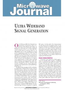
Fig. 1. OCEAN principles. OCEAN is an isothermal and homogeneous reaction based on the selective hybridization of two probes (an “amplifier” and an “anchor”) to the specific target nucleic acid (step 1). The target-anchor duplex forms with the specific (matched) amplifier a ternary structure, which contains the recognition site for a restriction endonuclease, while the structure is not stable for the nonspecific amplifier (step 2). Digestion with the enzyme produces a cleaved amplifier (step 3), which then dissociates (step 4), while the duplex target-anchor is recycled in the reaction. The amplifier is a dual labeled oligonucleotide containing an internal fluorophore and a quencher at 3⬘ (BHQ). During the reaction, as shown in step 4, the enzymatic cleavage separates the fluorophore and the quencher resulting in an increase of fluorescence. A phosphorothioate modification of the anchor probe confers resistance to endonuclease activity allowing the recycling of the targetanchor duplex in the reaction. The assay results in a sequence-specific signal amplification.
sequencing. We demonstrate that OCEAN combined with asymmetric PNA-PCR variant enrichment (see Fig. S1 in the online Data Supplement), can detect and simultaneously genotype KRAS codon 12 sequence alterations in human tissue specimens.
source and extraction of synthetic and genomic dna Synthetic DNA. To develop and optimize the OCEAN assays, the synthetic oligonucleotides GA61, GA62, GA93, GA94, and GA95 (see Table S1 in the online Data Supplement), representing DNA containing different genotypes of KRAS codon 12 (GGT-wild-type, GAT, GCT, GTT, and TGT), were synthesized by Sigma-Proligo. Plasmids. For the wild-type and the 4 most common KRAS codon 12 sequence alterations (GGT, GTT, GAT, GCT, and TGT), synthetic 166-bp fragments from 95 bp upstream to 68 bp downstream of this codon were cloned
Clinical Chemistry 52, No. 10, 2006
into pCR-Blunt plasmids (Invitrogen) previously digested with Stu I restriction endonuclease (New England Biolabs). The resulting recombinant plasmids were cultured in Escherichia coli strain DH5a and extracted with a QIAprep Spin Miniprep Kit (Qiagen). Control cell lines. To verify the specificity and selectivity of PNA-PCR/OCEAN, genomic DNA from cultured cell lines SW480 (homozygous GTT), PL45 (heterozygous GAT/GGT), and CALU 1 (heterozygous TGT/GGT), obtained from the American Type Culture Collection, (Manassas, VA) was extracted with the NucleoSpin™ Tissue method (Macherey-Nagel). Clinical samples. We used surgical specimens from 106 patients who underwent surgery for lung adenocarcinoma and non–small cell lung cancer (NSCLC). The specimens were obtained after we received internal review board approval from the Massachussets General Hospital Tumor Bank and from the Cooperative Human Tissue Network. Specimens were removed immediately after surgery and snap-frozen in liquid nitrogen. The specimens were accompanied by a pathology report that characterized them as normal lung tissue (NL) or tumor lung tissue (TL). Genomic DNA from lung samples was extracted at the Dana Farber Cancer Institute with a QIAamp™ DNA Mini Kit, (Qiagen Inc.). Typically, 30 mg lung tissue yielded 20 –30 g of purified genomic DNA.
ocean assays on synthetic oligonucleotides To validate the OCEAN assay for KRAS sequence variant detection, we used the synthetic oligonucleotides GA61, GA62, GA93, GA94, and GA95, the anchor, and amplifieroligonucleotides listed in Table S1 in the online Data Supplement. We designed all OCEAN probes with VisualOmp4 software (DNA Software, Inc.) with a custom made add-on for calculating melting temperatures of 3-way DNA junctions, based on the methods of SantaLucia et al. (26, 27 ). Each reaction contained 1 nmol/L anchor probe, 200 nmol/L amplifier internally labeled with fluorescein, and with a quencher (BHQ) at the 3⬘ end, 200 nmol/L unlabeled amplifier complementary to the wild-type, and 0.5 units/L BsoBI enzyme that digests the sequence 5⬘-CTCGGG-3⬘, in 20 L total volume. After 1 h incubation at 65 °C, the fluorescence signals from the amplifier cleavage fragments were detected in 15 L samples and quantified on a laser scanner (Typhoon 9200, Amersham Biosciences).
enrichment of variants using pna-pcr on plasmids and on cell lines with kras sequence variants PNAs that form a stable duplex with the wild-type sequence of KRAS codon 12 suppress wild-type amplification and prevent annealing and extension of the reverse
1857
primer during PCR (24 ). When a KRAS variant is present, the resulting mismatch reduces the stability of the PNADNA duplex; thus, amplification occurs as the primer competes with the mismatched PNA-DNA duplex. We designed and synthesized (Eurogentec) a PNA probe (see Table S1 in the online Data Supplement) to be used in combination with OCEAN-based identification of the genotype, and the preferential amplification of KRAS variant sequences was tested in recombinant plasmids and human cell lines containing KRAS sequence alterations. For plasmids, the PNA-PCR reaction was conducted on 500 pM of plasmid containing 1:1 KRAS codon 12 wild-type (GGT) or variant (GAT). The experiment was performed in the absence and the presence of 1 mol/L PNA probe complementary to the wild-type sequence. The reaction was performed in 20 L as follows: 120 s of initial denaturation at 94 °C, 60 s at 94 °C, 50 s at 70 °C, 60 s at 53 °C, and 60 s at 68 °C, for a total of 25 cycles followed by a 10-min final extension at 68°. For the human cell lines, 68 ng of genomic DNA was used in PNA-PCR reactions containing 1 mol/L PNA, 1 ⫻ BD Titanium™ Taq polymerase, PCR buffer (BD Biosciences), 200 mol/L dNTPs, 3% formamide, and 2.5 mmol/L MgCl2. The reaction was carried out at 95 °C for 5 min, followed by 35 cycles of 94 °C for 30 s (denaturation), 54 °C for 30 s (primers and PNA annealing), and 72 °C for 30 s (extension), with a final extension step of 72 °C for 5 min. The products were examined via ethidium-stained gel electrophoresis.
asymmetric pna-pcr-ocean We used PNA-PCR followed by OCEAN as a combined method for detection of the most common sequence variants of KRAS codon 12 in clinical lung cancer samples (see Fig. S1 in the online Data Supplement). An excess (5:1) of the forward primer was used in the PNA-PCR reactions to accumulate the upper DNA strand (singlestranded product), which is the substrate for the OCEAN amplifier and anchor. After PNA-PCR, we examined the sample via gel electrophoresis. For each possible type of KRAS codon 12 sequence variant, we used the doubly labeled amplifiers GA85QF-GA88QF (see Table S1 in the online Data Supplement) to perform a series of specific OCEAN reactions on 1:200 dilutions of the PCR product. The reactions were performed at 65 °C for 1 h in the presence of 200 nmol/L unlabeled wild-type amplifier, 200 nmol/L labeled amplifier specific for the sequence variant, 1 nmol/L anchor probe, and 0.5 units/L BsoBI enzyme. A 15-L aliquot from each OCEAN reaction was loaded onto 384-well microplates (OptiPlate-384 Black, Perkin-Elmer) and read on a fluorescence microplate reader (FluoDia T70, PTI). For each measurement we used the instrument software to calculate the mean of 5 fluorescence readings. The fluorescence values obtained were corrected by subtracting the mean value obtained from identical experiments carried out in parallel on wild-type
1858
Amicarelli et al.: Genotype-Specific Signal Generation
genomic DNA. After subtraction of the wild-type, we empirically selected a threshold of 1000 fluorescent units to distinguish wild-type from variant samples. Thus, negative signals or signals ⬍1000 a.u. were determined to be wild-type. Experiments were repeated in triplicate and the SDs were calculated.
rems-pcr protocol We adapted the REMS-PCR protocol to a TaqMan realtime PCR format and used the thermostable BstNI enzyme, which digests wild-type KRAS alleles (25 ). The REMS-PCR primers used were: 5⬘-ACT GAA TAT AAA CTT GTG GTA GTT GGA CCT-3⬘ (forward) and 5⬘-GCA TAT TAA AAC AAG ATT TAC-3⬘ (reverse). We used Oligo 6 software (Molecular Biology Insights) to design a Taqman probe sequence close to but not overlapping the KRAS codon 12 sequence variant region 5⬘-FAM-CAC TCT TGC CTA CGC CAC CAG-TAMRA-3⬘. We used REMS-PCR to detect sequence variants in codon 12 of the KRAS gene in the same samples analyzed by PNA-PCR/ OCEAN and compared the results. In each PCR reaction, we used 30 ng of genomic DNA as a template. Along with each test of clinical samples, we used 30 ng of human male genomic DNA (wild-type negative control), SW480 genomic DNA (variant control), and no-template as positive controls. We performed PCR for 55 cycles (94 °C for 15 s and 55 °C for 1 min) in a reaction containing PCR buffer (10 mmol/L Tris-HCl, pH 8.3, 50 mmol/L KCl, 0.01% gelatin, 1.5 mmol/L MgCl2), 0.2 mmol/L dNTPs, 0.26 mol/L of probe (from IDT Inc.), 0.2 mol/L each of KRAS forward and reverse primers, 0.625 units Taq DNA polymerase (Promega Corporation), and 10 units of BstNI (New England Biolabs). Experiments were repeated in triplicate and SDs were calculated.
Results validation of ocean principle on synthetic dna The theory behind the OCEAN method, that single nucleotide mismatches at carefully calculated temperatures and buffer conditions destabilize the hybridization of 3-way DNA junctions while the fully matched structure remains relatively stable, is consistent with experimental results (28 ) and a recently reported genotyping method (29 ). Our assays, designed using software that allows calculation of the thermal stability of 3-way DNA junctions (26, 27 ), generated signals for the wild-type sequence and for the 4 most common sequence variants of KRAS codon 12 (GTT, GAT, GCT, TGT), which represent the majority (⬎90%) of sequence variants found in lung cancer (9 ). We synthesized 5 oligonucleotide amplifiers (see Table S1 in the online Data Supplement) differing by a single nucleotide and corresponding to targeted KRAS sequence variant, a single anchor-oligonucleotide, and oligonucleotides representing wild-type or variant forms of KRAS codon 12 sequences (see Table S1 in the online Data Supplement). For each KRAS sequence, we performed 5 different OCEAN reactions in parallel, each with a different amplifier. The products were examined by gel electrophoresis on a fluorescent laser scanner. Fig. S3 (online Data Supplement) shows the expected 3-way DNA junction formed between a fully matched amplifier and the wild-type KRAS sequence (codon 12 is underlined). The data indicate that after a 1 h reaction, a strong fluorescent signal is generated from the cut amplifier (Fig. 2A). By quantifying the signals obtained, we observed that each amplifier produced the highest signal when incubated with its fully matched target. When the fluorescently labeled amplifier hybridized fully to the corresponding KRAS sequence, the signal generation was sub-
Fig. 2. (A) Specificity of OCEAN reaction. Fluorescence detected by a laser scanner after 1 h OCEAN reaction on 500 pM synthetic single strand target. Each target contains a different mutated sequence of KRAS codon 12 (GTT, GAT, GCT, TGT). For each target, 4 OCEAN reactions, each containing a different labeled amplifier specific for the sequence variant of interest, were performed. Each assay produced the highest fluorescent signal with its own specific target. (B) Time-dependent development of the OCEAN signal. Three OCEAN reactions specific for GAT sequence variant were performed on 500 pM synthetic single strand GAT, 500 pM synthetic single strand TGT or in the absence of target (sample B) in 30 L total volume. 6 L of each reaction were withdrawn every 30 min, and loaded on 15% polyacrylamide, 7 mol/L urea gel. The gel fluorescence was detected via a laser scanner (Typhoon™ system, Amersham Biosciences). The upper bands represent the uncleaved labeled amplifiers, while the lower bands represent the cleaved labeled amplifiers. In the specific OCEAN assay (on GAT target) the amount of cut-amplifier increases vs reaction time, while the amount of uncleaved amplifier decreases. The fluorescent signal values were calculated as percentage of cut amplifier using ImageQuant™ software available on the laser scanner system. The data demonstrate the time-dependent development of the OCEAN signal.
Clinical Chemistry 52, No. 10, 2006
stantially stronger than when a single base mismatch was present, and therefore we were able to determine both the presence and type of the KRAS sequence variant. We incubated a KRAS sequence corresponding to a GAT codon 12 sequence variant with either fully matched (GAT) or mismatched (TGT) amplifier and used fluorescence gel imaging to quantify the percentage of input amplifier digested by the enzyme as a function of time. The gradual increase of the fluorescent signal from the cut amplifier is depicted in Fig. 2B. For the fully matched sequence, the fluorescent signal from the digested amplifier increased with time, consistent with the expectation that after enzymatic digestion the cut amplifier dissociates and is replaced by a new, intact amplifier to propagate the reaction. In contrast, for the mismatched amplifier, the signal amplification was substantially weaker. After a 2 h enzymatic reaction, 81.2% or 1.7%, respectively, of the total input of matched or mismatched amplifier was digested.
pna-pcr detection of variant kras sequences with on plasmids and human cell lines Before OCEAN-genotyping of the sequence variants, we enriched the variant sequences by PNA to detect small concentrations of somatic KRAS codon 12 sequence alterations in the presence of an excess of wild-type sequences. After amplification from plasmids engineered to contain the wild-type (GGT) KRAS codon 12 sequence, the wildtype sequences were suppressed in the presence of PNA (Fig. 3A). In contrast, despite the presence of PNA plasmids containing a variant (GAT) KRAS codon, 12 sequences displayed efficient amplification of the KRAS sequence. Next, we amplified KRAS codon 12 sequences with genomic DNA from 4 human cell lines containing wild-type or variant KRAS (K562, wild-type GGT; SW480, homozygous GTT; PL45, heterozygous GAT/GGT; and CALU 1, heterozygous TGT/GGT), in the presence or the absence of PNA. The PNA-PCR reaction suppressed the amplification of the wild-type gene, leading to a selective amplification of KRAS variant templates (Fig. 3B). The selective amplification of variant KRAS in the presence of PNA was more prominent for the homozygous variant
1859
cell line SW480 than for the heterozygous cell lines PL45 and Calu 1. The latter 2 cell lines also contain wild-type sequences not expected to amplify efficiently in the presence of PNA.
pna-pcr/ocean on genomic dna from human cells To combine selective PNA-PCR amplification of KRAS codon 12 sequence variants with OCEAN-based genotyping, we carried out the PNA-PCR reaction with an unbalanced primer ratio (forward:reverse, 5:1) to preferentially generate the upper DNA strand, which acts as the template for the OCEAN reaction. After the asymmetric PNA-PCR reaction on the variant cell lines SW480, PL45, and CALU 1, we carried out OCEAN reactions with oligonucleotide amplifiers designed to screen for the most common KRAS codon 12 sequence alterations encountered in human lung adenocarcinoma and NSCLC, (GTT, GAT, GCT, TGT) for 1 h on 1:200 dilutions of the amplification product. We read the fluorescence on a fluorescence microplate reader and corrected the fluorescence values obtained by subtracting the mean value from identical experiments carried out in parallel on wild-type genomic DNA. The fluorescence signals obtained when oligonucleotide amplifiers matching the correct genotype were used were substantially stronger than for nonmatching amplifiers (Fig. 4A). The PNA-PCR/OCEAN assay detected the presence and genotype of sequence alterations correctly for the 3 variant human cell lines.
selectivity of pna-pcr/ocean To examine the selectivity of the combined PNA-PCR/ OCEAN assay, we serially diluted genomic DNA from the homozygous GTT variant cell line SW480 into wild-type KRAS codon 12 DNA from K562 cells to obtain decreasing ratios of variant-to-wild-type alleles, 100%, 30%, 10%, 3%, 1%, 0.3%, and 0%. We performed triplicate independent experiments with an asymmetric PNA-PCR reaction with a primer ratio of 2:1, 50 ng of total genomic DNA, 1 mol/L probe PNA complementary to the wild-type sequence, followed by a 5-h OCEAN assay containing the
Fig. 3. (A), selection of KRAS variant sequences on plasmids, using PNA-PCR. Ethidium bromide-stained gel showing a PCR reaction performed on plasmid containing the KRAS codon 12 wild-type (GGT) and variant (GAT) sequences. The experiment was performed in the absence or in the presence of 1 mol/L PNA probe. PNA-PCR results in selective amplification of the KRAS variant template. The protocol of Dabritz et al. (24 ), with minor modifications was used. (B), Selection of KRAS variant sequences on human genomic DNA, using PNA-PCR. Ethidium bromide-stained gel showing a PCR reaction performed on DNA from human cell lines containing KRAS codon 12 sequence variants in the presence or absence of 1 mol/L PNA. K562, wild-type GGT; SW480, homozygous GTT; PL45, heterozygous GAT/GGT; and CALU 1, heterozygous TGT/GGT.
1860
Amicarelli et al.: Genotype-Specific Signal Generation
rems-pcr screening for kras codon 12 sequence alterations The REMS-PCR method uses a thermostable restriction enzyme, BstN1, that destroys the PCR product from wild-type KRAS codon 12 sequences (25 ). REMS-PCR was performed essentially as described in (25 ), with the modification that a fluorescently labeled TaqMan probe was used to monitor the reaction in real time. Control experiments with genomic DNA from the KRAS-variant cell line SW480 and wild-type DNA demonstrate the presence and absence, respectively, of PCR product (see Fig S3A in the online Data Supplement). Dilution of the variant (SW480) into wild-type genomic DNA was then performed and REMS-PCR was applied. A linear change of the PCR threshold vs concentration of the variant DNA was observed down to 0.1% variant-to-wild-type ratio (see Fig. S3B in the online Data Supplement).
detection of kras sequence alterations in clinical samples using pna-pcr/ocean, pna-pcr/sequencing, or rems-pcr/sequencing
Fig. 4. (A), Validation of PNA-PCR/OCEAN on human cell lines with KRAS sequence alterations. After PNA-PCR, the product was split into 4 OCEAN reactions designed to screen for the 4 most common KRAS codon 12 sequence alterations encountered in human lung tumors: GTT, GAT, GCT, and TGT. Each OCEAN reaction consisted of a 1:200 dilution of a PNA-PCR product obtained from 10 ng of starting genomic DNA extracted from the ATCC cell lines SW480 (homozygous GTT), PL45 (heterozygous GAT/GGT) and CALU 1 (heterozygous TGT/GGT). The signal from 15 L samples was detected on a fluorescence microplate reader. The PNA-PCR/OCEAN assay correctly detected the sequence variant and its genotype in all 3 cases. (B), Selectivity of the combined PNA-PCR/OCEAN method in detecting KRAS sequence alterations. DNA from the homozygous variant cell line SW480 was serially diluted into DNA from a cell line with wild-type KRAS and screened via PNA-PCR/OCEAN. The combined PNA-PCR/OCEAN method was able to discriminate the variant from the wild-type down to a 0.3% variant-to-wild-type ratio.
labeled GTT amplifier, with fluorescence detected on a microplate reader. The combined PNA-PCR/OCEAN methodology enabled reproducible differentiation of the variant from the wild-type down to a 0.3% variant-towild-type ratio (Fig. 4B).
Fig. 5. Representative PNA-PCR/OCEAN signals obtained from KRAS sequence variant detection in clinical samples. Four OCEAN reactions, containing in each a different labeled amplifier specific for the possible sequence variants (GTT, GAT, GCT, TGT), were performed on a 1:200 dilution of the amplification product obtained after PNA-PCR from genomic DNA extracted from clinical samples. The results were obtained in microplate reader format. For each measurement the mean of 5 background-corrected fluorescence readings was obtained. Signals obtained from 5 variant samples (TL3, TL30, TL42, TL37, and TL39) and 5 wild-type samples (TL55, TL72, TL92, NL15, NL37) are depicted.
To examine the reliability of PNA-PCR/OCEAN in detecting KRAS codon 12 sequence variants in clinical samples, we screened 106 clinical lung surgical samples in triplicate independent experiments. We first performed a PNA-PCR reaction on these samples. When a product visible on gel electrophoresis was obtained, we processed the DNA for direct sequencing at the Dana Farber Cancer Institute sequencing Core facility to identify the sequence alteration, but when there was no product visible, we used regular PCR amplification followed by sequencing to verify the anticipated wild-type sequence. At the same time, we diagnosed the genotype of the clinical samples with PNA-PCR followed by fluorescence microplatebased OCEAN reactions with amplifiers addressing a different KRAS codon 12 sequence variant in each reaction. Representative results for clinical lung samples are depicted in Fig. 5. We screened 5 samples harboring KRAS codon 12 sequence variants (TL3, TL30, TL42, TL37, and TL39) and 5 wild-type samples (TL55, TL72, TL92, NL15, and NL37) with amplifiers corresponding to the 4 most common sequence alterations encountered in lung samples. In each case, OCEAN generated a genotype-specific signal corresponding to the correct KRAS codon 12 sequence in agreement with the sequencing results. The 106
1861
Clinical Chemistry 52, No. 10, 2006
Table 1. Summary of results comparing the 3 independent methods for KRAS sequence variant detection and genotyping. PNA-PCR/ OCEAN
REMS-PCR/ SEQUENCING
16/18
12/18
81/86
85/86
86/86
N/A
106
106
Identification of sequence variant and genotype Identification of wild-type (no gel used) Identification of wild-type (gel used) TOT samples analyzed
surgical samples were also screened via real-time REMSPCR in triplicate experiments. Samples generating a realtime PCR signal (sequence variant-positive) were then processed for sequencing. The detailed results of the 3 independent methods (PNA-PCR/sequencing, PNAPCR/OCEAN, and REMS-PCR/sequencing) are presented in Table S2 in the online Supplement Data, and a summary of results is presented in Table 1. There was generally an agreement among the 3 independent methods. Of the 3 methods, PNA-PCR/sequencing, which does not require enzymatic steps for sequence alteration identification and therefore is assumed to be more robust, was taken to be the calibrated method against which the 2 other methods were compared. The concordance rate between PNA-PCR/OCEAN and PNA-PCR/sequencing was 98% but dropped to 93% when OCEAN was conducted blindly after PNA-PCR, i.e., without prior examination of the presence or absence of a visible band on agarose gel electrophoresis after the PNA-PCR step. In 5% of cases, there may have been nonspecific signals that exceeded the selected threshold of 1 000 fluorescence units. The concordance rate of real-time REMS-PCR/ sequencing with PNA-PCR/sequencing was 93%. The 7% discrepancy could in part be because of failure of the restriction enzyme to digest efficiently during PCR, owing to the presence of impurities. In a few cases (e.g., sample TL60), both PNA-PCR/OCEAN and REMS-PCR scored KRAS sequence alterations, but the genotypes detected after sequencing were different, possibly because these clinical samples contained a mixture of wild-type and variant genomic DNA, and the selection efficiency of the 2 methods differed for the 2 genotypes, leading to distinct genotype calling. Overall, the 2 methods, PNA-PCR/ OCEAN and REMS-PCR/sequencing agreed in 89% of cases.
Discussion Many genotyping and sequence-variant detection methods have been reported (1, 2, 30 –37 ), including PCRbased methods for detection of KRAS codon 12 sequence alterations (38 – 45 ). Although the sensitivity and selectivity of these methods are often adequate for analyzing clinical surgical samples, a postassay sequencing step must be applied for validation of the results and identifi-
cation of the specific KRAS nucleotide changes, increasing the complexity and cost. In other cases, the genotype can be provided in a single postPCR step, but the expense of the associated instrumentation is prohibitive for small molecular diagnostics laboratories, limiting wide application of the method (e.g., MALDI-TOF (18 )). Genotyping technologies include methods with signal generation by the cleavage of branched DNA structures by the 5⬘ nuclease activity of enzymes, such as TaqMan assays (1 ), in which specific probes are cleaved by Taq DNA polymerase, and Invader assays (34 ), in which Cleavase (a thermostable flap endonuclease) recognizes and cleaves a 3-dimensional branched complex. In the OCEAN method, the specific reaction leading to enzymatic digestion of the amplifiers leads to a loss of quenching monitorable on a fluorescence microplate reader. Identification of KRAS sequence variant genotypes after PCR by OCEAN could circumvent the requirement for sequencing. Identification of specific genotypes of KRAS codon 12 sequence alterations in surgical tumor specimens or bodily fluids is necessary for differentiating benign from malignant tissues and determining disease prognosis. For example, in NSCLC patients, conversion of wild-type GGT to GTT or CGT has been associated with a poorer prognosis than conversion to TGT (8, 9 ). PNAPCR/OCEAN is a low-cost approach for genotyping that can replace expensive sequencing technologies in small or modest-sized molecular diagnostics laboratories. We envision adaptation of the 3-way DNA digestion principle for detecting other somatic sequence variants [such as specific epidermal growth factor receptor deletions and single-nucleotide substitutions (46 )] and genotyping blood-related disorders. In other applications such as single nucleotide variant genotyping, OCEAN does not require any additional selective procedures (unpublished data). In conclusion, OCEAN is novel and practical technology for rapid and reliable postPCR detection of DNA sequence changes in human specimens. Combined PNAPCR/OCEAN simultaneously detects the presence and type of KRAS sequence alterations in clinical samples. The method has high selectivity and sensitivity required for analysis of surgical/biopsy tumor samples and can be used for rapid diagnosis of KRAS sequence alterations in human tissue and, potentially, fluid specimens. We anticipate the use of this method for identification and genotyping of variants in other genes.
References 1. Luderer R, Verheul A, Kortlandt W. Rapid detection of the factor V Leiden mutation by real-time PCR with TaqMan minor groove binder probes. Clin Chem 2004;50:787– 8. 2. El Housni H, Heimann P, Parma J, Vassart G. Single-nucleotide polymorphism genotyping by melting analysis of dual-labeled probes: examples using factor V Leiden and prothrombin 20210A mutations. Clin Chem 2003;49:1669 –72.
1862
Amicarelli et al.: Genotype-Specific Signal Generation
3. Kohler T, Rost AK, Purschwitz K, Vondran S, Remke H, Wagner O, et al. Genotyping of human apolipoprotein E alleles by the new qualitative, microplate-based CASSI-detection assay. Biotechniques 1998;25:80 –5. 4. Merz JF, Cho MK. Testing for Alzheimer’s. Science 1998;281: 1288 –9. 5. Trent RJ, Le H, Yu B, Young G, Bowden DK. DNA testing for haemochromatosis: diagnostic, predictive and screening implications. Pathology 2000;32:274 –9. 6. Janne PA, Engelman JA, Johnson BE. Epidermal growth factor receptor mutations in non-small-cell lung cancer: implications for treatment and tumor biology. J Clin Oncol 2005;23:3227–34. 7. Eberhard DA, Johnson BE, Amler LC, Goddard AD, Heldens SL, Herbst RS, et al. Mutations in the epidermal growth factor receptor and in KRAS are predictive and prognostic indicators in patients with non-small-cell lung cancer treated with chemotherapy alone and in combination with erlotinib. J Clin Oncol 2005;23:5900 –9. 8. Le Calvez F, Mukeria A, Hunt JD, Kelm O, Hung RJ, Taniere P, et al. TP53 and KRAS mutation load and types in lung cancers in relation to tobacco smoke: distinct patterns in never, former, and current smokers. Cancer Res 2005;65:5076 – 83. 9. Hilbe W, Dlaska M, Duba HC, Dirnhofer S, Eisterer W, Oberwasserlechner F, et al. Automated real-time PCR to determine K-ras codon 12 mutations in non-small cell lung cancer: comparison with immunohistochemistry and clinico-pathological features. Int J Oncol 2003;23:1121– 6. 10. Shigematsu H, Gazdar AF. Somatic mutations of epidermal growth factor receptor signaling pathway in lung cancers. Int J Cancer 2006;118:257– 62. 11. Kobayashi S, Boggon TJ, Dayaram T, Janne PA, Kocher O, Meyerson M, et al. EGFR mutation and resistance of non-smallcell lung cancer to gefitinib. N Engl J Med 2005;352:786 –92. 12. Mukohara T, Engelman JA, Hanna NH, Yeap BY, Kobayashi S, Lindeman N, et al. Differential effects of gefitinib and cetuximab on non-small-cell lung cancers bearing epidermal growth factor receptor mutations. J Natl Cancer Inst 2005;97:1185–94. 13. Gorre ME, Mohammed M, Ellwood K, Hsu N, Paquette R, Rao PN, et al. Clinical resistance to STI-571 cancer therapy caused by BCR-ABL gene mutation or amplification. Science 2001;293: 876 – 80. 14. Roumiantsev S, Shah NP, Gorre ME, Nicoll J, Brasher BB, Sawyers CL, et al. Clinical resistance to the kinase inhibitor STI-571 in chronic myeloid leukemia by mutation of Tyr-253 in the Abl kinase domain P-loop. Proc Natl Acad Sci U S A 2002;99:10700 –5. 15. Branford S, Rudzki Z, Walsh S, Grigg A, Arthur C, Taylor K, et al. High frequency of point mutations clustered within the adenosine triphosphate-binding region of BCR/ABL in patients with chronic myeloid leukemia or Ph-positive acute lymphoblastic leukemia who develop imatinib (STI571) resistance. Blood 2002;99:3472–5. 16. Liu WH, Makrigiorgos GM. Sensitive and quantitative detection of mutations associated with clinical resistance to STI-571. Leuk Res 2003;27:979 – 82. 17. Mullis KB, Faloona FA. Specific synthesis of DNA in vitro via a polymerase-catalyzed chain reaction. Methods Enzymol 1987; 155:335–50. 18. Ehrich M, Bocker S, van den Boom D. Multiplexed discovery of sequence polymorphisms using base-specific cleavage and MALDI-TOF MS. Nucleic Acids Res 2005;33:e38. 19. Blondal T, Waage BG, Smarason SV, Jonsson F, Fjalldal SB, Stefansson K, et al. A novel MALDI-TOF based methodology for genotyping single nucleotide polymorphisms. Nucleic Acids Res 2003;31:e155. 20. Liu W, Smith DI, Rechtzigel KJ, Thibodeau SN, James CD. Denaturing high performance liquid chromatography (DHPLC) used in
21.
22. 23. 24.
25.
26.
27.
28.
29.
30.
31.
32.
33.
34.
35.
36.
37.
38.
39.
the detection of germline and somatic mutations. Nucleic Acids Res 1998;26:1396 – 400. Bortolin S, Black M, Modi H, Boszko I, Kobler D, Fieldhouse D, et al. Analytical validation of the tag-it high-throughput microspherebased universal array genotyping platform: application to the multiplex detection of a panel of thrombophilia-associated singlenucleotide polymorphisms. Clinical Chemistry 2004;50:2028 – 36. Tyagi S, Kramer FR. Molecular beacons: probes that fluoresce upon hybridization. Nat Biotechnol 1996;14:303– 8. Alajem S, Reinhartz A, Waksman M, Alizadeh AA, Staudt LM. US patent no. 6, 2005;887:662. Dabritz J, Hanfler J, Preston R, Stieler J, Oettle H. Detection of Ki-ras mutations in tissue and plasma samples of patients with pancreatic cancer using PNA-mediated PCR clamping and hybridisation probes. Br J Cancer 2005;92:405–12. Ward R, Hawkins N, O’Grady R, Sheehan C, O’Connor T, Impey H, et al. Restriction endonuclease-mediated selective polymerase chain reaction: a novel assay for the detection of K-ras mutations in clinical samples. Am J Pathol 1998;153:373–9. SantaLucia J, Jr. A unified view of polymer, dumbbell, and oligonucleotide DNA nearest-neighbor thermodynamics. Proc Natl Acad Sci U S A 1998;95:1460 –5. SantaLucia J, Jr., Allawi HT, Seneviratne PA. Improved nearestneighbor parameters for predicting DNA duplex stability. Biochemistry 1996;35:3555– 62. Duckett DR, Lilley DM. Effects of base mismatches on the structure of the four-way DNA junction. J Mol Biol 1991;221:147– 61. Yang W, Yaoi T, Huang S, Yang Q, Hatcher S, Seet H, et al. Detecting the C282Y and H63D mutations of the HFE gene by Holliday Junction-based allele-specific genotyping methods. Clin Chem 2005;51:210 –3. Castley A, Higgins M, Ivey J, Mamotte C, Sayer DC, Christiansen FT. Clinical applications of whole-blood PCR with real-time instrumentation. Clin Chem 2005;51:2025–30. Baris I, Koksal V, Etlik O. Multiplex PCR-RFLP assay for detection of factor V Leiden and prothrombin G20210A. Genet Test 2004; 8:381–3. Behrens M, Lange R. A highly reproducible and economically competitive SNP analysis of several well characterized human mutations. Clin Lab 2004;50:305–16. French C, Li C, Strom C, Sun W, Van Atta R, Gonzalez B, et al. Detection of the factor V Leiden mutation by a modified photocross-linking oligonucleotide hybridization assay. Clin Chem 2004;50:296 –305. Simundic AM, Topic E, Stefanovic M. Detection of factor V Leiden by PCR-SSCP using GMA precast Elchrom scientific gels. Clin Appl Thromb Hemost 2003;9:227–31. Ozkan D, Erdem A, Kara P, Kerman K, Meric B, Hassmann J, et al. Allele-specific genotype detection of factor V Leiden mutation from polymerase chain reaction amplicons based on label-free electrochemical genosensor. Anal Chem 2002;74:5931– 6. Hessner MJ, Budish MA, Friedman KD. Genotyping of factor V G1691A (Leiden) without the use of PCR by invasive cleavage of oligonucleotide probes. Clin Chem 2000;46:1051– 6. Evans JG, Lee-Tataseo C. Determination of the factor V Leiden single-nucleotide polymorphism in a commercial clinical laboratory by use of NanoChip microelectronic array technology. Clin Chem 2002;48:1406 –11. Suzuki Y, Orita M, Shiraishi M, Hayashi K, Sekiya T. Detection of ras gene mutations in human lung cancers by single-strand conformation polymorphism analysis of polymerase chain reaction products. Oncogene 1990;5:1037– 43. Mitsudomi T, Viallet J, Mulshine JL, Linnoila RI, Minna JD, Gazdar
Clinical Chemistry 52, No. 10, 2006
AF. Mutations of ras genes distinguish a subset of non-small-cell lung cancer cell lines from small-cell lung cancer cell lines. Oncogene 1991;6:1353– 62. 40. Kwiatkowski DJ, Harpole DH, Jr., Godleski J, Herndon JE, 2nd, Shieh DB, Richards W, et al. Molecular pathologic substaging in 244 stage I non-small-cell lung cancer patients: clinical implications. J Clin Oncol 1998;16:2468 –77. 41. McKinzie PB, Parsons BL. Detection of rare K-ras codon 12 mutations using allele-specific competitive blocker PCR. Mutat Res 2002;517:209 –20. 42. Fuery CJ, Impey HL, Roberts NJ, Applegate TL, Ward RL, Hawkins NJ, et al. Detection of rare mutant alleles by restriction endonuclease-mediated selective-PCR: assay design and optimization. Clin Chem 2000;46:620 – 4.
1863
43. Stork P, Loda M, Bosari S, Wiley B, Poppenhusen K, Wolfe H. Detection of K-ras mutations in pancreatic and hepatic neoplasms by non-isotopic mismatched polymerase chain reaction. Oncogene 1991;6:857– 62. 44. Haliassos A, Chomel JC, Grandjouan S, Kruh J, Kaplan JC, Kitzis A. Detection of minority point mutations by modified PCR technique: a new approach for a sensitive diagnosis of tumor-progression markers. Nucleic Acids Res 1989;17:8093–9. 45. Shi C, Eshleman SH, Jones D, Fukushima N, Hua L, Parker AR, et al. LigAmp for sensitive detection of single-nucleotide differences. Nat Methods 2004;1:141–7. 46. Paez JG, Janne PA, Lee JC, Tracy S, Greulich H, Gabriel S, et al. EGFR mutations in lung cancer: correlation with clinical response to gefitinib therapy. Science 2004;304:1497–500.