www.afm-journal.de www.MaterialsViews.com
Li Liu, Shaohua Jiang, Yue Sun, and Seema Agarwal*
FULL PAPER
Giving Direction to Motion and Surface with Ultra-Fast Speed Using Oriented Hydrogel Fibers
encapsulation, microfabrication, robotics, smart drug carrier, etc. In such actuators, poly(N-isopropyl acrylamide) (PNIPAM) is often used as the thermoresponsive active hydrogel layer undergoing swelling and shrinking below and above the lower critical solution temperature (LCST). The outcome is mostly reversible bending and rolling motions in the time range from many seconds to hours, depending upon the size and shape.[17,18] The instant actuation is not possible due to the use of bulk hydrogel films with slow fluid transport. Further, the bilayers change shape reversibly, mostly from flat objects to either curved (with varied degree of curvatures), circular, or bent structures, depending upon the dimensions and thickness of the two layers with hydrogel as outside layer. Control over the direction of deformation and surface side is not possible due to isotropic swelling and shrinking. The problem of control over the direction of deformation was tackled in a work by Studart and co-workers[12] by using constrained swelling/shrinking of hydrogels in water with or without a temperature trigger. For this purpose, aluminium oxide platelets (7.5 mm diameter, 200 nm thickness) coated with superparamagnetic iron oxide nanoparticles were fixed in different orientations in hydrogel forming materials such as alginate, gelatin, and PNIPAM using a magnetic field. The presence of oriented particles restricted swelling/shrinking in the direction of reinforcement providing different deformation patterns, such as bending and twisting, but lacked fast deformations. The reversible bending and twisting took several minutes to hours. Similar movements in multistrip hydrogels with compositional gradient also took several hours.[19] In another work, irrespective of the hydrogels, the selfassembled superparamagnetic nanoparticles were also fixed in other polymer matrix and actuation was achieved using a magnetic field. The actuator showed small-scale bending and recoiling in different directions.[7] Alignment of mesogenic units in a polymer matrix via rubbing or photoalignment also provides directional control to actuation.[20,21] Inspired by chiral seed pods, flat to helical transitions were achieved in a synthetic system by using the shrinkage of two layers in perpendicular directions. The uniaxial stretched layers along perpendicular directions cut at different angles will either bend or make helices, depending upon the orientation on the relief of strain.[22] Porous materials with an increased rate of solvent diffusion show faster swelling in comparison to the bulk materials.[23,24]
Thermoresponsive hydrogel fibrous membranes showing directionally controlled movements and surface change with ultra-fast speed are presented for the first time. They show reversible coiling, rolling, bending, and twisting deformations in different controllable directions for many cycles (at least 50 cycles tried) with inside-out change in surfaces and shapes. Speed, reversibility, large-scale deformations and, most importantly, control over the direction of deformation is required in order to make synthetic actuators inspired from natural materials or otherwise. A polymeric synthetic material combining all these properties is still awaited. This issue is addressed and provide a very simple system fulfilling all these requirements by combining porosity and asymmetric swelling/shrinking via orientation of hydrogel fibers at different angles in a fibrous membrane. Electrospinning is used as a tool for making membranes with fibers oriented at different angles.
1. Introduction The utility of external stimuli triggered movements of polymeric materials is undisputable in many fields of application. Speed, reversibility, large-scale deformations and, most importantly, the control over the direction of movement is desired in order to make synthetic replicas inspired from natural materials or otherwise. Although innovative concepts for polymeric actuation triggered by, for example, electrical stimulation,[1–3] light,[4–6] magnetic field,[7] pH,[8] humidity and water,[9–13] and many others[14,15] are shown covering the last few years, a polymeric synthetic actuator with control in direction of movement and reversible change in shapes at high speed is still awaited. The pioneering work of Hu et al.[16] regarding actuation based on differential swelling/shrinking of two layers triggered by water and temperature in a bilayer polymeric system remains the basis of thermoresponsive polymeric actuators, highly interesting for applications in tissue engineering, cell
L. Liu, Dr. S. Jiang, Y. Sun, Prof. S. Agarwal Macromolecular Chemistry II and Bayreuth Center for Colloids and Interfaces Universität Bayreuth Universitätsstraße 30, 95440 Bayreuth, Germany E-mail:
[email protected] This is an open access article under the terms of the Creative Commons Attribution-NonCommercial-NoDerivatives License, which permits use and distribution in any medium, provided the original work is properly cited, the use is non-commercial and no modifications or adaptations are made.
DOI: 10.1002/adfm.201503612
Adv. Funct. Mater. 2016, 26, 1021–1027
© 2015 The Authors. Published by WILEY-VCH Verlag GmbH & Co. KGaA, Weinheim
wileyonlinelibrary.com
1021
www.afm-journal.de
FULL PAPER
www.MaterialsViews.com
High porosity was utilized in providing fast actuation to polymeric actuators with acetone, camphor sulphonic acid, ethanol, and sodium hydroxide as solvents.[2,21,25] We recently showed the fastest temperature-triggered bilayer polymeric actuators, with a thickness of more than 100 µm and planar size of 25 × 5 mm, reversibly bending and rolling to tubes in less than 1 s.[26] But a still unresolved challenge is to control the direction of movement without sacrificing instant/fast reversible large-scale actuation. We addressed the issue and in this work, present a large size temperature-triggered polymeric actuator with control in the direction of movement at ultra-fast speed, i.e., ≈0.6–5 s depending upon the type of movement. There is a complete reversal in direction of movement providing not only switch between two shapes (rolls, tubes, helices) but also between two different surfaces with temperature.
2. Results and Discussion A fibrous bilayer system with thermoplastic polyurethane (TPU) and cross-linked PNIPAM fibers, oriented at different angles as passive and active layers, respectively, is used for directional controlled movement. The angle of fiber orientation controls the direction of movement. The bilayers are made by sequential electrospinning of TPU with a small amount of photo crosslinker (4-acryloylbenzophenone, ABP) and thermoresponsive copolymer of N-isopropyl acrylamide with 2 mol% of photo cross-linker acryloylbenzophenone (P(NIPAM-ABP)), followed by pressing at 300 bar for 20 min and photo cross-linking. The fibers were collected on a rotating disc (diameter 20 cm, disc rim 4 cm) at 850 rpm to obtain a parallel alignment of fibers. The alignment degree was ≈95% for P(NIPAM-ABP) and ≈82%
for TPU (Figure 1), and porosity was about 56%. The aligned fibers did not show any molecular orientation as seen by polarized FT-IR (Figure S1, Supporting Information). The average fiber diameters of TPU and P(NIPAM-ABP) were 501 ± 149 and 1368 ± 185 nm, respectively. The bilayers were cut at different angles (0°-fibers oriented parallel to the long axis; 90°-fibers oriented perpendicular to the long axis; 45°-fibers oriented 45° to the long axis) to get a varied orientation of P(NIPAM-ABP) fibers with respect to the long axis of the sample (Scheme 1) for controlling the direction of actuation. Pure TPU fibrous membrane with oriented fibers did not show any change in dimensions in water at different temperatures (Figure S2, Supporting Information) and acted as a passive layer in the present bilayer membrane system. By contrast, the P(NIPAM-ABP) fiber mat with oriented fibers shows reversible anisotropic swelling/shrinking in water with respect to the dry as-spun fibrous membrane with changes in temperature (Figure 2). The degree of anisotropic swelling/shrinking is dependent upon the angle of orientation of the P(NIPAM-ABP) fibers with respect to the long axis of the sample and made basis of directionally controlled movement. The as-spun fiber mat (length (L) × width (W) × thickness (t): 2.0 cm × 0.5 cm × 46 µm) with P(NIPAM-ABP) fibers aligned parallel to each other in the direction of the long axis (0° orientation) in water at 40 °C (above LCST; the LCST of P(NIPAM-ABP) fiber mat is 27 °C as measured by micro-DSC (differential scanning calorimetry) (Figure S3, Supporting Information); it is taken as the peak temperature in micro-DSC heat flow curve) showed a shrinkage in length by about 35%, but an increase in the width and thickness by 15% and 41%, respectively, in comparison to the dry state (Table 1). No change in dimensions is expected for hydrogel bulk films at 40 °C in comparison to the dry film. The
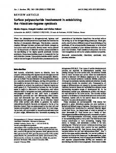
Figure 1. SEM images of a) P(NIPAM-ABP) side—as spun bilayer membrane, b) P(NIPAM-ABP) side—after treatment with hot water, c) TPU side—as spun bilayer membrane, and d) the cross section of the bilayer sample in water at 40 °C after drying shows a strong interface between TPU (left side) and P(NIPAM-ABP) (right side). Scale bar = 10 µm.
1022
wileyonlinelibrary.com
© 2015 The Authors. Published by WILEY-VCH Verlag GmbH & Co. KGaA, Weinheim
Adv. Funct. Mater. 2016, 26, 1021–1027
www.afm-journal.de www.MaterialsViews.com
FULL PAPER Scheme 1. Schematic showing the formation of bilayer actuators by sequential electrospinning of TPU and P(NIPAM-ABP) followed by UV crosslinking (steps 1 and 2). The samples were cut at different angles to get varied orientations of thermoresponsive P(NIPAM-ABP) fibers (steps 3 and 4). The TPU and P(NIPAM-ABP) solutions were mixed with rhodamine B (RB) and methylene blue, respectively, to observe the actuation phenomenon optically better.
significant shrinkage in the direction of fiber orientation on putting the fiber mat in water at 40 °C is due to the relaxation of the electrospun fibers to the lower energy coiled conformation (Figure 1). During fiber formation, the electrospun fibers are deposited in the stretched state due to very fast evaporation of solvent. This is a very well-known fact and this behavior is similar to the solvent-triggered shape-memory polymers and temperature-driven contraction of electrospun fibers.[2,27,28] Also, the oriented fibers are present in a layer-by-layer structure with many contact points between the fibers. The shrinkage in the direction of fiber orientation together with expulsion of water between the fibers lead to the slight expansion in the direction perpendicular to the fiber orientation. In dropping from 40 to 4 °C, the same mat in water below LCST is expected to be hydrophilically swollen in all directions. In fact, symmetric swelling in planar dimensions (length and width) was observed at 4 °C. The fiber mat showed about 38% expansion in length and width in comparison to the fiber mat
at 40 °C. If we compare the expanded dimensions at 4 °C to the as-spun fiber mat, the swelling is anisotropic. The increase in width was 58%, whereas the length reduced by about 9% in comparison to as-spun fibers. In brief, in going from the as-spun bilayer mat to the wet states, the shrinkage in length (direction of fiber orientation) dominates the increase in width above LCST, whereas expansion in width (perpendicular to the direction of fiber orientation) dominates the shrinkage in the length below LCST. The increase in thickness above and below LCST was 41% and 326%, respectively. The trend is just the opposite for P(NIPAM-ABP) membrane with fibers oriented perpendicular to the long axis of the sample (fiber orientation 90°). The membrane shrinks more in the width (direction of fibers) at 40 °C, whereas it swells more in the perpendicular direction at 4 °C (Table 1, Figure 2). The change in dimensions of a P(NIPAM-ABP) fibrous mat with fibers aligned at different angles in water at different temperatures was reflected in directionally controlled movement of a
Figure 2. Size change of cross-linked P(NIPAM-ABP) fibrous membrane (original size: 2.0 cm × 0.5 cm × 46 µm) in water at different temperatures.
Adv. Funct. Mater. 2016, 26, 1021–1027
© 2015 The Authors. Published by WILEY-VCH Verlag GmbH & Co. KGaA, Weinheim
wileyonlinelibrary.com
1023
www.afm-journal.de
FULL PAPER
www.MaterialsViews.com Table 1. Size change of pure P(NIPAM-ABP) fibrous membranes with fibers oriented at different angles (0°, 45°, and 90°) to the direction of the long axis of the sample in water at different temperatures (40 and 4 °C). L: length; W: width; D1 and D2: diagonal lengths. 0°
45°
40 °C
4 °C
As-spun
L [cm]
2.041
1.332
1.845
2.028
1.858
2.535
1.979
2.308
3.015
W [cm]
0.534
0.612
0.845
0.521
0.446
0.624
0.521
0.340
0.485
D1 [cm]
2.090
1.461
2.049
2.084
1.656
2.321
2.067
2.341
3.221
D2 [cm]
2.083
1.402
2.010
2.103
2.078
2.971
2.066
2.356
3.248
bilayer membrane with TPU. The thickness of the P(NIPAM-ABP) and TPU layers were 53 ± 9 µm and 18 ± 3 µm (Figure S4, Supporting Information), respectively, in bilayer membrane. TPU is used as a representative example for passive layer. It would also work with other hydrophobic polymers. At 40 °C (temperature above LCST), the P(NIPAM-ABP) layer with parallel orientation (0°) of fibers in the bilayer membrane shrinks in the direction of fiber orientation and, therefore, rolls sideways along the direction of fiber orientation with P(NIPAM-ABP) making inside surface of the rolls (Figure 3). On transferring the sample to water at 4 °C (below LCST), a directional change with opening of the rolls followed by re-rolling in the direction perpendicular to the fiber orientation with the P(NIPAM-ABP) layer now making the outside surface was observed. The rolling-re-rolling was reversible for many cycles without showing any sign of delamination. The bilayer interface as observed from the cross section of the sample is shown in Figure 1D. The actuation was ultra-fast, took ≈0.6 s from 4 to 40 °C, whereas the reversal of the process took ≈1.4 s
40 °C
90°
As-spun
4 °C
As-spun
40 °C
4 °C
(Figure 4, Movie S1, Supporting Information). Further experiments were carried out to study the actuation behavior at temperatures other than 40 and 4 °C. The bilayers showed sideways rolling along the direction of fiber orientation with P(NIPAM-ABP) making inside surface of the rolls at temperatures 25 °C and above. At 25 °C, the time required for rolling was about 15 s, whereas at temperatures 27 °C and above the process was very fast with complete rolling in less than 2 s (Figure S5, Supporting Information). The heat flow versus temperature transition in micro-DSC for P(NIPAM-ABP) was broad starting from 19 °C and the peak temperature (27 °C) was considered as LCST (Figure S3, Supporting Information). The opening of rolls followed by re-rolling in the direction perpendicular to the fiber orientation was possible at all temperatures below 19 °C. At temperature between 19 and 25 °C, the rolls simply open up but do not re-roll in another direction. The bilayer fibrous mat in hot water (above 27 °C) could lift ≈13.4 times mass of its own weight (55 mg was lifted by a bilayer membrane of 4.11 mg) (Figure S6, Supporting Information).
Figure 3. Fiber orientation-dependent actuation behavior of bilayer TPU (pink)/P(NIPAM-ABP) blue) fibrous membranes (length: 2.0 cm, width: 0.5 cm) in water at different temperatures. Black arrows show the fiber orientation direction, 0°, 45°, and 90° are angles between the fiber direction and the long axis of the sample, as indicated by a black dotted line on the sample. Scale bar = 0.5 cm. The TPU and P(NIPAM-ABP) were dyed to get a better contrast but in real photos and movies the contrast might not be visible as P(NIPAM-ABP) becomes transparent in contact with water.
1024
wileyonlinelibrary.com
© 2015 The Authors. Published by WILEY-VCH Verlag GmbH & Co. KGaA, Weinheim
Adv. Funct. Mater. 2016, 26, 1021–1027
www.afm-journal.de www.MaterialsViews.com
FULL PAPER Figure 4. Actuation time of the bilayer TPU/P(NIPAM-ABP) fibrous membrane with different orientations of fibers at 0°, 45°, and 90° with respect to the long axis of the sample (original length: 2.0 cm, width: 0.5 cm) in water at 40 and 4 °C, respectively. Scale bar = 0.5 cm.
When fibers were oriented at 90° (perpendicular to the long axis), similar control over direction, shape, and surface was observed. The difference was the sideways rolling (perpendicular to the direction of fiber orientation) at 4 °C with the P(NIPAM-ABP) layer on the outside (actuation time ≈4.0 s) and rolling along the direction of fibers at 40 °C with P(NIPAM-ABP) layer inside (actuation time ≈1.3 s) (Figures 3 and 4, Movie S2, Supporting Information). The P(NIPAM-ABP) mat with fibers oriented at 45° with respect to the long axis showed shrinkage along the fiber directions at 40 °C reflecting a very significant reduction in diagonal length D1 in comparison to D2 (Figure 2, Table 1). At 4 °C, the swelling took place in all directions almost symmetrically if we consider dimensions, in comparison to that of the sample at 40 °C. In comparison to the as-spun dry fiber, the swelling was asymmetrical, with more swelling in diagonal direction D2. Asymmetrical swelling and shrinkage was reflected in the twisting of the bilayer membrane with direction control. At 40 °C, the twisting followed shrinking parallel to the fiber orientation with right-handed twists leading to a tubular structure with P(NIPAM-ABP) as the inside layer. Whereas at 4 °C, swelling in a perpendicular direction led to the opening of the righthanded helix followed by rolling diagonally in the direction
Adv. Funct. Mater. 2016, 26, 1021–1027
perpendicular to the fiber orientation giving a helically rolled tubular structure, with P(NIPAM-ABP) as the outside layer. The complete inversion of the tube (inside out) took place in ≈0.7 s at 40 °C and ≈5 s at 4 °C (Figure 4, Movie S3, Supporting Information). Furthermore, fast actuation with similar directional control is possible for samples of different sizes as shown in Figure 5. The bigger sample requires more time for mass transport and therefore an increase in the time of actuation. Also, the curvature and the number of turns in a tubular coil can be controlled by changing the thickness ratio of the two layers. A thickness ratio (P(NIPAM-ABP):TPU) of 0.4 (sample size: 20 mm × 5 mm) for a sample with parallel arrangement of fibers (0° alignment) is not sufficient for making rolls/coils (Figure 6). The sample simply bended. The complete rolling took place at P(NIPAMABP):TPU thickness ratio ≈1.0. On increasing the thickness ratio, the coiling took place with increase in number of turns and curvature. Moreover, complex shapes can also be made by simply cutting the bilayer membranes into different patterns. As an example, one of the shapes provided a synthetic analogue of a Venus fly trap, mimicking only its movement pattern and actuation time in our case even with water vapors (Movie S4, Supporting Information).
© 2015 The Authors. Published by WILEY-VCH Verlag GmbH & Co. KGaA, Weinheim
wileyonlinelibrary.com
1025
www.afm-journal.de
FULL PAPER
www.MaterialsViews.com
Figure 5. Size-dependent actuation of the bilayered TPU/P(NIPAM-ABP) fibrous membrane (fibers are oriented at 90° with respect to the direction of long axis of the sample) (original size: length: 2.0 cm, width: 0.5 cm and length: 3.0 cm, width: 0.5 cm) in water at 40 and 4 °C, respectively. Scale bar = 0.5 cm.
3. Conclusion
actuators triggered by other stimuli in the future. The method is simple, versatile, and suitable for making large samples.
In conclusion, we show the first ever large size thermoresponsive polymeric actuator with directionally controlled reversible movements with very high speed. The movements led to the formation of tubes, coils, and helices, reversibly changing their shapes and surface sides with temperature. The inherent porosity, deposition of electrospun fibers in stretched state, and alignment in a simple way by collecting fibers on a rotating wheel provided the desired all-in-one actuators. Based on this concept, the appropriate combination of materials in bilayers can also provide polymeric
4. Experimental Section Materials: Cross-linkable PNIPAM (Mn = 12 × 104 g mol−1, Mw = 41 × 104 g mol−1) was made by free-radical copolymerization of NIPAM and photo cross-linker ABP, designated as (P(NIPAM-ABP) according to the previous report.[26] TPU (Desmopann DP 2590, Bayer Materials Science, Mn = 8.89 × 10⁴ g mol−1, Mw = 14.5 × 10⁴ g mol−1) and N,N′-dimethylformamide (DMF, 99.8%, Aldrich) were used as received.
Figure 6. Thickness ratio (PNIPAM-ABP/TPU) dependent bending of 0°-bilayered TPU/P(NIPAM-ABP) fiber mats (20 mm × 5 mm). a) Cross section of samples with different thickness ratio (pink side is TPU; scale bar = 50 µm); b) in water at different temperatures (scale bar = 5 mm); c) determination of radius (r) (scale bar = 2 mm); and d) plot of curvature against thickness ratio of P(NIPAM-ABP)/TPU. The curvature is taken as 1/r. The bended arch is assumed as an imaginary circle for the determination of r and for coils the radius of the inner most circle is taken for comparison purpose.
1026
wileyonlinelibrary.com
© 2015 The Authors. Published by WILEY-VCH Verlag GmbH & Co. KGaA, Weinheim
Adv. Funct. Mater. 2016, 26, 1021–1027
www.afm-journal.de www.MaterialsViews.com
dFa =
3 cos 2 θ − 1 2
(1)
where θ is the angle the individual fiber forms with the preferred direction controlled by the rotating collector. The values given are an average of a minimum of 100 fibers. The porosity (P) of the electrospun nanofiber mat was calculated using Equations (2)–(4)
ρ mat =
massmat area mat × thicknessmat
(2)
ρ film =
mass film area film × thickness film
(3)
ρ P = ⎛⎜ 1− mat ⎞⎟ × 100% ⎝ ρ film ⎠
(4)
where ρmat and ρfilm are the density of the pressed UV cross-linked P(NIAPM-ABP) electrospun fibrous membrane (mat) and film, respectively.
Supporting Information Supporting Information is available from the Wiley Online Library or from the author.
Acknowledgements L.L. and S.J. contributed equally to this work. The authors would like to acknowledge financial support from Deutsche Forschungsgemeinschaft (DFG). The work was carried out in the frame of SFB 840. L.L. thanks
Adv. Funct. Mater. 2016, 26, 1021–1027
China Scholarship Council for awarding fellowship for carrying out Ph.D. in Germany in the lab of Prof. Seema Agarwal. Received: August 26, 2015 Revised: November 8, 2015 Published online: December 28, 2015
FULL PAPER
Preparation of Fibrous Membranes: The pure TPU, P(NIPAM-ABP), and bilayer fibrous membranes were prepared by electrospinning of the corresponding polymers from DMF. The spinning conditions are given in Table S1 (Supporting Information). The TPU spinning solution had 4 wt% of UV cross-linker ABP, with respect to the weight of TPU, to cross-link the two layers at the interface in bilayer membranes. An amount of 0.4 wt% of Rhodamine B (RB) and methylene blue (MB), with respect to the weight of the corresponding polymer, were added to the TPU and P(NIPAM-ABP) solutions, respectively, to obtain color contrast in the bilayer. A rotating disc (diameter 20 cm, disc rim 4 cm) with a rotation speed of 850 rpm was used as a collector and provided parallel-aligned fibers. The bilayer TPU/P(NIPAM-ABP) aligned-fiber mat was made by sequential spinning of the two polymer solutions, i.e., first, pure TPU was spun, followed by the spinning of P(NIPAM-ABP). Subsequently, the bilayer nanofiber mats were pressed at 300 bar for 20 min at room temperature and cross-linked by UV light (Honle UVAHAND 250 GS) for 4 h for each side. Characterization: A scanning electron microscope (SEM) (Zeiss Leo 1530) was used to observe the morphology and diameter of fibers (Image J software). Before scanning, 3.0 nm of platinum (Pt) coating was applied to increase the conductivity of the samples. All the photos of the actuation process were captured by Video Remaker software from the corresponding videos. Micro-DSC was measured on a Setaram Micro-DSC III at a heating/cooling rate of 0.25 °C min−1. The molecular weight of P(NIPAM-ABP) was determined by gel permeation chromatography using DMF as the eluent at a flow rate of 0.5 mL min−1 at 25 °C. The degree of fiber alignment (dFa) was calculated according to the previous literature using the following equation[29]
[1] Y. Osada, H. Okuzaki, H. Hori, Nature 1992, 355, 242. [2] Q. Zhao, J. W. C. Dunlop, X. Qiu, F. Huang, Z. Zhang, J. Heyda, J. Dzubiella, M. Antonietti, J. Yuan, Nat. Commun. 2014, 5, 4293. [3] S. Taccola, F. Greco, E. Sinibaldi, A. Mondini, B. Mazzolai, V. Mattoli, Adv. Mater. 2015, 27, 1668. [4] S. Iamsaard, S. J. Aßhoff, B. Matt, T. Kudernac, J. J. L. M. Cornelissen, S. P. Fletcher, N. Katsonis, Nat. Chem. 2014, 6, 229. [5] X. Zhang, Z. Yu, C. Wang, D. Zarrouk, J.-W. T. Seo, J. C. Cheng, A. D. Buchan, K. Takei, Y. Zhao, J. W. Ager, J. Zhang, M. Hettick, M. C. Hersam, A. P. Pisano, R. S. Fearing, A. Javey, Nat. Commun. 2014, 5, 2983. [6] H. Yu, T Ikeda, Adv. Mater. 2011, 23, 2149. [7] J. Kim, S. E. Chung, S.-E. Choi, H. Lee, J. Kim, S. Kwon, Nat. Mater. 2011, 10, 747. [8] S. J. Kim, M. S. Kim, S. I. Kim, G. M. Spinks, B. C. Kim, G. G. Wallace, Chem. Mater. 2006, 18, 5805. [9] K. Zhang, A. Geissler, M. Standhardt, S. Mehlhase, M. Gallei, L. Chen, C. M. Thiele, Sci. Rep. 2015, 5, 11011. [10] M. Ma, L. Guo, D. G. Anderson, R. Langer, Science 2013, 339, 186. [11] A. Sidorenko, T. Krupenkin, A. Taylor, P. Fratzl, J. Aizenberg, Science 2007, 315, 487. [12] R. M. Erb, J. S. Sander, R. Grisch, A. R. Studart, Nat. Commun. 2013, 4, 1712. [13] F. Liu, S. Jiang, L. Ionov, S. Agarwal, Polym. Chem. 2015, 6, 2769. [14] E. T. Roche, R. Wohlfarth, J. T. B. Overvelde, N. V. Vasilyev, F. A. Pigula, D. J. Mooney, K. Bertoldi, C. J. A Walsh, Adv. Mater. 2014, 26, 1200. [15] C. Ohm, M. Brehmer, R. Zentel, Adv. Mater. 2010, 22, 3366. [16] Z. Hu, X. Zhang, Y. Li, Science 1995, 269, 525. [17] L. Ionov, e-Polymers 2014, 14, 109. [18] G. Stoychev, S. Turcaud, J. W. C. Dunlop, L. Ionov, Adv. Funct. Mater. 2013, 23, 2295. [19] Z. L. Wu, M. Moshe, J. Greener, H. Therien-Aubin, Z. Nie, E. Sharon, E. Kumacheva, Nat. Commun. 2013, 4, 1586. [20] L. T. de Haan, C. Sánchez-Somolinos, C. M. W. Bastiaansen, A. P. H. J. Schenning, D. J. Broer, Angew. Chem., Int. Ed. 2012, 51, 12469. [21] W. E. Lee, Y. J. Jin, L. S. Park, G. Kwak, Adv. Mater. 2012, 24, 5604. [22] S. Armon, E. Efrati, R. Kupferman, E. Sharon, Science 2011, 333, 1726. [23] D. Kuckling, J. Hoffmann, M. Plötner, D. Ferse, K. Kretschmer, H.-J. P. Adler, K.-F. Arndt, R. Reichelt, Polymer 2003, 44, 4455. [24] L.-W. Xia, R. Xie, X.-J. Ju, W. Wang, Q. Chen, L.-Y. Chu, Nat. Commun. 2013, 4, 2226. [25] C. O. Baker, B. Shedd, P. C. Innis, P. G. Whitten, G. M. Spinks, G. G. Wallace, R. B. Kaner, Adv. Mater. 2008, 20, 155. [26] S. Jiang, F. Liu, A. Lerch, L. Ionov, S. Agarwal, Adv. Mater. 2015, 27, 4865. [27] C. Azra, D. Alhazov, E. Zussman, Polymer 2015, 58, 162. [28] R. Xiao, J. Guo, D. L. Safranski, T. D. Nguyen, Soft Matter 2015, 11, 3977. [29] R. Dersch, T. Liu, A. K. Schaper, A. Greiner, J. H. Wendorff, J. Polym. Sci., Part A: Polym. Chem. 2003, 41, 545.
© 2015 The Authors. Published by WILEY-VCH Verlag GmbH & Co. KGaA, Weinheim
wileyonlinelibrary.com
1027