www.nature.com/scientificreports
OPEN
received: 16 March 2016 accepted: 13 July 2016 Published: 10 August 2016
Gradient moduli lens models: how material properties and application of forces can affect deformation and distributions of stress Kehao Wang, Demetrios Venetsanos, Jian Wang & Barbara K. Pierscionek The human lens provides one-third of the ocular focussing power and is responsible for altering focus over a range of distances. This ability, termed accommodation, defines the process by which the lens alters shape to increase or decrease ocular refractive power; this is mediated by the ciliary muscle through the zonule. This ability decreases with age such that around the sixth decade of life it is lost rendering the eye unable to focus on near objects. There are two opponent theories that provide an explanation for the mechanism of accommodation; definitive support for either of these requires investigation. This work aims to elucidate how material properties can affect accommodation using Finite Element models based on interferometric measurements of refractive index. Gradients of moduli are created in three models from representative lenses, aged 16, 35 and 48 years. Different forms of zonular attachments are studied to determine which may most closely mimic the physiological form by comparing stress and displacement fields with simulated shape changes to accommodation in living lenses. The results indicate that for models to mimic accommodation in living eyes, the anterior and posterior parts of the zonule need independent force directions. Choice of material properties affects which theory of accommodation is supported. The eye lens together with the cornea are the optical elements of the eye and refract light to the retina. Whilst the cornea provides two-thirds of the refractive power, the lens is able to adjust its power enabling the eye to focus over a range of distances. This is accomplished by the lens changing shape, a process referred to as accommodation. With age, this accommodating ability gradually diminishes, reducing the near range of focus so that by the sixth decade of life, the eye is no longer able to focus on close objects; this loss of function is termed presbyopia1–3. There remains a paucity in the understanding of accommodation and hence of how the relevant components of the eye contribute to the functional loss. Currently there are two opposing theories that purport to explain the accommodative mechanism1–3. The significant difference between these theories lies in the mediation of forces in the zonule and the tensions imparted on the different parts of the zonule during accommodation1–3. The zonule encircles the equator of the lens and is composed of fibrils that run from the capsule, within which the lens is located, to different parts of the ciliary muscle and are broadly divided into anterior, posterior and equatorial sections1–3. The oldest and more established theory, is that of Helmholtz1, who postulated that as the lens changes shape to focus on distant objects, the increased tension in the zonular fibres stretches the lens to a flatter shape to decrease optical power. Conversely, when the lens focusses on objects at close range, decreased tension in zonular fibres allows the lens to assume a more curved shape providing an increase in optical power; the latter is considered a more relaxed form for the lens1. Schachar2,3 hypothesised that the accommodated lens (the form the lens assumes for near vision) requires an increase in tension in the equatorial zonule that causes the lens to assume a spindle shape as the central surfaces become more curved and the peripheral surfaces flatten2,3. As it is not possible to examine the zonular movements in a living human eye because the iris blocks the view of the equatorial region of the lens, computational modelling, using advanced methods based on measurements of human lenses, offers a means of studying the action of the zonule during accommodation. Comparing the results of such models with those obtained from clinical measurements on live eyes allows an optimisation of model parameters to obtain a Faculty of Science Engineering and Computing, Penrhyn Road, KT1 2EE, Kingston-upon-Thames, UK. Correspondence and requests for materials should be addressed to B.K.P. (email:
[email protected])
Scientific Reports | 6:31171 | DOI: 10.1038/srep31171
1
www.nature.com/scientificreports/ physiologically relevant model that will provide a greater understanding of zonular behaviour during accommodation and what may determine the functional loss with age. The accuracy of any computational model relies on the data used to construct it, in this case on both geometric parameters of the components of the eye involved in accommodation and on the mechanical properties of the lens. The geometrical parameters of curvature and thickness can be obtained from live eyes using clinical biomicrosopic means4–7. Since the lens grows throughout life, with age, its thickness increases and the central anterior and posterior radii of curvature decrease4–7. Material properties have been much harder to ascertain. In vitro studies, either with respect to age or across the lens, differ greatly8–14. The seminal work of Fisher provides values of Young’s moduli for both the lens substance and the lens capsule over a wide age range8,15. Subsequent studies have reported different trends and values9–14. Such divergence in measurements may arise from different measurement techniques and inherent assumptions. There is a varying protein distribution across the lens resulting in the gradient refractive index that contributes to the high degree of image quality16. The linear relationship between protein concentration and refractive index is well understood16,17. The material properties across the lens also vary, as experimental studies9,11 have shown, but the correlation between these changes and the protein concentration is still unclear. Recently, in vivo studies using Brillouin optical microscopy have reported profiles of elasticity along the lens central axis18,19 that mimic the gradient of refractive index16,17,20, suggesting linear relationships may also exist between mechanical and optical properties. Finite Element Analysis (FEA) models can be used to test the veracity of measured material properties and a number of FEA models have been suggested21–28. The present study proposes the most biologically accurate FEA lens models to date, using lens curvatures and material gradients based on experimental data20 to test different material properties proposed in previous work8,12,15. The effect of changes in elastic moduli of the lens substance, different levels of stretching forces provided by zonular fibres and various stretching angles of zonular fibres are analysed to determine how they affect the anterior and posterior curvatures of the lens approximated by spheres and conicoids of revolution to determine which theory of accommodation is supported, and how the model results compare to studies on living eyes.
Results
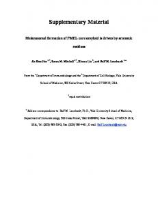
Stress patterns. Models incorporating Fisher’s data. Stress patterns for the three different ages of lens mod-
els with single values for the cortical modulus are shown in the Fig. 1(a–c). The corresponding stress values of each model are indicated using colour coding (as von Mises stress in MPa) with the colour bar on the left side. The lens nucleus indicated in dark blue has minimal stress values for each lens model. There is a high stress region indicated in green and yellow colours concentrated near the nuclear region of the equator, which is especially marked in the 16-year-old lens. The anterior stress is higher than the posterior stress for all three models. Figure 1(d–f) shows the three aged models in which the lens cortex has been divided into layers of different moduli based on the gradient index measurement of Bahrami et al.20. The cortex in the smallest of these lens models, based on the 16-year-old lens20, is divided into four layers; the cortical regions in the other two lenses have five layers. Young’s moduli for each model increases by 0.6 MPa increment from the innermost to the outermost layer, maintain a mean value of all layers that is in accordance with the values reported by Fisher8; Young’s moduli of the nucleus is constant. Layered stress patterns are observed in the anterior and posterior cortical regions of the 16-year-old lens, and largely in the posterior cortical region for the 35-year-old and the 48-year-old lenses. The stresses at the equatorial cortical region are more evenly distributed than in the single cortex models. Dividing the cortex further into more layers whilst maintaining the same mean value as reported by Fisher8: ten layers for the 16-year-old lens model, twelve layers for the 35-year-old lens model and eleven layers for the 48-year-old lens model, in accordance with the contours of refractive index20 gives results shown in Fig. 1(g–i). The stresses are more evenly distributed in all three lens models from this set than in the models with the single value of cortical modulus (Fig. 1a–c) and in those with fewer layers (Fig. 1d–f). Models incorporating Wilde et al. ’s data. The stress patterns for lens models incorporating material properties reported by Wilde et al.12 are displayed in Fig. 2. For single cortex models, the 16-year-old and the 35-year-old lenses display similar stress patterns to those using material properties of Fisher8 (Fig. 2a,b). The 48-year-old lens models, however, shows greater differences depending on the moduli used; the model using the elastic moduli of Wilde et al.12 has a high stress region in the equatorial region of the nucleus with minimal stresses in dark blue colour appearing in the posterior cortex (Fig. 2c). For models with multiple layers (following the same layered format used to apply the material properties measured by Fisher8) the resulting stress patterns are shown in the Fig. 2(d–i). The 16-year-old and 35-year-old lens models do not show the jagged edged stress patterns in the posterior cortex that are seen in the models with the material properties of Fisher8 (Fig. 1d–i). The stresses are lower and more diffuse in the models with the material properties of Wilde et al.12 compared to those of Fisher8. Conversely, the two 48-year-old lens models with multiple cortical layers have regions of higher stress particularly in the nuclear equatorial region compared to the respective models using the properties measured by Fisher8.
Lens deformations. The models were subjected to simulated stretching forces of 0.08 N. The sagittal defor-
mations of the nucleus and cortex are listed in Table 1 for the single cortex models as the representative forms since, in terms of deformation, multi-layer models demonstrate similar changes to corresponding single cortical modulus models. Changes in thickness of the nucleus are larger than those of the cortex for all listed models. All three aged lens models using material properties of Fisher8 and the two younger lens models using material properties of Wilde et al.12 have lower elastic moduli in the nucleus than in the cortex. The deformation of the nucleus
Scientific Reports | 6:31171 | DOI: 10.1038/srep31171
2
www.nature.com/scientificreports/
Figure 1. Stress patterns (as von Mises stress in MPa = N/m2 × 106) for human lens models aged 16, 35 and 48 based on optical measurements20 and using material properties of Fisher8.
in these models is almost three to six times larger than that of the cortex. The 48-year-old model using material properties of Wilde et al.12 has higher elastic moduli in the nucleus than in the cortex; compared to the other age models, this oldest model shows the least deformation. The displacements of the anterior pole, posterior pole and equator (as illustrated in Fig. 3) for all the lens models are listed in Table 2. With age, the displacements of the poles and the equator decrease. For the two younger lenses, the anterior pole has a greater displacement than the posterior pole for both sets of material properties in single and multi-layer models. There are no significant changes in displacement values when comparing single to multi-layer models (Table 2). Table 3 gives the central radii of curvature of the lens anterior and posterior surfaces for all the models, including both the anterior and posterior surfaces for the undeformed state and for a deformation caused by a cumulative force of 0.08 N. For the spherical surface approximation, the two younger lens models constructed with the material properties of Fisher8 and the youngest lens model with the material properties of Wilde et al.12 show an increase in anterior central radii of curvature with stretching. For all other models the anterior lens surface becomes steeper as the model is stretched. The posterior surfaces for all the cases become flatter with stretching. When a conicoid of revolution is used to represent the lens surfaces, only the youngest lens model shows an increase in anterior central radius of curvature with stretching with both sets of material properties. The two older lens models have a decrease in anterior radii of curvature with stretching. The posterior central radii of curvature of all the three aged models using the material properties of Fisher8 increase with stretching. Using the material porperties of Wilde et al.12, the two younger lens models show a decrease and the oldest lens model an increase in posterior central radii of curvature with stretching. Except for the posterior surface of the 48-year-old lens which has a conic constant